

 PDF
PDF ePub
ePub Citation
 Print
Print


Introduction
Inflammation is a complex biological response to pathogens and damaged cells in the human body [1]. Chronic and uncontrolled inflammation can be common in various diseases, such as cardiovascular diseases, autoimmune rheumatoid arthritis, systemic lupus erythematosus, cancer, and Alzheimer's or Parkinson's diseases [2-5]. Many transcriptional factors, inflammatory cytokines, enzymes, and other mediators have been shown to be related to these effects [6]. Macrophages participate in the autoregulatory loop of the inflammatory process. Therefore, therapeutic interventions controlling various inflammatory diseases target macrophages and their products [7].
Lipopolysaccharide (LPS) is a major component of the cell walls of Gram-negative organisms. LPS activates immune cells to upregulate inflammatory responses and causes the production of pro-inflammatory cytokines [8]. Nitric oxide (NO) reflects the degree of LPS-induced inflammation in macrophages [9]. During the inflammatory process, NO is produced by nitric oxide synthase (iNOS), which plays a regulatory role in the expression of pro-inflammatory mediators [10,11]. NO is also reported to affect the activity of cyclooxygenase-2 (COX-2) [12]. Prostaglandins (PGs), the product of COX-2, are stimulated by LPS and cytokines and can contribute to the pain and swelling associated with inflammation [13]. Furthermore, LPS-stimulated inflammation can be identified by pro-inflammatory cytokines, including tumor necrosis factor (TNF)-α, interleukin 6 (IL-6), and interleukin 1β (IL-1β), which are related to the induction of insulin resistance and the development of Type 2 diabetes mellitus (T2DM) [14].
Hawthorn fruit is a traditional medicine used in treating the cardiovascular diseases, and angina pectoris of coronary heart disease [15,16]. Recent reports have shown that hawthorn fruit exhibits anti-oxidant, anti-atherosclerotic, and hypolipidemic activities [17-20]. We previously reported on the antioxidant and anti-inflammatory properties of the methanolic extract of hawthorn fruit [21,22]. In this study, our objective was to investigate the mechanisms behind the anti-inflammatory activities of the water fractionated portion of hawthorn fruit on RAW 264.7 cells.
Materials and Methods
We collected hawthorn fruits (Crataegus pinnatifida Bunge var. typica Schneider) from native trees in Chuncheon, Korea in autumn, 2008. The fresh fruits were air-dried and then ground into fine powder. Fifty grams of powder was extracted twice with one liter of 70% methanol. The extract was filtered with vacuum filtration (100-mm; Whatman, Maidstone, UK). The solvent was evaporated under reduced pressure using a vacuum rotary evaporator (CCA-1110; Eyela, Tokyo, Japan). Coarse extract was successively extracted with dichloromethane, ethyl acetate, n-butanol, and water in a continuous Soxhlet extractor. Among the extracted fractions, the water fraction exhibited significantly more anti-inflammatory activity than the others (data not shown), and so the water fraction of the hawthorn fruit was used for further analysis.
Cell line and cell culture
Murine macrophage RAW 264.7 cell lines were purchased from the Korean Cell Line Bank (Seoul, Korea) and grown in Dulbecco's modified Eagle's medium (DMEM) supplemented with 10% fetal bovine serum, 100 U/ml penicillin, and 100 µg/ml streptomycin. Cells were cultured in a humidified atmosphere and incubated at 37℃ in 5% CO2.
Treatment of cells with hawthorn fruit
Exponentially growing RAW 264.7 cells were plated at a density of 105 cells/well in 6- and 96-well microplates in culture medium and allowed to adhere for 16 h before treatment. Hawthorn fruit water fraction was added to the cultures at final concentrations of 200, 400, and 600 µg/ml with 2 µg/ml lipopolysaccharide (LPS) for 24 h.
MTT (1-(4,5-dimethylthiazol-2-yl)-3,5-diphenylformazan) assay
Cell viability was evaluated with a conventional MTT assay. At 24 h prior to culture termination, 20 µl of the MTT solution (4 mg/ml in a phosphate-buffered saline (PBS), pH 7.4) was added to each well, and the cells were continuously cultured for 4 h. Then 200 µl of dimethyl sulfoxide (DMSO) was added for solubilization. Absorbance was measured at 550 nm using an enzyme-linked immunosorbent assay (ELISA) plate reader (Bio-Tek, Winooski, VT, USA).
Nitric oxide (NO) determination
The level of NO production in cell culture supernatants was determined using a colorimetric assay based on the Griess reaction [23]. Aliquots of 100 µl of supernatants were mixed with 100 µl Griess reagent (50 µl of 1% sulfanilamide in 5% phosphoric acid and 50 µl of 0.1% naphthyl-ethylenediamine dihydrochloride). After 10 min, the absorbance was determined at 550 nm with an ELISA.
Total RNA extraction and reverse transcription polymerase chain reaction (RT-PCR)
The total RNA from the LPS-stimulated RAW264.7 cells was prepared with a TRIzol RNA isolation kit (Invitrogen, Carlsbad, CA, USA) and stored at -80℃ until required. One µg of the RNA was reverse-transcribed into cDNA and used as a template for RT-PCR amplification. The primers and the amplification conditions are listed in Table 1. PCR was performed with a DNA gene cycler, and the amplification was followed by denaturation at 94℃ for 30 s, annealation at 55℃ for 30 s, and primer extension a 72℃ for 45 s. PCR products were analyzed on 1% agarose gels, and bands were visualized with ethidium bromide staining and a Mini BIS image analysis system (DNR Bio-Imaging Systems Ltd., Kiryat Anavim, Israel). A densitometric analysis was preformed with image analysis software (Quantity one; Bio-Rad, Hercules, CA, USA).
Statistical analysis
All tests were carried out independently in triplicate (n = 3), and the data are expressed as the mean ± standard derivation (SD). Statistical significance was evaluated with analysis of variance (ANOVA) using SPSS 16.0 (SPSS Inc., Chicago, USA). Results were considered to be significant when P < 0.05.
Results
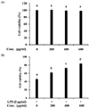
To investigate the cytotoxicity of the hawthorn fruit water fraction, we incubated RAW 264.7 cells with different dosages of the hawthorn fruit water fraction for 24 h. Cell viability did not seem to be affected for hawthorn fruit water fraction concentrations up to 600 µg/ml, as observed with an MTT assay (Fig. 1A). On the contrary, the hawthorn fruit water fraction protected the RAW 264.7 cells from LPS-induced apoptosis (Fig. 1B). 45.7% of cells were viable after stimulation with LPS for 24 h. However, after treatment with 200, 400, and 600 µg/ml of the hawthorn fruit water fraction, the viability of the RAW cells increased to 61.8, 72.7, and 83.4%, respectively.
Inhibition NO production in LPS-stimulated RAW 264.7 cells
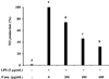
The effect of the hawthorn fruit water fraction on NO inhibition was determined by treating the RAW264.7 cells with it in the presence or absence of LPS stimulation. The cells that were stimulated with LPS produced significant levels of NO in conditioned medium (Fig. 2). The level of NO production induced by LPS in RAW cells decreased significantly in a dose-dependent manner when treated with different concentrations of the hawthorn fruit water fraction.
Suppression of iNOS, COX-2, TNF-α, IL-1β, and IL-6 mRNA expression in LPS-stimulated RAW264.7
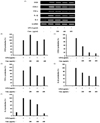
To determine if the above effect on NO production was related to the changes in the levels of iNOS, the expression of iNOS mRNA was measured with RT-PCR (Fig. 3A). Glyceraldehyde-3-phosphate dehydrogenase (GAPDH), the most commonly used housekeeping gene, was used to normalize gene expression in this study. The cells treated with hawthorn fruit water fraction exhibited stronger iNOS presentation than the control (Fig. 3B). However, cells treated with LPS along with the hawthorn fruit water fraction significantly inhibited COX-2 mRNA expression in a dose-dependent manner. Expression levels of COX-2 declined by 45.5, 80.2, and 85.7% when LPS-stimulated cells were treated with 200, 400, and 600 µg/ml of the hawthorn fruit water fraction, respectively (Fig. 3C).
We also examined the expression of several pro-inflammatory cytokines, such as TNF-α, IL-1β, and IL-6 mRNA. High concentrations of the hawthorn fruit water fraction inhibited TNF-α mRNA. Cells exhibited 60.2% of TNF-α mRNA when incubated with 600 µg/ml of the hawthorn fruit water fraction, as compared to the LPS-stimulated cells (Fig. 3D). A hawthorn fruit water fraction concentration of 200 µg/ml did not inhibit TNF-α mRNA expression.
The hawthorn fruit water fraction also attenuated the LPS-stimulated expression of IL-1β and IL-6 mRNA. At a dosing rate of 400 µg/ml of the water fraction of hawthorn fruit, control group cells showed reductions in expression of IL-1β and IL-6 by 44.8 and 34.4%, respectively, as compared to the LPS-stimulated group (Fig. 3E and Fig. 3F). When the dosage of hawthorn fruit water fraction was increased to 600 µg/ml, no further reduction in the expression of IL-16 was noted; contrastingly, the expression of IL-6 decreased sharply.
Discussion
Hawthorn fruit, used in traditional medicine, is considered to be safe in existing pharmacological and toxicological studies [15]. In previous studies, we showed that the methanolic extracts of hawthorn fruit possess anti-inflammatory properties, achieved through the inhibition of LPS-induced NO production in RAW 264.7 cells [22]. In the present study, we examined the potential anti-inflammatory effects and underlying molecular mechanisms of hawthorn fruit water fraction from 70% methanolic extract in an LPS-stimulated inflammatory model using RAW 264.7 cells.
NO is an important mediator and regulator of inflammatory responses [24]. In an inflammatory response, the overproduction of NO, a highly reactive molecule, reacts with superoxide, creating cytotoxicity and tissue damage in an organism [25]. NO-induced oxidative stress is associated with many diseases, such as septic shock, atherosclerosis, Alzheimer's disease, diabetes, and Parkinson's disease [26,27]. The down-regulation of NO has been used to treat such diseases. In the current study, hawthorn fruit water fraction was shown to inhibit the production of NO in LPS-stimulated RAW 264.7 cells in a dose-dependent manner. The hawthorn fruit water fraction also protected the RAW 264.7 cells against NO-induced toxicity and damage. In addition, the hawthorn fruit water fraction was quite safe at different concentrations. Thus, we considered the hawthorn fruit water fraction to have potential properties for preventing NO-related diseases.
Macrophages, important components in the human immune defense system, respond actively to inflammation by releasing pro-inflammatory cytokines, such as iNOS, COX-2, TNF-α, IL-12, IL-1β, and IL-6, and other inflammatory factors, such as NO and PGE2 [28]. These cytokines are playing a principal role in inflammatory diseases and processes [29]. Usually, activated inflammatory cells produce high quantities of NO, which in turn produce iNOS. In the present study, the water fraction from hawthorn fruit inhibited NO production but did not change the iNOS mRNA levels which produced by LPS stimulation. These unaffected levels of iNOS mRNA could be attributed to the state of macrophage differentiation, the complex of stimuli, and tissue location, which are dependent on iNOS expression [30]. Previously, our group reported that hawthorn fruit is a NO free radical scavenger [31]. The reduction of iNOS-catalyzed NO production could attribute to the NO free radical scavenging activity of the hawthorn fruit water fraction. NO is necessary for maintaining prolonged COX-2 gene expression, a central mediator in inflammation [32]. All of the steps that involve COX-2 gene expression are considered to be potential therapeutic targets [33]. In the present study, hawthorn fruit water fraction suppressed COX-2 mRNA expression, supporting its anti-inflammatory potential.
TNF-α is a key mediator in defense responses and the induction of apoptosis [34]. It can stimulate the production or expression of IL-6, IL-1β, PGE2, collagenase, and adhesion molecules, eliciting a number of physiological effects, including septic shock, inflammation, and cytotoxicity [35]. IL-6 is a well-known pro-inflammatory cytokine, regarded as an endogenous mediator of LPS-induced fever [36]. IL-1β is considered to be another pivotal pro-inflammatory cytokine, primarily released by macrophages, and plays a role in the pathophysiology of rheumatoid arthritis [37]. In this study, TNF-α, IL-1β, and IL-6 production in LPS-stimulated RAW 264.7 cells was significantly inhibited when the cells were incubated with increasing concentrations of hawthorn fruit water fraction. The decrease in production of TNF-α, IL-1β, and IL-6 seemed to attenuate cytokine-mediated host-destructive processes in inflammatory tissues.
In conclusion, our present findings indicate that hawthorn fruit water fraction is a potent inhibitor of LPS-induced NO production and COX-2, TNF-α, IL-1β, and IL-6 gene expression in RAW 264.7 cells. The hawthorn fruit water fraction did not inhibit iNOS mRNA expression, and it had relatively different mechanisms compared to its methanolic extract [22,38,39]. The difference may relate to the respective active mechanism of diverse compounds. Additional studies of the active compounds in hawthorn fruit water fraction are currently underway.

 XML Download