

 PDF
PDF ePub
ePub Citation
 Print
Print


Introduction
Among natural antioxidants, flavonoids are most abundant in our daily foods. The common structure of flavonoids is the flavan nucleus, which consists of 15 carbon atoms arranged in three rings (phenylchromanone structure, C6-C3-C6). Rings A and B are benzene rings and ring C is a heterocyclic pyran or pyrone.
The flavonols kaempferol, myricetin, and quercetin are members of the flavonoid subclass and are found in many foods, including onions and tea (Miean & Mohamed, 2001; Ross & Kasum, 2002). Although the predominant component of these major flavonols in edible plants is quercetin, followed by myricetin and kaempferol (Miean & Mohamed, 2001), all three have antioxidant capacity (de Vries et al., 1998; Marfak et al., 2003; Park et al., 2006). Nevertheless, kaempferol has been relatively unnoticed in comparison with quercetin or myricetin, and a few in vitro studies have assessed it.
Recently, it was found that the flavonoids possessing antioxidant properties offer new, possible strategies for cancer chemotherapy. It has been reported that antioxidant enzyme activation by kaempferol may play an important role in apoptosis in human lung non-small carcinoma H460 cells (Leung et al., 2007). In addition, many studies have reported that kaempferol significantly inhibits cancer cell growth in vitro (Bestwick et al., 2007; Casagrande & Darbon, 2001; Knowles et al., 2000) and that this growth inhibition stimulates the signal transduction pathway leading to apoptosis. It has been shown to cause apoptosis in various cells, such as leukemia cells, lung cancer cells and glioblastoma (Chen et al., 2005; Leung et al., 2007; Nguyen et al., 2003a; Nguyen et al., 2003b), although the cellular mechanisms underlying the action of kaempferol- induced cell cycle arrest and apoptosis are still unclear.
Therefore, the present study was performed to investigate the effects of kaempferol on cell proliferation and apoptosis and explored the mechanism for these effects in human breast carcinoma MDA-MB-453 cells.
Materials and Methods
The cell culture and kaempferol treatment
Human breast cancer MDA-MB-453 cells were purchased from the KCLB (Korean Cell Line Bank, Korea). Cells were routinely maintained in RPMI 1640 (Invitrogen [Molecular Probes], Gibco, Carlsbad, CA, USA), supplemented with 10% FBS and antibiotics (50 U/ml of penicillin and 50 µg/ml streptomycin, Gibco) at 37℃ in a humidified atmosphere containing 5% CO2. Cells were treated with kaempferol ranging from 1 to 200 µM and incubated for 24 and 48 hrs. Kaempferol was purchased from Sigma and dissolved in DMSO (final concentration 0.1% in medium).
Cell viability assay
Cell viability was determined using the MTT assay. At 24 and 48 hrs point, the cells exposed to kaempferol were added to methyl thiazolyl tetrazolium (MTT). Four hours later, DMSO was added to each well to dissolve the resulting formazan crystals and then absorbance was recorded at 540 nm in a microplate reader (SpectraMax Plus; Molecular Devices).
Cell cycle distribution
Cells were then harvested, washed with cold PBS, and processed for cell cycle analysis. Briefly, the cells were fixed in absolute ethanol. The fixed cells were centrifuged at 1,000 rpm and washed with cold PBS twice. RNase A (20 µg/ml final concentration) and propidium iodide staining solution (50 µg/ml final concentration) were added to the cells and incubated for 30 min at 37℃ in the dark. The cells were analyzed with a FACS Calibur instrument (BD Biosciences, San Jose, CA, USA) equipped with CellQuest 3.3 software. ModFit LT 3.1 trial cell cycle analysis software was used to determine the percentage of cells in the different phases of the cell cycle.
Immunoblotting assay
Cells were lysed in RIPA buffer (1% NP-40, 150 mM NaCl, 0.05% DOC, 1% SDS, 50 mM Tris, pH 7.5) containing protease inhibitor for 1 hrs at 4℃. The supernatant was separated by centrifugation, and protein concentration was determined by Bradford protein assay kit II (Bio-Rad). Proteins (25 µg/well) denatured with sample buffer were separated by 10% SDS-polyacrylamide gel. Proteins were transferred onto nitrocellulose membranes (0.45 µm). The membranes were blocked with a 1% BSA solution for 3 hrs and washed twice with PBS containing 0.2% Tween-20, and incubated with the primary antibody overnight at 4℃. Antibodies against p53, phosphorylated-p53 (serine 6, 9, 15, 20, 37, 46, or 392) and β-actin were purchased from Cell Signaling (Cell Signaling Inc., MA, USA) and Santa Cruz (Santa Cruz Biotechnology, Inc., CA, USA), respectively, and used to probe the separate membranes. The next day, the immunoreaction was continued with the secondary goat anti-rabbit horseradish-peroxidase-conjugated antibody after washing for 2 hrs at room temperature. The specific protein bands were detected by Opti-4CN Substrate kit (Bio-Rad Laboratories, CA, USA).
Statistical analysis
All data were expressed as percentage compared with vehicle-treated control cells, which were arbitrarily assigned 100%. Data were analyzed by one-way analysis of variance followed by Dunnett's multiple comparison test (Sigma Stat, Jandel, San Rafael, CA, USA). For all comparisons, differences were considered statistically significant at P<0.05.
Results and Discussion
Recently, a great deal of attention has been focused on how flavonoids function in relation to their biological properties, especially in terms of their anticarcinogenic activity (Ren et al., 2003; Galati & O'Brien PJ, 2004). In particular, kaempferol shows specific inhibitory activity for cancer cell growth (Nguyen et al., 2003a; Nguyen et al., 2003b; Yoshida et al., 2008; Zhang et al., 2008), but the mechanisms underlying the effects of kaempferol in the induction of cell cycle arrest and apoptosis in human breast cancer cells are still unknown. Although it has been reported that kaempferol has estrogen activity (Prouillet et al., 2004; Hung, 2004), estrogen-negative MDA-MB-453 cells were used because we wanted to investigate the estrogen-independent effects of kaempferol.
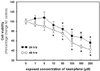
We first examined the antiproliferative effect of kaempferol on human breast cancer MDA-MB-453 cells at various concentrations (ranging from 1 to 200 µM) and exposure times (24 and 48 hrs) using the MTT assay (Fig. 1). The observed antiproliferative activity of kaempferol was consistent with reports that kaempferol inhibits cancer cell growth in various cells (Bestwick et al., 2007; Casagrande & Darbo, 2001; Knowles et al., 2000). A statistical difference was first seen at 50 and 10 µM for 24 and 48 hrs, respectively, compared with controls.
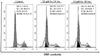
To determine whether the anti-proliferative activity of kaempferol would lead to cell cycle arrest, we analyzed the cell cycle distribution of MDA-MB-453 cells treated with kaempferol using fluorescence-activated cell sorting (FACS) under the treatment conditions at which kaempferol had a significant result (50 and 10 µM for 24 and 48 hrs, respectively). As shown in Fig. 2, a significant decrease in the number of G2-phase cells was seen with kaempferol treatment at concentrations of 50 and 10 µM for 24 and 48 hrs, respectively (8.75 and 9.84% of the cell population compared with control value 16.75%, p<0.05). These accumulations of the cell population in the G2/M phase were accompanied by a concomitant increase in the cell population in the S phase (28.12 and 30.90% of the cell population compared with control value 21.8%, p<0.05).
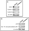
The cell cycle is tightly mediated through a complex network of positive and negative cell-cycle regulatory molecules such as cyclin-dependent kinases (CDKs) and CDK inhibitors (CKIs). Cyclin the down-regulation of CDK1 (Cdc2), as well as cyclin A and cyclin B, by kaempferol may be a main cause of the G/M phase arrest. CDK1 is a catalytic subunit of the M-phase promoting factor, which is activated at the G2/M transition and controls the onset of mitosis. Several investigators have shown that CDK1 in combination with cyclin A and cyclin B is critical in the G2/M phase transition (Jessus & Beach, 1992; Morla et al., 1989). In the present study, CDK1, which is combined with cyclin A/B in the control of the G2/M phase, was markedly decreased by kaempferol (Fig. 3A). Thus, kaempferol inhibits the cell cycle at the G2/M phase via down-regulation of CDK1 in human breast carcinoma MDA- MB-453 cells.
In addition, we observed a higher percentage of apoptosis in sub-G0 phase exposed to kaempferol. Compared to the vehicle-treated cells, the apoptotic cells exposed to kaempferol were observed 23.12 and 31.90 % at 10 and 50 µM for 24 and 48 hrs, respectively. To scrutinize these results further, we investigated the apoptotic pathway in MDA-MB-453 cells exposed to 50 µM kaempferol for 24 hrs (Fig. 3).
Apoptosis is an important series of events leading to programmed cell death that is also essential for development and tissue homeostasis. Recently, the regulation of apoptosis has been proposed as a promising target for cancer chemotherapy (Fesik, 2005; Sun et al., 2004; Zhang et al., 2005). The activation of p53 mediates coordinated antiproliferative effects, including effects on cell cycle and apoptosis (Guimaraes & Hainut, 2002).
In this study, kaempferol increased the expression of p53, and then clearly induced the phosphorylation of p53 at serine 15, but not at serine 6, 9, 20, 37, 46, or 392 (data not shown). Several studies have shown that p53 phosphorylated at serine 15 is associated with apoptosis in cancer cells (Ito et al., 2004; Jiang et al., 2004; Mota et al., 1968). In addition, it has been reported that quercetin increased the levels of total p53 and p53 phosphorylated at serine 15 in A549 cells (Kuo et al., 2004). Thus, kaempferol-induced apoptosis may be related to p53 accumulation-mediated phosphorylation at serine 15 residues.
Consequently, we suggest the existence of multiple pathways by which kaempferol results in G2/M-phase cell cycle arrest via downregulated CDK1 and apoptotic cell death via modulation of the p53 pathway. Therefore, the anticancer activity of kaempferol may be useful for developing anticancer medicine.
 XML Download