

 PDF
PDF Citation
 Print
Print


Lipomas are benign tumors of adipose tissue. They are relatively common in older dogs and are rarely symptomatic [1]. Most lipomas occur at subcutaneous sites, but intrathoracic, intra-abdominal, and intrapelvic sites have also been described. Within body cavities, they can remain undetected for a long time and may become quite large before they result in clinical signs. Typically, the clinical signs of such tumors are related to the organs that they compress or entrap [28].
Lipomas can be hard to differentiate from obesity-related fat deposition on computed tomography (CT) examination. Usually, lipomas appeared homogeneous fat-density lesion on CT images and demonstrate only mild or no enhancement on post-contrast enhanced images [4]. In this report, an uncommon case of lipoma in the abdominal cavity is presented.
A 13-year-old, 5 kg, castrated male mixed-breed dog with a history of diarrhea, dyschezia, and hematochezia was referred to the Veterinary Medical Teaching Hospital, Seoul National University, Korea. Physical examination revealed increased abdominal pressure and pain when palpated. The patient had a caudal abdominal mass that had been identified on sonography by a regional doctor. Hematological examination revealed an increased white blood cell count (21,830/µL), and thrombocytosis (platelet count 76 × 103/µL). Serum biochemistry results were within normal ranges.
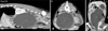
On abdominal radiographs, an oval-shaped, soft-tissue opacity mass was identified in the mid- to caudal abdomen. The mass displaced the descending colon dorsally with compression, the intestines cranially and peripherally, and the bilateral kidneys dorsally. The urinary bladder (UB) was indistinct because of the mass, and serosal detail around the mass was slightly decreased. Abdominal ultrasonography showed a cystic mass closely adjacent to the prostate, and the inside of the mass was partially septated. Because the mass was too large to be evaluated by ultrasound only, for further characterization of the mass for surgical planning, helical CT scans of the thoracic, abdomen, and pelvic cavities were performed. The CT revealed a large fluid-filled structure which displaced the UB, prostate, and descending colon. The left kidney and caudal vena cava (CVC) were compressed by the mass, and the mass had continuity with the ventral aspect of the right lobe of the prostate. Prostatic parenchyma was irregularly contrast-enhanced and contained multiple cysts and mineralization (Figs. 1 and 2). In addition, there were thickened and marked contrast-enhanced bilateral ureters.
The mass showed almost homogeneous fluid-attenuation values (−4 to 40 HU; HU, Hounsfield unit) and had a potential anatomic relationship with the prostate gland. Based on these observations, the tentative CT-based diagnosis was a paraprostatic cyst.
After CT examination, ultrasound-guided fine-needle aspiration biopsy of the mass was performed and gross results showed a turbid, serosanguineous to brown to red fluid. Cytologic examination indicated a non-inflammatory cyst. Two weeks later, cyst resection and omentalization were conducted. The lesion was adhered to the omentum, UB, bilateral ureters, descending colon, and prostate. Biopsy of the prostate was also performed.
Histopathologic diagnosis of the mass indicated a necrotic, inflamed, partially calcified cystic lipoma. The cyst lining was comprised partially of necrotic fat accompanied by fibrin exudation and hemosiderin pigmentation, surrounding by a thick fibrous capsule. Some calcification of the capsule wall was evident. Inflammation was lymphoplasmacytic and relatively mild. Diagnosis of the prostate was lymphoplasmacytic interstitial prostatitis with epithelial hyperplasia.
Simple lipomas, infiltrative lipomas, and liposarcomas are three types of adipose tumors described in veterinary literature [2]. Simple lipomas are well demarcated soft masses within a thin capsule and can occasionally grow to a remarkable size. Infiltrative lipomas, poorly circumscribed tumors with the macroscopic appearance of mature fat, are regarded as benign and do not metastasize. However, they are locally aggressive and usually invade adjacent muscle, fascia, nerve, myocardium, joint capsule, and even bone. Meanwhile, liposarcomas are malignant mesenchymal tumors and regionally invasive with a low metastatic potential. Metastatic sites include the lungs, liver, spleen, and bone [136].
Lipomas can be differentiated from liposarcomas based on morphologic and histologic appearance. Histologically, lipomas have indistinct nuclei and cytoplasm resembling normal fat, whereas liposarcomas are characterized by increased cellularity, distinct nuclei, and abundant cytoplasm with one or more droplets of fat. Infiltrative lipomas cannot be readily distinguished from the more common simple lipoma by examining cytology or small biopsy specimens; typically, CT is used to better delineate these tumors [1].
Paraprostatic cysts are relatively unusual compared with other prostatic diseases and are commonly seen in older, large breed, intact male dogs. The etiology of paraprostatic cysts is insufficiently described. Several mechanisms have been proposed for the development of these cysts, including anomalous enlargement of the Müllerian duct remnant, abnormal retention of prostatic secretions caused by ductal obstruction, and as a sequel to prostatic hematoma [5101214]. CT of paraprostatic cysts reveals large, fluid-filled cavitary lesions arising from the prostate gland. Such cysts cause mass effects in the region of the UB. They may have thickened enhancing walls, non-enhancing fluid-filled centers, and occasionally mineralization. Also, they may displace the UB cranially and predispose to cystitis and ureteritis [13].
On CT examination, lipomas can be hard to be differentiated from obesity-related fat accumulation. Simple lipomas on CT are homogeneous fat-density lesions (−40 to −150 HU), which distinguishes them from soft-tissue masses [911]. Infiltrative lipomas are locally aggressive and infiltration of muscles and fibrous tissue can be observed, however, there is no metastasis. On the other hand, liposarcomas are heterogeneous and may exhibit fat and/or soft-tissue density values [715].
Initially, the patient was considered to have a paraprostatic cyst with high diagnostic priority based on the following. First, the mass was located in the mid- to caudal abdomen and had displaced the descending colon dorsally and the small intestine craniolaterally. Because 98% of lipomas occur at subcutaneous sites in dogs [2], this location was considered unusual. The cystic lesion showed a potential anatomic relationship to the prostate gland, and because the adhesion was severe, it was thought to be a paraprostatic cyst at the time of surgery. Furthermore, the mass had an unusually high HU results (−4 to 40 HU), higher than those of normal fat tissue (−135 to −105 HU). Regardless, as a result of the histopathologic examination, the mass was identified as a cystic lipoma, and the main reason for the high HU results for the cystic content was inflammation and necrosis.
Based on the results of this case, it is suggested that intra-abdominal lipomas can have a cyst-like appearance and produce high HU values due to necrosis. Therefore, the possibility of HU value changes of a structure being associated with histopathologic changes should be considered during abdominal CT evaluations.
 XML Download