

 PDF
PDF ePub
ePub Citation
 Print
Print


Introduction
Anthrax is a zoonotic and infectious disease that occurs around the world and affects a wide range of species including humans and herbivorous mammals [1,21]. Bacillus (B.) anthracis, a Gram-positive and spore-forming bacterium, is the etiological agent of anthrax [16]. Virulent strains of B. anthracis contain two virulence plasmids: pXO1 and pXO2. These plasmids contain genes that enable toxin production and capsule synthesis in the bacterium, and are thus vital factors that influence the full virulence of B. anthracis [7,17,21].
B. anthracis cells that encounter an inhospitable environment form spores that can survive in the environment for an extended period of time and resist a wide variety of severe stresses. B. anthracis has an almost worldwide geographic distribution (World Health Organization, 2003) and causes sporadic outbreaks of anthrax in diverse locations [12]. In Korea, the first case of anthrax was recorded in 1905, and additional cases have occasionally occurred between 1905 and 2008 [6]. The most recent Korean anthrax outbreaks [6] include two cases that were reported in 1995 (one in Kyungju and another in Hongseong), one case in 1995 (in Hongseong), two other cases in 2000 (in Changnyung), and one case in 2008 (in Yeongcheon).
Depending on the route of infection, anthrax can be transmitted to humans as inhalational, gastrointestinal, or cutaneous forms of the disease. B. anthracis infectiousness and survivability has led to the use of this bacterium for bioterrorism. Because of both natural anthrax outbreaks and potential bioterrorism use, many studies have focused on the identification, detection, and molecular subtyping of B. anthracis [23]. B. anthracis strains are genetically monomorphic microbes with low levels of sequence diversity, and this bacterium is considered to be evolutionarily "young" [19,23]. Characterization of closely related B. anthracis strains is necessary for epidemiological studies, examination of biological contamination through environmental isolation, and forensic investigation of bioterrorism events [3]. A variety of discriminatory approaches have been examined for their potential use in identifying and subdividing microbial pathogen groups. In particular, a high-resolution genotyping system introduced by Keim et al. [11] is suitable for the detailed analysis of closely related isolates. This system involves the examination of single nucleotide polymorphisms (SNPs), the performance of multiple-locus variable-number tandem repeat analysis (MLVA), and the assessment of single nucleotide repeats (SNRs). These three genotyping methods have been used to identify effective genetic markers and understand broader genetic relationships among B. anthracis strains.
SNPs are rare in B. anthracis, but they have been identified and used as genetic markers for multiple whole-genome sequencing [23]. The mutation rates of SNPs are very stable (10-10 changes per nucleotide per generation), and a set of canonical SNPs from eight different strains can be used to discriminate among major clusters of B. anthracis [11,25,26]. MLVA has been performed to examine several loci (MLVA-8, MLVA-15, and MLVA-25) that have higher mutation rates than SNPs. The combination of MLVA and SNP analysis can therefore be used to evaluate B. anthracis genotypes [23]. SNRs that display very high mutation rates (as high as 6.0 × 10⁻⁴ mutations per generation) are a type of variable-number tandem repeat (VNTR) [10]. Repeated stretches of a single type of nucleotide are more likely to occur in SNRs than in other types of simple sequence repeats (SSRs). DNA containing VNTRs can undergo strand separation and base pair slippage, thereby increasing the chances of slipped-strand mispairing that produces mutations at the SNR locus [15,23]. Due to the high mutation rate, SNR analysis can provide additional powerful genetic resolution when examining B. anthracis strains with the same MLVA genotype. Typical methods for this type of analysis have been described by Kenefic et al. [13] and Stratilo et al. [23]. Several strains of B. anthracis have already been studied using the aforementioned methods. For example, investigations have performed in the United States (47 isolates), Italy (53 isolates), and Canada (19 isolates), and established genotype reference markers [3,13,23]. Subsequently, genotyping was recently performed to assess and identify several candidate strains in Namibia and Bangladesh [1,2].
Several B. anthracis isolates from Korea have also been analyzed with various molecular typing techniques such as amplified fragment length polymorphism (AFLP), SNP analysis, and MLVA [20]. In a study by Ryu et al. [20], eight MLVA markers were assessed to elucidate the evolutionary pattern of nucleotide differences among B. anthracis isolates from 12 different regions in Korea, and nine MLVA types were identified. Among these nine MLVA types, three (M1 to M3) were classified as pathogenic B. anthracis isolates and assigned to new genotypes belonging to the A4 and B3 clusters. In a study by Jung et al. [6], B. anthracis isolates were arranged into A3a, A3b, and B1 clusters using MLVA and subdivided into four canSNP subgroups (B.Br.001/002, A.Br.005/006, A.Br.001/002, and A.Br.Ames). In the present study, we isolated new Korean strains of B. anthracis from the soil and measured the genetic diversity between strains that were obtained from different areas of the Korean peninsula. Furthermore, we typed these strains using SNR analysis and compared our findings with previously published MLVA and canonical SNP (canSNP) results.
Materials and Methods
Bacterial strains
B. anthracis strains that were used in this study are listed in Table 1. Five strains were from different regions of the world (ATCC14185, ATCC14578, Pasteur, delta Sterne, and Sterne). ATCC14185 and ATCC14578 stains obtained from American type culture collection (ATCC, USA), and Pasteur, delta Sterne, and Sterne strain obtained from Animal and Plant Quarantine Agency (QIA, Korea). Four strains isolated in Korea were obtained from the Korean Center for Disease Control and Prevention (KCDC, Korea). Three were new isolates from different regions of Korea (HS, Bch, and KJ) and 14 other new isolates were acquired from one region in Korea (CH). The seventeen new isolates from Korea were collected from the soil and characterized with Gram staining, motility testing, and catalase analysis.
Extraction of B. anthracis DNA
All B. anthracis strains were cultured in brain heart infusion broth (BHI; Difco; Becton, Dickson and Company, USA) and grown for 8 h at 37℃ with shaking. Total genomic DNA was extracted using a modified variant of Hunter's method [4]. To isolate the DNA, cells were harvested by centrifugation at 20,215 g for 10 min at 4℃, and then resuspended in 1 mL Tris-ethylenediamine tetraacetic acid (TE) containing lysozyme (0.1 mg/mL), 2% sodium dodecyl sulfate (SDS), and 10 µg/mL proteinase K at 55℃ for 3 h. After this incubation, DNA was extracted three times with 500 µL phenol-chloroform, precipitated with 1 mL isopropanol, and dissolved in 100 µL TE buffer with RNase (20 µg/mL).
B. anthracis screening by PCR
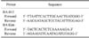
BA-813 and BA Kim were used as specific primers to amplify a portion of B. anthracis chromosomal DNA for identification [14,18]; the primer sequences are listed in Table 2. Template DNA (10 ng) and 0.5 pmol of each primer were added to a PCR mixture tube containing 2X EF-Taq Premix II (Solgent, Korea). The final volume was adjusted to 30 µL with distilled water. The following thermal cycling (Bio-Rad, USA) conditions were used: an initial denaturation step at 94℃ for 5 min followed by 30 cycles of denaturation at 94℃ for 1 min, annealing at 40℃ for 1 min, and primer extension at 72℃ for 1 min. The final extension step was performed at 72℃ for 5 min. The PCR products were electrophoretically separated on 1% agarose gel and stained with ethidium bromide (Sigma-Aldrich, USA).
Strain-specific SNP analysis with a TaqMan minor groove binding (MGB) allelic discrimination assay
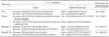
Strain-specific SNP analysis was performed for the four different SNP loci that were identified by Van Ert et al. [25] with a TaqMan MGB allelic discrimination assay. TaqMan MGB allelic discrimination assays are designed based on chromosomal and pXO1 SNPs that were previously reported [25]. The TaqMan MGB probes and primers were designed with ABI Primer Express software (Life technologies, USA) in accordance with the manufacturer's guidelines. Sequences of the primers and probes used in the current study are listed in Table 3. The reaction mixture contained 2× ABI Universal Master Mix (Life technologies), 250 nM of each probe, 600 nM each of forward and reverse primer, and 400 pg/µL of genomic DNA. The reaction was initially incubated at 50℃ for 2 min and 95℃ for 10 min followed by 40 cycles of denaturation at 95℃ for 30 sec, and annealing and extension at 60℃ for 1 min. Real-time and endpoint fluorescence data were collected with an ABI 7500 Real-Time PCR system (Life technologies).
SNR analysis of the B. anthracis strains
For the SNR analysis, we used primers that amplified two chromosomal SNR loci and two pXO2 SNR loci previously reported by Stratilo et al. [23]. The PCR mixtures contained 2X EF-Taq Premix II (Solgent), forward and reverse primers (0.5 pmol each), and genomic DNA (400 ng/µL). After an initial incubation at 95℃ for 5 min, 30 cycles of PCR were performed at 95℃ for 1 min, 60℃ for 1 min, and 72℃ for 1 min. The PCR products were separated in a 1% agarose gel and stained with ethidium bromide. For sequencing, the PCR products were cloned using a TA cloning vector (RBC Bioscience, Taiwan), purified with a Hi-Yield Plasmid Mini Kit (RBC Bioscience), and sequenced with the M13F primer in Gnotech (Korea). Using MEGA 4.0.2 software program, SNR data were aligned and analyzed with the UPGAMA clustering method [24].
Results
Screening of the B. anthracis isolates
Based on the Gram staining (positive), motility testing (non-motile), and catalase analysis (positive) results, we confirmed that the new bacteria strains isolated in Korea were B. anthracis. PCR amplification with a set of specific primers in chromosome (BA-813 and BA Kim) successfully produced a product [14], further proving that the new isolates were B. anthracis (data not shown). We also performed another round of PCR amplification with specific primers (pXO1: pag1, pag2, cya1, cya2, lef1, lef2, pXO2: cap1, cap2) in plasmid (Table 4) to confirm whether the isolated strains possessed toxin plasmids [18]. Based on these results, we subsequently performed SNR analyses of four SNR loci that are positioned in the bacterial chromosome and pXO2.
Strain-specific SNP analysis of the B. anthracis isolates
A collection of B. anthracis isolates including strains derived from the Ames reference strain was analyzed to generate SNP data for strain-specific discrimination. This SNP analysis was performed using a TaqMan MGB allelic discrimination assay. In 2007, Van Ert et al. [25] reported that SNPs specific for the Ames strain include Branch1-7, Branch1-26, Branch1-28, Branch1-31, PS-1, and PS-52. We therefore used the following four Ames-strain-specific SNPs for our analysis: PS-52, Branch1-7, Branch1-26, and PS-1. The Ames strain-specific PCR assay with these SNPs enabled the discrimination of B. anthracis isolates with a high degree of specificity. The Ames strain-specific SNP assay (Fig. 1 and Table 5) was able to discriminate between Ames and non-Ames strains with 100% accuracy using one chromosomal (Branch1-31) and two plasmid (PS-1 and PS-52) SNPs. However, we could not detect any differences among the non-Ames strains that were evaluated, indicating that analyses based on SNP loci alone have a limited resolution for assessing genetic diversity among B. anthracis strains with low levels of genetic variation.
SNR analysis of the isolated B. anthracis
To evaluate the genetic diversity of B. anthracis strains that were isolated in Korea, we performed an SNR analysis to characterize 26 strains, including four that were obtained from the KCDC. We used four primers previously described by Stratilo et al. [23]. Two of these primers (CL10 and CL12) were specific for regions on the chromosome whereas the remaining two (CL33 and CL35) were specific for the pXO2 plasmid.
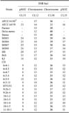
PCR analysis of the four SNR loci demonstrated that the isolates differed by a maximum of three base pairs at each locus: CL33 (9 and 10 bp), CL12 (12, 13 and 14 bp), CL10 (35 and 36 bp), and CL35 (10, 11, 12, and 13 bp). The 26 B. anthracis samples were divided to three groups according to source of the strains (reference strains, Korean strains from the KCDC, and other Korean strains), and the SNR analysis results were evaluated according to these groups (Table 6). Greater variation among the strains was observed according to region and similarity patterns were calculated (Fig. 2).
CH strains were newly isolated in Korea. Similar candidate strains were obtained by conducting several different extractions from the same soil samples. After confirming that the isolated bacteria were B. anthracis, a total of 14 new B. anthracis strains were used for the study. We named these strains and labeled them with their date of extraction. To determine whether genetic variation can be observed among B. anthracis isolates from the same soil source, an SNR analysis of four loci was performed. Several differences (numbers of base pairs) among the newly isolated strains were observed with this SNR analysis, and these strains were therefore divided into 10 different subgenotypes (SGTs; Fig. 3).
Discussion
Strain-to-strain variation analysis is important for understanding pathogenesis, immune mechanisms, bacterial evolution, and host adaptation of pathogenic bacteria. In situations involving natural or terrorism-related outbreaks of B. anthracis, powerful marker systems are necessary to appropriately address the crisis by correctly identifying the B. anthracis strain that is involved despite the low levels of diversity that are characteristic of this bacterium. Molecular typing that focuses on hypervariable VNTR loci has been used to successfully perform strain genotyping and phylogenetic analysis of many bacterial species [3,5,8]. However, this technique has proven to have extremely limited applicability for classifying B. anthracis due to the homogeneous nature of all of known strains [10]. Past studies have typed and differentiated isolates of this pathogen using SNP analysis and MLVA [6,8-12].
The SNR marker system is used to analyze genomic regions with very high mutation rates [23]. More importantly, SNR analyses can differentiate between selected strains with extremely low levels of genetic diversity. These high mutation rates render SNR markers unsuitable for determining the phylogenetic relationships among diverse isolates but permit the detection of lower levels of genetic diversity. In fact, even isolates from the same epidemic can be distinguished with an SNR-based approach [11].
In the present study, we isolated B. anthracis strains (CH, HS, Bch, and KJ) from soil in Korea, and analyzed the genotypes of these strains using SNR markers (CL33, CL12, CL10, and CL35) specific for the chromosome and pXO2 plasmid. These B. anthracis isolates were divided into two groups based on the region of origin. Variations in the SNR analysis results were observed not only between the Korean isolates and reference strains (non-Korean isolates) but also among the different types of Korean isolates. For example, strains from the KCDC (S0303, S0304, S0307, and S0308) differed by one or two base pairs in the four examined SNR loci. However, results for the KCDC strains greatly differed from those obtained for strains that were collected in other regions, including other locations in Korea.
In contrast to the MLVA-canSNP data that were previously obtained for strains used in this study [6], most of the SNR analysis results from the present investigation were similar for the various strains that were examined. These B. anthracis isolates were subdivided into the major A and B sublineages, using MLVA and canSNP findings to calculate simple matching coefficients. Notably, CH and KJ strains existed separately within each branch according to the MLVA and canSNP data, but were more closely related to each other than strains that were isolated from other regions. In the SNR analysis, the KJ strain appeared to be genetically close to the CH strains with respect to three of the examined SNR loci (all of the SNR loci except for CL33). Thus, the CH and KJ strains were combined into the same group (Fig. 2). All of the strains were compared to other strains that were previously analyzed based on the same SNR loci [23], and no similarities were observed. For identifying phylogenetic relationships, the MLVA-canSNP is considered to be more reliable than the SNR analysis. Therefore, these strains may be regarded as distinct.
We obtained several bacteria colonies from the soil samples from the same region (CH) and isolated the B. anthracis strains. Based on the results for the four SNP marker loci, we classified the 14 CH strains into 10 SGTs. SGT3, SGT6, SGT7, and SGT9 each include two isolates, and the other SGTs each contained one isolate. Compared to the MLVA-canSNP analysis, the SNR assay revealed more distinctions among the examined strains. In particular, we did not identify any differences among these strains based on the MLVA-canSNP results, but the SNR analysis findings indicated a number of genetic variations among the studied isolates. At the CL10 locus, there were differences of up to ± 5 base pairs in amplicon size for these isolates. Thus, we were able to validate the high resolution power of SNR analysis for differentiating strains with low levels of genetic variation. Given these results, we confirmed that the MLVA-canSNP analysis is suitable for global-scale studies to identify major bacteria groups. Additionally, the SNR analysis is appropriate for fine-scale focusing to identify minor variations among similar strains.
We did not have enough isolates to identify the ancestral strain of the examined samples. Relationships among the identified SGTs were therefore not important in the context of this study. However, it is interesting to note that these variations were observed for strains that were obtained from the same soil samples. This result might reflect the survivability of B. anthracis through several passages in inhospitable environments. Similarly, SNR genotypic differences have been observed for B. anthracis samples obtained from blood and tissue of infected animals during anthrax outbreaks; this diversity might also reflect the passage of bacterium in the affected animals [22]. Thus, an important conclusion based on our results is that genetic variations in B. anthracis could occur in a single original strain after several passages in the same soil.
Several molecular typing methods, which are called progressive hierarchical resolving assays using nucleic acids (PHRANA) by Keim et al. [9,10], demonstrate high resolving power when identifying phylogenetic relationships. To prevent and manage natural or bioterrorism-related bacterial outbreaks, PHRANA must be routinely performed with a several types of strains [13]. Among the PHRANA-related methods, SNP-MLVA analysis can be employed to investigate genetic relationships whereas SNR analysis can be utilized for fine-scale classification [22]. In particular, SNR analysis of loci has unsurpassed resolving power for detailed tracking of outbreaks and can be used for forensic studies [12,13]. In the present study, we obtained SNR analysis results for four loci of new Korean B. anthracis isolates. These findings may help identify markers specific for this pathogen.
 XML Download