

 PDF
PDF ePub
ePub Citation
 Print
Print


Introduction
Tracheal collapse is a potentially life threatening and commonly occurring respiratory disease in small breeds of dogs. It is a form of tracheal obstruction that is caused by cartilage flaccidity and flattening, and it is characterized by a chronic, paroxysmal honking cough [5,8-10]. In the affected animals, the cartilage usually collapses in a dorsoventral direction with the cervical trachea collapsing during inspiration and the thoracic trachea collapsing during expiration [5,10-12].
Conventional radiography has been used to evaluate tracheal collapse. The lateral radiographs of the neck and thorax are diagnostic in approximately 60% of dogs with severe tracheal collapse, and using only these lateral radiographs can result in false negative and false positive diagnoses due to inadequate positioning, poor radiographic technique or the superimposition of the esophagus or the cervical muscles [8,12,13]. Fluoroscopic and tracheoscopy are considered to be the most sensitive methods for diagnosing tracheal collapse [1,5,8,11,14], but these techniques are not widely available in standard veterinary practice.
Ultrasonography may also be used to assess the dynamic movement of the trachea, as can fluoroscopy, but ultrasonography doesn't expose the animal to radiation, and it can be performed with minimal or no sedation [9,14]. An ultrasonographic examination has been used to diagnose tracheal collapse by identifying the simple changes in the shape of the tracheal margin so as to characterize the lesion at the time of collapse [14].
The purposes of this study were to describe the ultrasonographic appearance of tracheal collapse and to assess the thoracic inlet tracheal ring width (TITRW) and the first tracheal ring width (FTRW) for establishing a cut-off level of TITRW/FTRW ratio for making diagnosis and evaluating the severity of tracheal collapse. We also wanted to evaluate the feasibility of performing ultrasongraphic measurement for identifying a tracheal collapse.
Materials and Methods
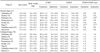
Tracheal ultrasonography was performed in 56 normal dogs and 84 dogs with tracheal collapse in the thoracic inlet region. There were 6/10 (normal/tracheal collapse) Chihuahuas, 8/11 Maltese, 6/13 Pekingese, 4/10 Pomeranians, 6/12 Miniature Poodles, 5/6 Pugs, 10/15 Yorkshire Terriers and 11/7 small mixed breed dogs. The mean age, body weight, tracheal ring width and TITRW/FTRW ratio of each breed are listed in Table 1.
Tracheal collapse was diagnosed based on the following criteria: having a chronic honking cough on the history taking and physical examination, and having a distinctly narrowed tracheal lumen identified on the radiographic examinations. Fifty six dogs that had a radiographically normal trachea and no evidence of respiratory problems made up the control group for making comparison with the tracheal-collapsed dogs.
The animals were positioned in right lateral recumbency for the cooperative dogs and in the standing position for the uncooperative dogs. To minimize the tracheal pain or compression, their head and neck were maintained in the neutral position. The tracheal ring was scanned using an ultrasound machine with a 7-MHz linear transducer (Aspen; Acuson, USA). After shaving the hairs perpendicular to the tracheal surface and applying an acoustic coupling gel, the probe was positioned in a transverse direction over the trachea. With using particular caution so as not to distort or compress the trachea, the probe was slowly pivoted around the trachea to evaluate the general tracheal ring shadow. To minimize a potential artifactual lengthening caused by the tracheal displacement and asymmetrical measurement, the tracheal ring shadow width was measured when an extension line, with both end points of a continuous tracheal ring shadow without any intercartilaginous gap on the image, was maximally parallel to the probe surface [14]. The acquisition time of all the images was 6.4 ± 1.1 min for the cooperative dogs and this was 14.2 ± 2.8 min for the uncooperative dogs.
The maximum width of the first tracheal ring was measured at immediately caudal to the cricoid cartilage. The maximum width of the thoracic inlet tracheal ring was measured at immediately cranial to the manubrium. The tracheal ring widths at both expiration and inspiration were measured 3 times and then they were averaged. The TITRW/FTRW ratio was calculated for each phase of respiration in all the dogs.
The recorded data, including age, weight, the tracheal ring width and the TITRW/FTRW ratio, was computed using software SPSS (version 12.0; SPSS, USA) and these values were expressed as means ± SD. The correlation between weight and the first tracheal ring width was compared using Pearson's coefficient test. The significance of the differences of the first tracheal ring width and TITRW/FTRW ratio, which depended on the respiration variance, was evaluated by paired t-tests, and unpaired t-tests were used for comparing between the normal dogs and the tracheal collapse dogs. The width and TITRW/ FTRW ratio between breeds were compared using the one-way ANOVA test and Duncan's post hoc test. In addition, a cutoff level was statistically analyzed according to the receiver operating characteristic curve, which was derived from the comparison of the tracheal widths of the normal and tracheal collapse dogs. All the analyses were performed using a level of significance of p ≤ 0.05.
Results
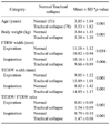
The mean age, body weight, tracheal ring width and the TITRW/FTRW ratio were measured and the results are listed in Table 1. The mean age of the tracheal collapse dogs (5.53 ± 1.82 years) was older (p = 0.001) than that of the normal dogs (3.85 ± 1.04 years). The mean weight of the tracheal collapse dogs (5.38 ± 1.30 kg) was higher (p = 0.01) than that of the normal dogs (3.84 ± 1.43 kg). However, there was no statistical correlation between weight and the tracheal ring width (Table 2).
Ultrasonographic measurements could be carried out in 129 of 140 dogs without the need for chemical sedation. However, it was not possible to obtain images from 5 normal dogs that became quite aggressive, and it was also not possible to obtain the images in 6 dogs with a collapse trachea due to their uncontrolled dyspnea and panting. These dogs were excluded from the data analysis. Although respiratory distress, panting or coughing was present in some animals, there were no serious problems related to the experimental procedure.
Radiographic finding
All the abnormal dogs displayed moderate to severe tracheal collapse in the cervicothoracic region. A flattened and collapsed trachea was located along with the midline or it was rotated to the left side. Left caudal bronchial collapse that was caused by left side heart enlargement was present along with tracheal collapse in 15 dogs (2 Chihuahuas, 3 Maltese, 5 Pomeranians, 2 Miniature Poodles and 3 Yorkshire Terriers).
Ultrasonographic appearance of the tracheal rings
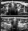
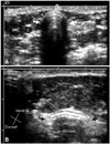
Ultrasonography showed the tracheal ring as a semicircular shape with a hyperechoic shadow in the normal dogs (Fig. 1), whereas ultrasonography demonstrated the tracheal ring as a flat shape in dogs with a collapsed trachea (Fig. 2B). The tracheal ring shadow created a reverberation artifact by the air filled tracheal lumen and this made the dorsal image unclear and this made the dorsal image unclear.
The first tracheal ring was centrally located under the sternohyoid muscle and medial to the sternothyroid and sternocephalicus muscles. The thoracic inlet tracheal ring was located under the sternocephalicus muscle and it was surrounded by the sternohyoid and longus capitis muscles (Figs. 1 and 2). However, these relationships could not be discriminated when the trachea was laterally displaced and collapsed (Fig. 2B).
Measuring the ultrasonographic tracheal ring widths
The TITRW/FTRW ratios ranged from 0.6 to 0.9 in the normal dogs and from 1.4 to 1.6 in the tracheal collapsed dogs. The normal dogs, the Pugs and Pekingese together, as compared with all other breeds, had a significantly narrow (p = 0.05) thoracic inlet tracheal ring and a low TITRW/ FTRW ratio (Table 1).
In expiration, the TITRW of the normal dogs (9.05 ± 1.52 mm) was greater (p = 0.001) than that of the tracheal collapsed dogs (15.89 ± 1.01 mm). In inspiration, the TITRW of the normal dogs (8.02 ± 1.42 mm) was greater (p = 0.001) than that of the tracheal collapsed dogs (14.85 ± 1.17 mm). When comparing between the normal dogs and the tracheal collapse dogs, the TITRW/FTRW ratio of the tracheal collapsed dogs was recorded as high (p = 0.001) in both expiration and inspiration. The expiration TITRW/ FTRW ratio of the normal dogs (0.81 ± 0.09) was higher (p = 0.001) than that of the tracheal collapsed dogs (1.54 ± 0.09). The expiration TITRW/FTRW ratio of the normal dogs (0.79 ± 0.10) was higher (p = 0.001) than that of the tracheal collapsed dogs (1.47 ± 0.08) (Table 2). From the above results, the cutoff levels of the TITRW/FTRW ratio for the evaluation of tracheal collapse could be set at 1.16 in expiration and at 1.13 in inspiration.
Discussion
The tracheal ring was easily identified as a hyperehoic curved and crescent-shaped structure with a reverberation [14]. The width between either ends of the tracheal ring could be easily measured from the shadow formed by the cartilage. These procedures could be carried out in 129/140 (93%) dogs without the need for chemical restraint.
There have been several quantitative studies that have reported on evaluating the tracheal diameter. In one study, the mean ratio of tracheal diameter to thoracic inlet diameter was evaluated, and the diameter of the normal trachea is approximately one fifth of the depth of the thoracic inlet [7]. A previous study using cadavers showed that the actual measurement of the mean tracheal width indicates that collapsed tracheas are generally wider than those in the normal toy breeds [2]. In another study, the ratio of the width to the height of the trachea has been reported to be 1:1 in a normal trachea and 4:1 in a collapsed trachea [3,4]. In these studies, a comparison of the first tracheal ring with the thoracic inlet tracheal ring revealed that the mean TITRW/FTRW ratios were significantly increased in dogs with tracheal collapse. These results suggest that the thoracic inlet tracheal ring widths are significantly widened and flattened compare with the first tracheal ring and the results are similar to previous study [2,4,7]. It may indicate that there is a loss of the normal tracheal conformation and also the loss of rigidity of the tracheal muscles [2,4-7].
Both Pug and Pekingese dogs had a relatively narrow thoracic inlet tracheal ring width compared to other breeds. These breeds of dog are known as breeds with the characteristics of a brachycephalic syndrome, including a hypoplastic trachea [11,14-16].
The cutoff level of the TITRW/FTRW ratio for the evaluation of tracheal collapse was set 1.16 in expiration and 1.13 in inspiration. Because the trachea has a variable diameter depending on the phase of respiration [1,9], ultrasonographically measuring the tracheal width that corresponds to the phase of respiration was also difficult in this current study. To enhance the ultrasongraphic measuring procedure for practical usage, selecting a cutoff value without it having a relationship with the phase of respiration is most suitable. When all the TITRW/FTRW ratios of the normal and tracheal collapse dogs were analyzed without regard to the respiration phase, the cutoff level of tracheal collapse was 1.14. If a TITRW/FTRW ratio is higher than 1.14, it could be indicative of tracheal collapse.
The aim of the present study was to evaluate the feasibility of performing US for evaluating tracheal collapse. However, it was not possible to perform an independent, 'blind' evaluation because the ultrasonographers were informed about the inclusion and exclusion criteria. It was also not possible to perform double blind ultrasonographic measurement in all the dogs with respiratory distress and in the aggressive or frightened dogs. Because the study was not performed prior the ultrasonographic evaluation and was limited to the dogs with significant tracheal collapses, the findings may not be generalizable to dogs with a mild to moderated stage of tracheal collapse. Further studies should address these limitations and they may need to evaluate dogs with various degrees of tracheal collapse.
The first tracheal ring can be easily defined and measured because the distal cricoid cartilage gives a landmark for the beginning of the first tracheal ring. However, there are several pitfalls to obtain an accurate thoracic inlet tracheal ring width: it is not possible to repeatedly locate the probe at the same tracheal ring, the possibility of a lengthening effect induced by obliquely scanning can not be ruled out, the width is difficult to measure when a dog is panting and the phase of respiration is difficult to determine. Therefore, a restrained manner that soothes the dog and calms it and a scan technique ensuring the maximum width are considered to be the most important factors for obtaining a more accurate measurement. The standing position for a dog that resists being placed in lateral recumbency also permitted a favorable condition for measurement in this study.
Tracheal ultrasonography was previously shown to have distinct advantages for demonstrating tracheal collapse in dogs as it is noninvasive and safe [9,12,14] and it could be accomplished within a few minutes. In addition, measurement of the TITRW/FTRW ratio can play a role in evaluating tracheal collapse. We think that ultrasonographic examination of the trachea, although limited to the cervical tracheal collapse, is a useful supplementary technique to discriminate an ambiguous radiographic diagnosis of tracheal collapse.
 XML Download