

 PDF
PDF ePub
ePub Citation
 Print
Print


INTRODUCTION
Right double inferior vena cava (RDIVC) with retrocaval ureter is an extremely rare anomaly with only a few cases reported in the literature. Preoperative identification of this IVC anomaly is essential before embarking on ureteric repair, else serious surgical mishap can occur. To the best of our knowledge, surgical repair of retrocaval ureter with RDIVC by a single-incision laparoscopic technique has not yet been described in the literature. This case report highlights the embryological basis and the surgical technique of repair by using the single incision multiple port laparo-endoscpic (SIMPLE) technique. Because one of the major hurdles in single-incision laparoscopic surgery is the limited maneuverability and difficult anastomotic suturing, we performed end-to-end ureteric anastomosis by using the Santosh Postgraduate Institute (PGI) ureteric tacking fixation technique [1].
CASE REPORT
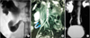
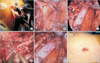
A 23-year-old male presented in the urology department with complaints of dull aching right flank pain. He had no episodes of hematuria, graveluria, fever, or other urinary symptoms. The results of a general physical and abdominal examination were unremarkable. His serum creatinine was 0.9 mg%. Preoperative urine analysis was unremarkable with pH of 6.5, specific gravity of 1.010, and no pus cells or red blood cells. Ultrasound of the abdomen revealed right-sided hydroureteronephrosis involving the upper ureter with RDIVC. Because the patient had a prior history of contrast allergy, noncontrast magnetic resonance imaging was performed, which showed a dilated pelvicalyceal system. The upper ureter was dilated with medial deviation and coursed behind the lateral IVC at the level of the third lumbar vertebra with abrupt narrowing at that level. The upper ureter then passed anteriorly through the space between the duplicated right IVC. The middle and distal ureter was normal in caliber (Fig. 1B). The results of a preoperative ethylene-dicysteine scan revealed a split function of 43% for the right kidney with significant tracer retention at the end of 3 hours of study. The diagnosis of symptomatic retrocaval ureter with significant obstruction and RDIVC was supported, and the patient underwent single-incision multiport laparoendoscopic repair with the Santosh PGI ureteric tacking fixation technique (Fig. 2).
With the patient under general anesthesia, retrograde pyelography was performed, which showed the fishhook appearance of the right ureter (Fig. 1A). A 6-Fr DJ stent was placed. Subsequently, the patient was placed in a modified flank position. A nearly 3-cm incision was made at the level of the umbilicus and three conventional laparoscopic ports were inserted: two 12-mm and one 5-mm ports. The colon was medially mobilized, and the gonadal vein was divided. After soft tissue dissection, the RDIVC was clearly seen with the ureter coursing in between the double IVC. Following mobilization, the stenotic ureteric segment was excised and both ends were brought lateral to the IVC and spatulated. To make anastomotic suturing easier, both ureteric ends were brought close and their adventitia was tacked to the adjacent abdominal wall by using hemostatic clips. This maneuver stabilized both ureteric margins, and an end-to-end anastomosis was performed with Vicryl 3-0 interrupted sutures followed by release of the clips. An 18-Fr abdominal drain was placed. The patient had an uneventful postoperative recovery. The operating time was 95 minutes. Blood loss was minimal and the hospital stay was for 3 days.
At the 8-month follow-up, the magnetic resonance urogram was repeated and showed preserved renal parenchyma with a normal-caliber ureter (Fig. 1C). The postoperative diuretic scan revealed split function of 45% for the right kidney with adequate tracer clearance by the end of 3 hours of study.
DISCUSSION
First described by Horchstetter in 1893 [2], retrocaval ureter is a rare anomaly that occurs in 1 out of 1,000 live births [3]. It is caused by persistence of the posterior cardinal vein during embryonic development [4]. The incidence of IVC anomalies varies from 0.2% to 8.7% [5]. IVC anomalies are a result of abnormal regression or persistence of three pairs of embryonic vessels, namely, the posterior cardinal, subcardinal, and supracardinal veins [6]. Infrarenal IVC anomalies were classified by Chuang et al. [7] in 1974 into four types: persistent right posterior cardinal vein (retrocaval ureter), persistent right supracardinal vein (normal IVC), persistent left supracardinal vein (left IVC), and persistent right and left supracardinal veins (double IVC). IVC duplication is of two principal types: double-sided (with one IVC on either side of the aorta) or single-sided (both IVC on one side of the aorta) [8]. The reported incidence of double-sided IVC is around 0.2% to 3% [5], whereas less than a dozen cases of right single-sided double IVC (or RDIVC) have been documented in the literature. Our patient is among the rarest of the cases that have been reported to have retrocaval ureter with RDIVC [8]. RDIVC may or may not be associated with retrocaval ureter depending on its pathogenesis [6,8]. Persistence of the right posterior cardinal with the supra- or subcardinal vein will result in RDIVC with retrocaval ureter, whereas persistence of sub- and supracardinal veins with regression of the posterior cardinal vein will result in RDIVC without a retrocaval ureter [8]. The patient in this case underwent surgical repair because he was symptomatic with hydroureteronephrosis. Small case series and reports exist describing transperitoneal and retroperitoneal approaches for laparoscopic repair of retrocaval ureter. The transperitoneal approach has the advantage of familiar anatomy, a larger working space, and easier suturing compared with the retroperitoneoscopic approach [9,10].
This is the first reported case of retrocaval ureter with RDIVC managed by the SIMPLE technique utilizing the Santosh PGI ureteric tacking fixation technique. As published in a previous report [1], this technique is easily reproducible, is technically feasible, provides a good aesthetic outcome, utilizes conventional endoscopic ports, and has minimal morbidity when performed by expert laparoscopic surgeons. The SIMPLE technique also helps to reduce the cost burden on patients, because it does not require specialized single-incision port devices or special laparoscopic instruments. One of the limitations of single-incision laparoscopic surgery is the limited maneuverability of the instruments, thereby increasing the operating time. The Santosh PGI ureteric tacking fixation technique helps in stabilizing and maintaining alignment of the two ends of the ureter and showed a reduction in mean operating time compared with other reports [1]. Laparoscopic repair of the right retrocaval ureter with RDIVC poses technical challenges. The ureteric dissection is performed between the two IVC instead of between the IVC and the aorta. Because the vessel walls of the IVC are thinner than the walls of the aorta, inadvertent injury can easily occur owing to mechanical tension or thermal damage by electrosurgical or harmonic shear devices leading to profuse bleeding. This IVC anomaly also highlights the importance of preoperative cross-sectional imaging instead of relying solely on intravenous urography or retrograde pyelography. Major surgical mishaps can occur should the lateral component of the RDIVC be confused for a dilated right ureter or the medial component be confused for the aorta during a laparoscopic upper urinary tract reconstruction or nephrectomy.
In summary, retrocaval ureter with RDIVC is an extremely rare anomaly reported in the literature. Preoperative identification can theoretically reduce the risk of intraoperative major surgical mishap during upper urinary tract surgery. To the best of our knowledge, this is the first case report of managing such a scenario with single-incision multiple-port laparoscopic surgery. The SIMPLE technique along with the Santosh PGI ureteric tacking fixation technique, with its inherent advantages over conventional single-incision or multiple-incision laparoscopic surgery, is a feasible option for the management of such cases.
 XML Download