

 PDF
PDF ePub
ePub Citation
 Print
Print


INTRODUCTION
Prostate cancer (PC) is the most common malignancy among men in Western nations and is the second leading cause of cancer-related death in the United States, with increasing rates being reported in Korea [1,2]. However, the etiology of PC is not yet clearly identified. Among the many causes of cancer, it is well established that the link between chronic inflammation and cancer involves cytokines and mediators of inflammatory pathways, which act during the various steps of tumorigenesis [3]. In addition, histologic, molecular genetic, and epidemiologic evidence strongly suggests that inflammation and infection are important in the etiology of PC [3-5].
The cyclooxygenases (COXs) are a family of enzymes that catalyze the rate-limiting step of prostaglandin biosynthesis. COX-2 may play a key role in the tumorigenesis of a variety of human malignancies by stimulating cell proliferation and angiogenesis, inhibiting epithelial differentiation, enhancing cell invasiveness and tumor metastasis, inhibiting apoptosis, mediating immune suppression, and increasing the production of mutagens [6-8]. The expression of COX-2 in prostate tissues or PC has been the subject of many recent studies [4,5,9]. Several such have examined the expression of COX-2 in post-atrophic hyperplasia, proliferative inflammatory atrophy, and prostate intraepithelial neoplasia, but the results are still controversial [9-13].
To the best of our knowledge, there has been little research carried out on the role of COX-2 expression in the chronic inflammation of normal prostatic glands. We therefore compared the overexpression of COX-2 in noninflammatory benign prostatic hyperplasia (NI-BPH), inflammatory benign prostatic hyperplasia (I-BPH), and PC. In addition, we investigated the relationships between COX-2 overexpression and both angiogenesis and apoptosis.
MATERIALS AND METHODS
We evaluated apoptosis by two immunohistochemical methods. We assessed Bcl-2 expression, which exerts an anti-apoptotic effect in the intrinsic pathway of apoptosis. Second, we determined the apoptotic index by the terminal deoxynucleotidyl transferase nick end labeling (TUNEL) assay, which revealed DNA fragmentation. Similarly, we evaluated angiogenesis by two immunohistochemical indexes: vascular endothelial growth factor (VEGF), which is the most potent tumor angiogenic factor, and microvascular density (MVD), which reflects the angiogenic vasculature present.
1. Tissue samples
The tissue samples used in this study were obtained from the files of the Department of Urology, Keimyung University Dongsan Medical Center, during the period of 2005 to 2009. A total of 121 BPH and PC patients were included in this study. Specimens were obtained from 57 PC patients who underwent radical prostatectomy and 64 BPH patients who underwent transurethral resection of prostate (TURP). The BPH histopathologies were classified by the presence of chronic inflammation (infiltration of macrophages, lymphocytes, and plasma cells) as follows: NI-BPH (n=23) and I-BPH (n=41).
2. Tissue microarray
Tissue microarray was constructed from formalin-fixed paraffin-embedded prostate tissue specimens from 121 cases. In each sample, the part comprising NI-BPH, I-BPH, or PC was selected by light-microscopic examination and was used for tissue microarray. Cores measuring 0.3 cm in diameter were taken from the donor paraffin blocks and were then rearranged in the recipient paraffin blocks by using a manual tissue arrayer (MTA-1, Beecher Instruments, Sun Prairie, WI, USA).
3. Immunohistochemical analysis
Conventional 4 µm sections were obtained from the tissue microarray blocks and incubated in an oven at 60℃ overnight. Sections were then dewaxed in xylene for 10 minutes and rehydrated through graded alcohol to distilled water. The activity of endogenous peroxidases was blocked with 3% hydrogen peroxide in methanol for 15 minutes. Subsequently, sections were subjected to antigen retrieval by microwaving at high power in 10 mM citrate buffer (pH 6.0) for 15 minutes. The primary antibodies used were COX-2 (160112; Cayman Chemical Co., Ann Arbor, MI, USA; 1:400), Bcl-2 (18-0193; Zymed, San Francisco, CA, USA; 1:800), and VEGF (SC-7269; Santa Cruz Biotechnology, Santa Cruz, CA, USA, 1:1,000). All slides were stained by use of a Lab Vision Autostainer 360 (Lab Vision, Fremont, CA, USA).
4. TUNEL assay
In the histologic sections, fragmented nuclear DNA associated with apoptosis was labeled by TUNEL [14] with an ApopTag® peroxidase in situ apoptosis detection kit (S7100, Chemicon, Temecula, CA, USA) according to the manufacturer's instructions. Briefly, after deparaffinization and blocking of endogenous peroxidase with 3% H2O2 in distilled water for 5 minutes, sections were incubated with 20 g/ml proteinase K for 20 minutes. After prehybridization treatment, sections were incubated with TdT and dUTP-digoxigenin for 60 minutes at 37℃. Incubation with anti-digoxigenin antibody-peroxidase for 30 minutes was used to detect dUTP-digoxigenin labeling, followed by color development with a solution containing 3,3'-diaminobenzidine. Hematoxylin was used for counterstaining.
5. Interpretation
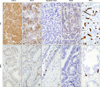
Immunohistochemical staining for COX-2, VEGF, and Bcl-2 was interpreted as positive if any portion of cancer cells or epithelial cells within a prostate gland showed cytoplasmic immunoreactivity. Scoring was performed by 2 independent pathologists, after reaching consensus, by using a multi-headed microscope. To highlight tumor microvessels, endothelial cells were stained for CD34. The densest vascular areas were identified by scanning tumor sections on a low-power field (×40 and ×100). After identification of the densest vascularization, a vessel count was performed on a high-power field (×400). For interpretation of TUNEL staining, we counted apoptotic epithelial cells on a high-power field after determination of the densest region.
6. Statistical analysis
Statistical analysis was carried out by using SPSS ver. 17.0 (SPSS Inc., Chicago, IL, USA). The significance of COX-2, Bcl-2, and VEGF overexpression among various pathologic patterns was assessed by using either the Pearson chi-square or Mantel-Haenszel chi-square test, as appropriate. The Mann-Whitney U-test or Kruskal-Wallis test was used for the comparisons of MVD and apoptotic index. Tukey's test was used for post-hoc multiple comparisons. The strength of association between each immunohistochemical score was assessed by using the Spearman correlation analysis. Statistical analysis was performed, with p<0.05 considered significant.
RESULTS
1. Immunohistochemical expression in BPH and PC
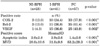
The majority of BPH samples (79.7%) were negative for COX-2. In contrast, the PC samples were positive for COX-2 in 57.9% of cases (p<0.001). Similarly, an overexpression of Bcl-2 and VEGF was observed in PC compared with BPH samples (p<0.001 and p=0.035, respectively). Furthermore, the apoptotic index was lower (1.4±2.0; p<0.001) and MVD was higher (53.2±29.3; p=0.001) in PC samples than in BPH samples (Table 1, Fig. 1).
2. Immunohistochemical expression in NI-BPH, I-BPH, and PC
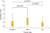
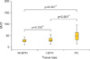
We divided the BPH specimens into NI-BPH and I-BPH subgroups. Overexpression of COX-2 was gradually increased in cases of NI-BPH, I-BPH, and PC (13.0%, 24.4%, and 57.9%, respectively; p<0.001). The overexpression of Bcl-2 and VEGF also gradually increased (p<0.001 and p=0.002, respectively). The apoptotic index and MVD showed significant differentiation in each group (p<0.001, respectively) (Table 2). Although the apoptotic index was higher in I-BPH (p<0.001), there was no significant difference between NI-BPH and I-BPH (p=0.050) or between NI-BPH and PC (p=0.641) (Fig. 2). MVD was significantly higher in PC (p<0.001), but there was no significant difference between NI-BPH and I-BPH (p=0.530) (Fig. 3).
3. Correlation between COX-2 overexpression and apoptosis
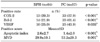
The mean apoptotic index of all cases studied was 2.0±2.5. The correlation between COX-2, Bcl-2, VEGF immunoreactivity, MVD, and the apoptotic index is summarized in Table 3. There was a significant correlation between the overexpression of COX-2 and Bcl-2 (p=0.001). However, the apoptotic index was not correlated with the overexpression of COX-2 or Bcl-2 (p=0.344 or p=0.927).
DISCUSSION
The COXs are a family of myeloperoxidases, located on the luminal side of the endoplasmic reticulum and nuclear membrane, that catalyze the rate-limiting step of prostaglandin biosynthesis from arachidonic acid [15]. Prostaglandins, specifically, are important for physiological functions such as vasodilatation (PGD2, PGE2, and PGI2), gastric cytoprotection (PGI2), the maintenance of renal homeostasis, and platelet aggregation. Prostaglandins also play a major role in mediating fever (PGE2), in pain sensitivity, and in inflammation [15]. Overexpression of COX-2 has been demonstrated to contribute to carcinogenesis by stimulating cell proliferation, inhibiting apoptosis, and enhancing angiogenesis; all of these effects are thought to be mediated via PGE2 [16-18].
At present, COX-2 overexpression has been identified in many tumor tissues. As well, COX-2 overexpression is well-established in PC [5,9]. Madaan et al found expression of COX-2 in 74% of 82 cases of PC [5], Kirschenbaum et al reported expression of COX-2 in 63% of 31 cases [4], and Yoshimura et al found increased expression in 28 cases of PC [9]. Yet, research on the expression of COX-2 in human PC is still controversial. Zha et al conducted a large study analyzing 144 samples of PC as large sections or tissue-microarray cores and found them to be negative for COX-2 [10]. On the other hand, COX-2 expression in a variety of pathologic prostate conditions has been studied. An increased expression of COX-2 in atrophic areas (post-atrophic hyperplasia or proliferative inflammatory atrophy), which were associated with inflammation, has been described in several publications [10-12]. Denkert et al observed an overexpression of COX-2 in PC (44.7%) and prostate intraepithelial neoplasia (72.1%) samples when compared with benign prostatic tissue [13].
In this study, overexpression of COX-2 was observed in PC compared with benign prostatic tissue, and the gradual expression of COX-2 was identified in NI-BPH, I-BPH, and PC. This is in line with the above-mentioned studies, which suggest that chronic inflammation may trigger COX-2 expression in the early stages of prostate carcinogenesis.
Apoptosis is important for the development and maintenance of tissue homeostasis of multicellular organisms [19,20]. An important factor in tumorigenesis is the balance between the proapoptotic and antiapoptotic members of the Bcl-2 family [21]. In a tumor cell, a mutation of the Bcl-2 gene, which results in increased expression, will suppress the normal function of the proapoptotic proteins Bcl-2-associated X protein (BAX) and Bcl-2 homologous antagonist killer (BAK). On the other hand, if a mutation of the BAX or BAK genes causes a down-regulation of expression, then the cell will also lose its ability to regulate apoptosis-again causing tumorigenesis.
In this study, a significant correlation between the overexpression of COX-2 and Bcl-2 was identified, and their gradual overexpression was linked to chronic inflammation and carcinoma. We also found that the apoptotic index was high in BPH compared with PC. These data suggest that COX-2 overexpression in PC is associated with a decrease in apoptosis. In previous studies, the overexpression of COX-2 was found to increase the cellular levels of Bcl-2; therefore, it may cause resistance to apoptosis of premalignant cells [21]. However, the mechanism by which COX-2 inhibition induces apoptosis is not well understood. Studies have suggested that decreased cellular PGE2 and increased arachidonic acid levels might be involved in the inhibition of cell proliferation and the induction of apoptosis [22].
We divided the BPH cases into NI-BPH and I-BPH. The apoptotic index was higher in I-BPH than in NI-BPH or PC, although there were no significant differences between NI-BPH and I-BPH. Therefore, the apoptotic index was not correlated with overexpression of COX-2 and Bcl-2. Apoptosis is usually increased in inflammatory cells as a result of cell death mechanisms induced by cytotoxic lymphocytes [23]. Cell death can be caused by either necrosis or apoptosis. Cytotoxicity is an activity performed by specialized cells such as natural killer cells and CD8+ T cells and is a highly organized multifactor process performed by different cells from the immune system [23]. We suggest that this multifactor process was a cause of the discordant apoptotic index in this study.
Cancer-induced angiogenesis is the result of increased expression of angiogenic factors, decreased expression of anti-angiogenic factors, or a combination of both events [24]. One of the mechanisms by which COX-2 acts as a tumor promoter is through the stimulation of angiogenesis [25]. COX-2 was expressed in newly formed blood vessels within tumors grown in animals, whereas under normal physiological conditions the quiescent vasculature expresses only the COX-1 enzyme [26]. COX-2 modulates angiogenesis by augmenting the release of angiogenic peptides, such as VEGF, thymidine phosphorylase, basic fibroblast growth factor (bFGF), and nitric oxide, by the tumor cells [25,27]. VEGF, a highly specific mitogen of vessel endothelial cells, is the most potent tumor-angiogenic factor and is capable of promoting the proliferation and migration of endothelial cells as well as increasing vascular permeability [28].
In this study, overexpression of VEGF and high MVD were shown in PC compared with BPH. Furthermore, a significant correlation between the overexpression of COX-2 and VEGF was identified, and this gradual expression was associated with inflammation and carcinoma. We also found that MVD was higher in tissues positive for COX-2 and VEGF.
These data suggest that COX-2 may play a critical role in tumor angiogenesis. Recent studies demonstrate that COX-2 overexpression correlates with VEGF mRNA or protein production [29,30]. COX-2 overexpression has been detected in the angiogenic vasculature present within the tumors and preexisting vasculature adjacent to cancer lesions, suggesting that COX-2 may induce newly formed blood vessels to sustain tumor cell viability and growth.
CONCLUSIONS
We found that COX-2 overexpression in PC was correlated with a decrease in apoptosis and an increase in angiogenesis. Chronic inflammation of BPH causes the overexpression of COX-2, resulting in an increase in expression of Bcl-2 and VEGF. It is likely that chronic inflammation plays a role in the intermediate step of carcinogenesis in the prostate.

 XML Download