

 PDF
PDF ePub
ePub Citation
 Print
Print


INTRODUCTION
Testicular torsion, an abnormal twisting or rotation of the testis and spermatic cord, is a pathologic condition resulting in acute, severe scrotal pain. The twisted spermatic cord leads to a decrease or even complete loss of blood flow to the affected testis or other scrotal contents [1]. The peak incidence of testicular torsion occurs in males between the ages of 12 and 18 years, yet it can be seen in any age group, and it is estimated that 1 out of every 4,000 men below the age of 25 will have torsion of the testis [2]. The risk of bilateral synchronous or metachronous testicular torsion is 2 percent. Testicular torsion is also common in undescended or cryptorchid testes. As a medical-legal issue, litigation against physicians for outcomes after testicular torsion repair far exceeds its proportionate representation among urological cases.
CLINICAL ASPECTS
1. Signs and symptoms
Acute, severe scrotal pain is the consistent presentation of testicular torsion, which is sometimes associated with nausea, vomiting, and low-grade fever. Other conditions such as epididymoorchitis and torsion of the testicular appendages are also associated with scrotal pain and therefore need to be considered when diagnosing testicular torsion. On physical examination, the hemiscrotum of the affected side is typically swollen, tender, and inflamed, with an absent cremasteric reflex [3], and with pain unrelieved by elevation of the scrotum [4]. The torsed testicle will also be tender, elevated, and may have a horizontal lie.
Testicular torsion can occur intermittently, because episodic twisting of the spermatic cord can occur followed by spontaneous resolution. Patients presenting with acute scrotal pain followed by rapid resolution should be treated with bilateral orchiopexy to prevent further torsion [5]. Patients with prior orchiopexy may still develop testicular torsion, but such cases are rare [6].
High-frequency transducer sonography including pulsed and color Doppler vascular imaging is the primary imaging modality of choice for assistance in evaluation of the acute scrotum [7]. Diagnosis of testicular torsion based on clinical findings without the use of imaging has a false-positive rate of 50% [8]. To estimate intratesticular blood flow, color Doppler ultrasound is very useful; in fact, color Doppler was 97% accurate in diagnosing testicular torsion in patients with a painful scrotum when detection of intratesticular blood flow was the only criterion used [9]. Another imaging modality that has been used to evaluate patients with testicular torsion is subtraction dynamic-contrast enhanced MRI. This imaging modality can determine decreased testicular perfusion and hemorrhagic necrosis [10]; however, scrotal MRI is not time- or cost-effective in the diagnosis of the acute scrotum.
2. Types of testicular torsion
Generally, testicular torsion is classified into two types according to its relation to the tunica vaginalis. The first type, intravaginal torsion, occurs within the tunica vaginalis and is the most common. If the tunica vaginalis completely encircles the testis, epididymis, and the distal part of the spermatic cord, instead of covering the posterolateral aspect of the testis only, the patient has a deformity called "bell-clapper." In this condition, the testis tends to lie horizontally and is more within the tunica vaginalis. A "bell-clapper" deformity is bilateral in 80% of males with intravaginal torsion, thus putting them at greater risk for torsion [11,12].
The second type of testicular torsion occurs outside the tunica vaginalis and is sometimes termed extravaginal torsion. It occurs exclusively in newborns when the testis and gubernaculum can rotate freely [13]. Its incidence is one in 7,500 newborns. Complicated pregnancies and vaginal deliveries are associated with higher risk for neonatal testicular torsion. Neonates with testicular torsion present with scrotal swelling, discoloration, and with a firm painless mass in the scrotum [14] because the testis is usually necrotic from infarction at the time of birth. Sonographic features will include thickened skin, an ipsilateral hydrocele, and an enlarged heterogeneous testicle without color Doppler flow to the testis or the spermatic cord [15].
3. Torsion of a cryptorchid testis
Acute left scrotal pain along with an empty hemiscrotum raises the suspicion of testicular torsion in the cryptorchid testis. In 73% of cases of torsion of a cryptorchid testis, it was reported on the left side. This often presents with erythremia along with a tender and firm mass in the groin area. Doppler ultrasound is useful to confirm the diagnosis. A delay in presentation, diagnosis, or referral to the urologist often leads to very poor rates of surgical testicular salvage in such cases [16].
4. Torsion of the testicular appendix
Torsion of the testicular appendix is the most common cause of acute scrotum in children. In 91% to 95% of appendiceal torsions, the appendix testis is involved and this is most commonly seen in boys between 7 and 14 years of age [7]. Both testicular torsion and torsion of testicular or epididymal appendages have the same presentation clinically except that the onset of pain is more gradual in torsion of epididymal appendages. In these case, there is a palpable small, firm nodule on the top of the testis on physical examination, with the bluish discoloration of the appendix "blue dot sign," which is visible through the overlying skin [17].
Ultrasound is only useful in cases of appendiceal torsion without a definitive diagnosis based on clinical findings. Typical ultrasound findings in torsion of the appendix include a circular mass adjacent to the testis or epididymis, with variable echogenicity [17]. There may be peripheral flow by color Doppler around the torsed appendage and a reactive hydrocele with skin thickening may be present [18]. Ultrasound cannot demonstrate blood flow or the lack thereof in the torsed or in the normal appendix testis, and there is no difference in echogenicity between the normal and the torsed appendix testis [19]. No surgical treatment is needed for the torsed testicular or epididymal appendage. Conservative treatment with analgesics is recommended.
5. Effects of ipsilateral torsion on the contralateral testis
For many years, there has been clinical concern about the contralateral testis in cases of unilateral testicular torsion, which was supported by early laboratory studies. Careful examinations of severe damage to the seminiferous epithelium after repair of unilateral torsion have never shown a significant contralateral effect on spermatogenesis [20] or even on a number of other parameters [21]. Specific studies of the contralateral human testis now support the conclusion that the so-called contralateral effects of ipsilateral torsion are actually manifestations of preexisting contralateral lesions [22].
6. Treatment of testicular torsion
The onset of acute, severe scrotal pain begins the countdown before testicular infarction occurs. Immediate surgical exploration and bilateral orchiopexy is the treatment of choice in acute testicular torsion, although orchiectomy is required for infarcted or necrotic testicles with delayed presentation [23]. The ability to salvage a testicle depends on the duration and degree of torsion. Surgical intervention within these 6 hours may have a testis salvage rate as high as 100% [24]. If the intervention is made within 6 to 12 hours, testicular salvage rates decrease to 70% and from 12 hours to 24 hours it is diminished to 20% [24]. Salvage of the testis reported by most urologists means the testis is anatomically in its site without gross evidence of infarction. This, however, should not be taken to mean that the testis is functionally or physiologically normal, because ample data in animal models suggest significant defects in testicular function can occur even with a normal palpable testis. Age is a significant predictable risk factor for orchiectomy in patients with testicular torsion, due to the delay in seeking medical attention in older males [25].
Elective bilateral orchiopexy should be done for patients with intermittent testicular torsion. Otherwise, they will be at risk for developing complete testicular torsion with its sequelae of subsequent infarction and testicular loss [26]. Up to 97% of patients treated with prophylactic bilateral orchiopexy have complete resolution of their symptoms with a high likelihood of preventing future infarction [5].
In the case of acute neonatal testicular torsion, some authors think that urgent surgical exploration and concomitant contralateral orchiopexy is the best treatment [27,28]. In this scenario, the risks and benefits to the neonate must be considered; the risk of anesthesia on one hand and the risk of testicular loss in neonates on the other. Extravaginal testicular torsion of the testes at the time of delivery is never salvageable and hence it can be argued that emergent exploration may not be indicated. Occasionally, there is a chance for salvage of the testicle in neonatal torsions, which are first noted after birth and before one month of age, with emergent exploration [29]. Other authors advocate emergent exploration and contralateral orchiopexy in all newborns with intrauterine testicular torsion to decrease their risk of anorchia following contralateral torsion [28].
PATHOPHYSIOLOGY OF TESTICULAR TORSION
1. Animal models of testicular torsion
Experimentally, the testis can be rendered ischemic via two different methods. One method is to clamp the spermatic cord or artery and the other method is to twist the spermatic cord. Both procedures have been used in animal models; however, because the twisting of the spermatic cord more closely resembles clinical testicular torsion, this review will focus on reports that have used that animal model.
In experimental testicular torsion, the degree and duration of torsion can be set and the compromise of blood flow can be monitored. It is important to point out that the injury to the tissue occurs due to both the ischemic period and by the reperfusion. If the ischemic period is long, the added injury by reperfusion will be minimal [30]. Possible mechanisms of testicular dysfunction due to ischemia include the impairment of oxygen and nutrient supplies and the build-up of waste products via the vasculature. Decreases in the intracellular content of ATP and glycogen and an increase in intracellular Ca2+ may also play important roles in testicular injury [31]. This points to the importance of the time between the onset of torsion and its repair in evaluating the testicular injury. Prolonged testicular ischemia will lead to an infarcted testis that should be removed; however, in cases where blood flow returns, testicular function may still be affected. Spermatogenesis after clinical torsion repair is rarely assessed due to ethical concerns; however, testes of laboratory animals with atrophied testes show that testes of half the mass of a normal companion are completely aspermatogenic [20]. Thus, it is possible that significant numbers of "salvaged" human testes with >6 hours of torsion (approximately 65% of all torsions [32]) are not salvaged biologically, and those with torsions of increasing duration are even less likely to be so [33].
Using animal models of testicular torsion, our lab and others have focused on the minimal duration of ischemia followed by reperfusion needed to cause testicular dysfunction. In a number of species from rats [20] to pigs [34], a 1-hour, 720° torsion followed by torsion repair induces an ischemia-reperfusion injury (IR-injury) sufficient to disrupt the seminiferous epithelium. The method of inducing torsion used in our lab has been shown to cause a consistent, severe obstruction of testicular blood flow [20,21,34]; yet, it is interesting that a 1-hour, 720° torsion disrupts spermatogenesis in the rat [20], but a 2-hour, 720° torsion is required to induce the same effect in the mouse [35]. The IR-injury is associated with activation of neutrophils, upregulation of inflammatory cytokines and endothelial cell adhesion molecules (CAMs), increased thrombogenicity, release of massive intracellular Ca2+, and generation of oxygen-derived free radicals [31]. These are discussed in detail below.
2. Vascular consequences of torsion
It is known from studies in the rat that the permanent loss of spermatogenesis seen after repair of 1-hour, 720° torsion is not due to failure of average blood flow values to return to normal [21]. On the other hand, the pattern of testicular microvascular perfusion does remain altered in the hours and days after torsion repair [36]. In the normal testis, variation in microvascular blood flow is due to vasomotion or cyclic vascular contraction and relaxation under complex regulation [37,38]. Vasomotion is significantly altered after torsion repair and only returns days later [36]. The inhibition of the vascular contractions necessary for vasomotion is likely due to the vascular relaxing effects of NO, which is increased in the testis after torsion repair [39,40]. NO is also active in cell processes other than vascular relaxation and may be a key signaling molecule for processes in the testis leading to IR-injury. For example, NO has been reported to be a regulator of the expression of CAMs [41,42]. CAMs play a key role in IR-injury in the testis and other tissues because they are key modulators of leukocyte recruitment, and leukocyte recruitment is the forerunner of much of the IR pathology [42,43]. It is also true that testicular vascular permeability is increased after IR of the testis [44]. The association between this and neutrophil diapedesis across the vascular endothelium is possible but has not been explored.
3. The endocrine consequences of torsion
Endocrine function of the rat testis after repair of 1-hour, 720° torsion is significantly reduced; however, testosterone levels do return to normal values days after the repair of torsion [45,46]. The severe disruption of Sertoli cells is not a primary feature of testicular IR-injury, at least as assessed by overall synthesis and secretion of proteins in vivo [47]. That is not to say that Sertoli cell protein synthesis is completely unaltered after an IR event, it only says that Sertoli cells survive after torsion repair and continue production of a broad panel of proteins. Indeed, the transcription factor NFκB is activated after torsion repair and is localized to Sertoli cells [48].
4. Apoptotic consequences of testicular torsion
Cells die by either necrosis or apoptosis. Necrosis can be caused by a variety of extracellular factors and results in cell swelling, lysis, and an inflammatory-like response [49]. Apoptosis is the result of the activation of an intracellular program that leads to cell death without induction of an inflammatory response [49,50]. There are numerous paths to apoptosis depending on proximate causes and the tissue involved, but there are two major divisions of the pathway within the cell; the intrinsic or mitochondrial pathway and the extrinsic or "death domain"-mediated pathway [51,52]. The intrinsic pathway is induced by cell stresses such as irradiation, toxin exposure, or reactive oxidant injury. The primary effect of these stresses is on the mitochondria, where associations between pro- and anti-apoptotic members of the Bcl-2 family (e.g., Bax and Bcl-XL, respectively) either stabilize or destabilize the mitochondrial membrane [53]. Membrane destabilization leads to release of mitochondrial cytochrome c into the cytoplasm, its binding to Apaf-1, and activation of a cascade of cytoplasmic caspases that eventually activate a caspase-activated DNAse. This DNAse then degrades DNA into regular, approximately 185 bp fragments characteristic of true apoptosis [51]. The extrinsic pathway is activated by extracellular ligands [e.g., Fas Ligand (FasL), TNF-α] binding to specific cell membrane receptors (e.g., Fas, TNFR). These receptors initiate an intracellular process also leading to activation of the caspase cascade, but which largely bypasses the Bcl-2 family relationships in the mitochondria [51,52]. There is cross-talk between the two pathways, e.g., via Bid, and much more complexity than can be presented here.
Apoptosis, a significant process in normal spermatogenesis [54], is massively upregulated in a number of conditions disrupting spermatogenesis [55,56], including testicular torsion [57,58]. A major goal of our laboratory is to understand the molecular pathway to germ cell apoptosis (GCA) induced by testicular torsion and its repair or IR-injury of the testis. Our previous results have demonstrated that Bax is the predominant pro-apoptotic molecule in the rat testis and it exhibits increased expression after torsion [57]. Furthermore, results using the mouse testis have demonstrated the presence of Bax as well as the movement of Bax to the mitochondria after IR [59]. Other recent results have shown for the first time the presence of caspase-2 in the testis and its activation with GCA [59,60]. The role of caspase-2 in apoptosis in not well understood in any tissue, and whether it acts upstream or downstream of Bax remains controversial [61,62]. Upregulation of FasL gene expression is detectable after torsion repair with RT-PCR; nevertheless, it appears that testicular torsion induces rat GCA largely through the mitochondrial pathway where Bax and Bcl-XL are the predominant pro- and anti-apoptotic genes expressed. Furthermore, spermatogonia in stages II and III of the rat seminiferous tubule are the primary target cells in the earlier stages of this injury [30].
5. Torsion and the extratubular events leading to apoptosis
Data from our laboratory have demonstrated that cellular and molecular processes are initiated in the vascular endothelium by IR-injury and that they are important in the etiology of the lesion including GCA [35,63]. Thus, it is important to consider all aspects of testicular IR-injury, from endothelial cells to germ cells, to fully understand the pathology.
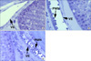
Contemporaneous with GCA in the rat is an increase in adhesion and diapedesis of leukocytes in the testicular vasculature and an increase in testicular oxidative stress [58]. These are the hallmarks of classic IR-injury studied in other tissues. IR-injury includes production of reactive oxygen species (ROS) by invading leukocytes, by resident cells themselves, or both [64]. We have demonstrated that mouse testicular torsion followed by repair induces a loss of spermatogenesis, an increase in GCA, an increase in testicular leukocytes, and an increase in oxidative stress [35,46,59,63]. Furthermore, mouse testicular IR-injury appears to mimic that in the rat testis, and in the mouse, testicular vascular endothelial CAMs, the selectins, are important in the recruitment of neutrophils in the inflammatory response (Fig. 1). We have demonstrated the key role of E-selectin in the recruitment of neutrophils into the testis after torsion repair and the subsequent increase in GCA [35]. Thus, GCA is largely induced via the intrinsic pathway by oxidative stress arising from invading leukocytes requiring E-selectin expression in the testicular vasculature. Enhanced expression of TNF-α and IL-1β has been shown after IR-injury in other cell types [65]. Recent research in other endothelia has provided evidence that TNF-α and IL-1β can stimulate E-selectin expression through an NFκB nuclear localization pathway or phosphorylation of JNK [66], but cytokine up-regulation and its source in the testis after IR, the role of NFκB or of JNK isoforms, the transcription factors phosphorylated by JNK, and which upstream kinases function most prominently in the torsion pathology remain open questions. It is clear that signaling pathways are very complex and often interactive. In this regard, it has recently been demonstrated that JNK signaling is important in inducing the intrinsic pathway to apoptosis in cultured fibroblasts [67], and TNF-α has been implicated in the induction of oxidative stress in certain cells [68] rather than being a response to it. When the neutrophils transmigrate through the endothelium to the interstitium of the testes, they are poised to release factors such as ROS [58] or FasL [57] that may directly cause apoptosis in the germ cell.
During the late phase of the reperfusion, there is an increased number of hematogenous macrophages. Macrophages can secrete IL-8 [69] as well as 25-hydroxycholesterol [70], a compound that is involved in spermatogenesis, but which at high concentrations can induce apoptosis in a number of cell types [71,72]. Thus, resident testicular macrophages may play a role both in the recruitment of neutrophils and in the observed germ cell apoptosis seen after IR of the testis. This is a possibility yet to be examined.
6. Therapies targeting specific discovered steps in the pathway to germ cell apoptosis
Understanding testicular IR-injury involves deciphering the effects on key cell types in the testis; this will give insights into the injury to the organ as a whole. An important reason for determining the molecular chain of events from the vasculature through to GCA is to identify potential target sites for therapeutic applications. For example, we have already determined that the testis after IR suffers oxidative stress amenable to treatment with oxygen radical scavengers [73,74]. It has been reported that treatment with the antioxidants allopurinol, superoxide dismutase, and catalase caused significant rescue of testes function after testicular detorsion. Other antioxidants, metal chelators, and Ca2+ channel blockers (e.g., vitamin E, deferoxamine, and diltiazem) have been also used to prevent IR-injury in testis. Treatment with dexamethasone also causes significant reduction in germ cell apoptosis and neutrophil adhesion in testicular subtunical veins and this reduction was reduced markedly by mifepristone, the glucocorticoid receptor antagonist [75].
The reduction of nitric dioxide (NO2) to nitric oxide (NO-) is promoted by ischemia-induced acidosis as well as enzymatic catalysis [28]. Peroxidation of lipids can result after the production of NO, and the end product of lipid peroxidation is malondialdehyde, which can be used as an estimate for the degree of lipid peroxidation. Numerous studies have demonstrated that decreasing the levels of NO reduces tissue damage. For example, mice pretreated with N-mono methyl L-arginine (L-NMMA), which acts by inhibiting the synthesis of NO, showed less histological damage than controls [76]. By contrast, mice pretreated with L-arginine (a precursor of NO) showed increased histological damage. Interestingly, preconditioning of the testis by exposing it to brief periods of transient ischemia (5, 10, and 15 minutes) followed by 720° torsion for 180 minutes showed significantly decreased levels of NO [77]. More recently, other compounds, including Vardenafil and Ginkgo biloba, have been associated with reduction of eNOS and iNOS, two enzymes critical for NO synthesis [78].
It has also been reported that molsidomine (MO), which is a precursor of a NO donor, has a protective effect against IR-injury in rat testes. MO treatment reduced the levels of malondialdehyde and increased the expression of Sonic hedgehog [79]. Sonic hedgehog is a neurogenic morphogen and is necessary for embryonic arterial differentiation and angiogenesis, possibly through the upregulation of vascular endothelial growth factor (VEGF), angiopoietins, and HIF1-α. Interestingly L-NG-nitroarginine methyl ester (L-NAME), the nitric oxide synthase inhibitor, reversed the protective effect of MO against IR-injury. The effects of MO may be related to reducing the effects of oxidative stress in the testes.
CONCLUSIONS
Torsion of the testis is a medical emergency occurring primarily in adolescent males and young men. Testicular torsion must be treated to avoid loss of the ipsilateral testis. IR of the testis results in germ cell apoptosis and subsequent decrease in testis weight and daily sperm production. Deciphering the pathways that lead to decreased spermatogenesis will aid in finding treatment options for affected patients.
 XML Download