

 PDF
PDF ePub
ePub Citation
 Print
Print


Abstract
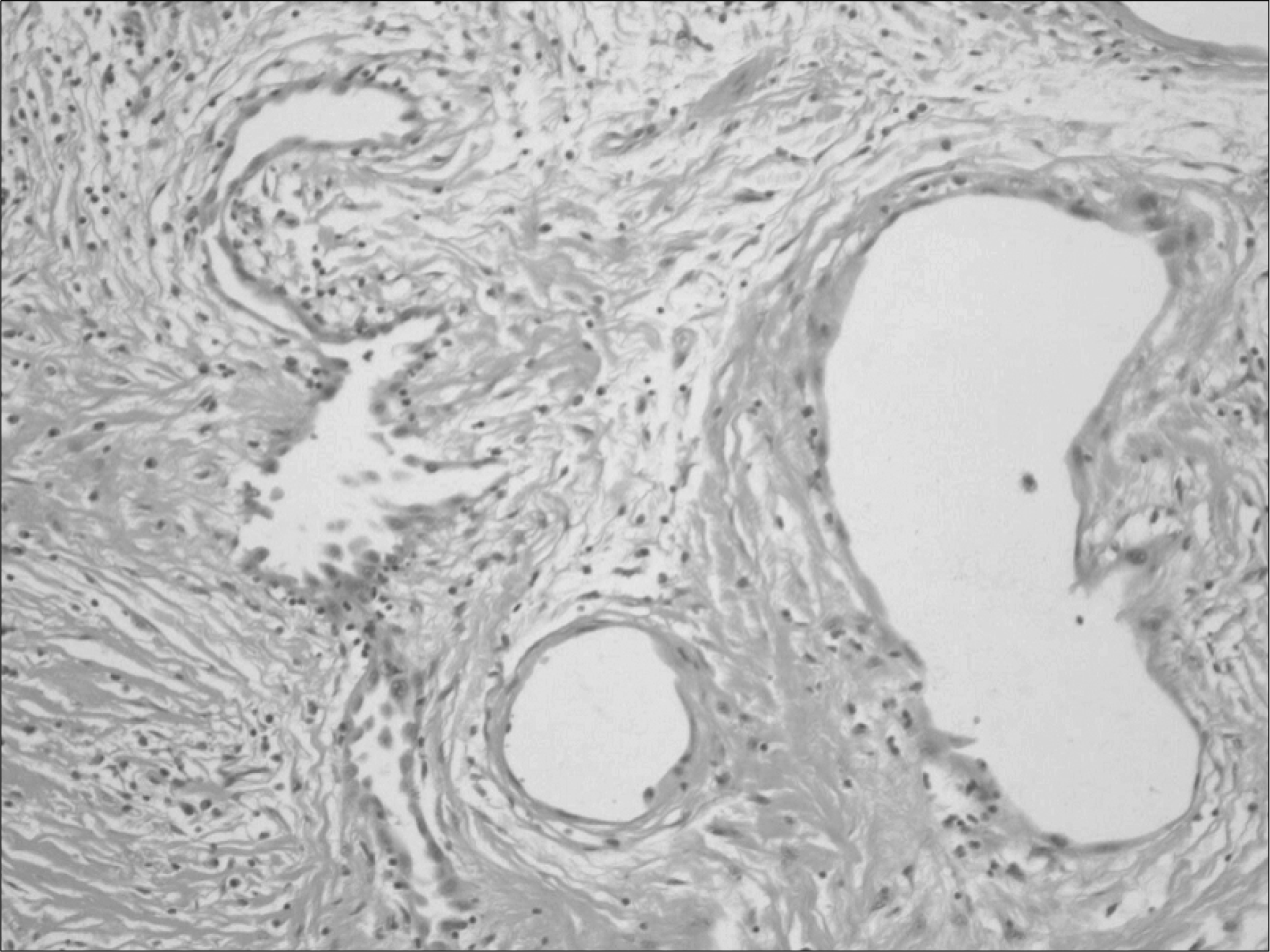
Adenomatoid tumors are rare benign neoplasms thought to be of mesothelial origin. Although most reported cases developed from the epididymis, rare cases have been reported in the testicular tunica, spermatic cord and ejaculatory ducts. Because of the benign nature of this tumor, the treatment of choice is local excision. We report a rare case of adenomatoid tumor of the spermatic cord treated by local excision.
REFERENCES
1.Tammela TL., Karttunen TJ., Makarainen HP., Hellstrom PA., Mattila SI., Kontturi MJ. Intrascrotal adenomatoid tumors. J Urol. 1991. 146:61–5.

2.Kwak H., Jung S., Park M., Chung J. Adenomatoid tumor of the testis with infiltration to the seminiferous tubules. Korean J Urol. 2006. 47:1127–9.
3.Park SM., Jun IS., Hwang CH., Heo C., Lee TH., Hong SJ, et al. Adenomatoid tumor of the tunica vaginalis testis. Korean J Urol. 2004. 45:1171–3.
4.Golden A., Ash JE. Adenomatoid tumor of the genital tract. Am J Pathol. 1945. 21:63–79.
5.Svanholm H., Paulsen S. Immunohistochemical and electron microscopic study of twelve adenomatoid tumors. Tumori. 1985. 71:141–5.
6.Stephenson TJ., Mills PM. Adenomatoid tumors: an immunohistochemical and ultrastructural appraisal of their histogenesis. J Pathol. 1986. 148:327–35.
7.Han CH., Sun IC., Kwak KM., Chung WK., Ha IS., Shin OR, et al. Adenomatoid tumor of the epididymis. Korean J Urol. 2002. 43:256–8.
8.Lioe TF., Biggart JD. Tumours of the spermatic cord and paratesticular tissue. A clinicopathological study. Br J Urol. 1993. 71:600–6.
9.Choi YH., Chae SW., Ahn HK., Lee MC., Park YE. Giant cystic adenomatoid tumor of the uterus. Korean J Pathol. 1993. 27:85–7.
10.Feuer A., Dewire DM., Foley WD. Ultrasonographic characteristics of testicular adenomatoid tumors. J Urol. 1996. 155:174–5.
Fig. 1.
Scrotal ultrasonogram revealing an approximately 2.5x2.0 cm, oval shaped, well demarcated, heterogeneous echogenic mass in the spermatic cord.
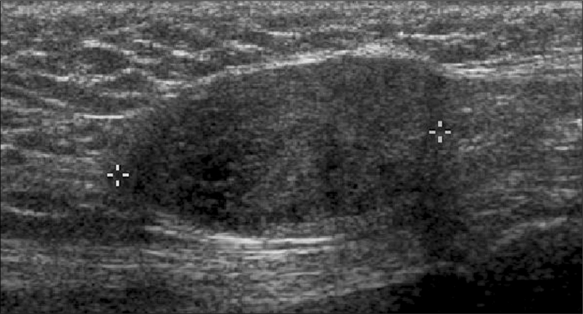
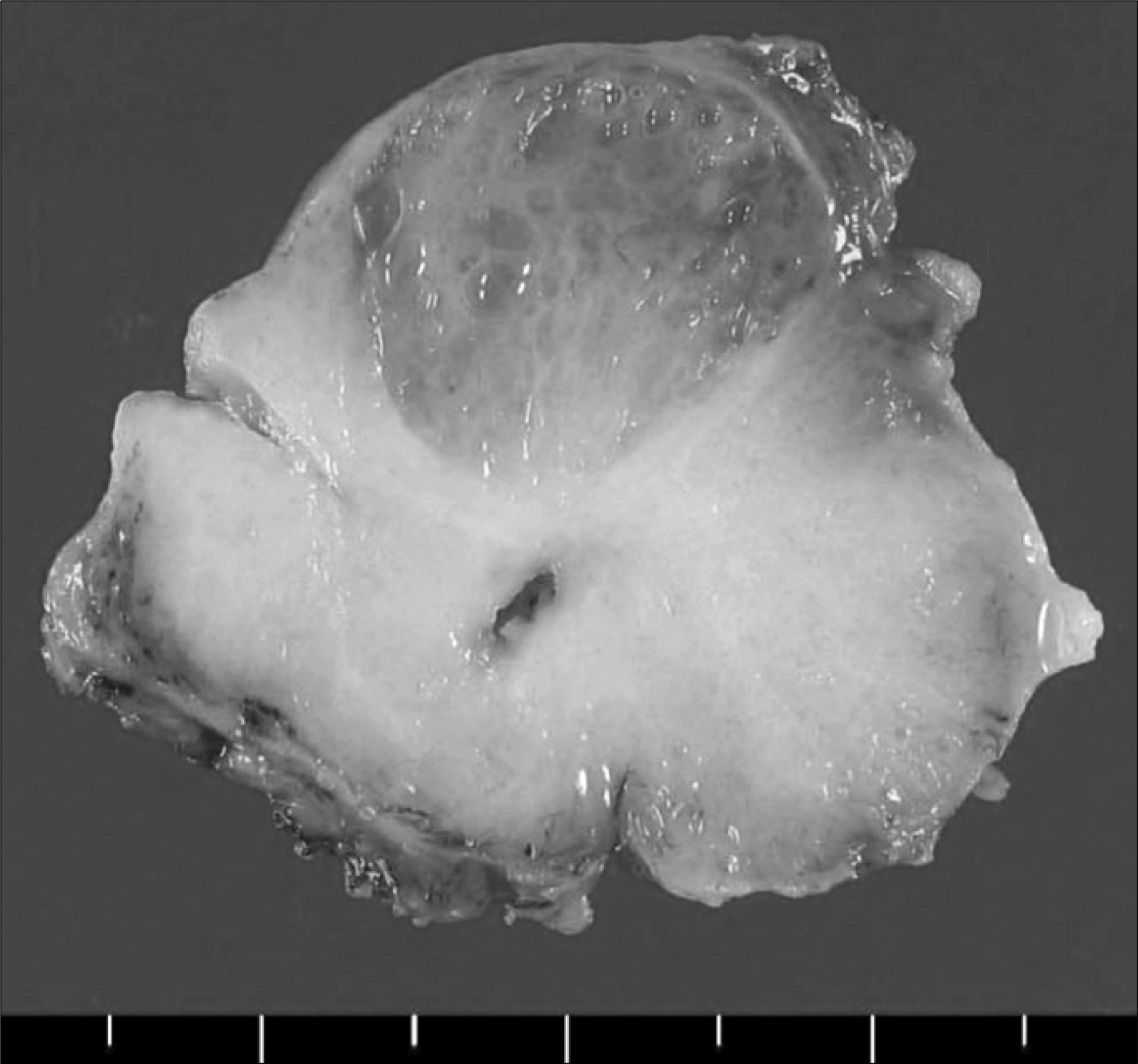
Fig. 2.
Grossly, a well-defined mass (2.8x2.5x2.0cm) was found with a grayish white, slightly myxoid, cut surface.

 XML Download