

 PDF
PDF Citation
 Print
Print


Abbreviations
AID
activation-induced cytidine deaminase
BCR
B cell receptor
CMV
cytomegalovirus
CR
caloric restrictions
CSR
class switch recombination
DAMP
damage-associated molecular pattern
DC
dendritic cell
EV
extracellular vesicle
IFV
Influenza virus
NET
neutrophil extracellular traps
NLRP3
NACHT, LRR and PYD domains-containing protein 3
SASP
senescence associated secretory phenotype
SA-T
senescence-associated T
SHM
somatic hypermutation
VZV
Varicella Zoster virus
INTRODUCTION
Aging, which is a complex biological process, results in profound alterations in the immune system. And these changes can accumulate to produce a progressive deterioration in the ability to respond to pathogens and develop proper and durable immunity after vaccination. Consequently, a higher mortality rate is associated with the elderly population. The multifaceted, age-associated changes of the immune system can be described by the term immunosenescence, which commonly refers to the diminished ability of the immune system. Although immunosenescence has gained increasing attention from the scientific community, its underlying molecular mechanisms remain to be fully investigated. Given a dramatic increase in the global life expectancy, identifying and understanding the mechanisms by which aging affects the components of the immune system will be crucial for the development of effective age-targeted vaccines and immunotherapies.
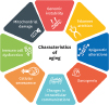
The cellular and molecular hallmarks of aging have been previously described to include the following: genomic instability, telomere attrition, epigenetic alterations, sarcopenia, change in intracellular communications, cellular senescence, immunosenescence, and mitochondrial dysfunction (1) (Fig. 1). In addition to these features, chronic inflammation is considered as the key underlying mechanism leading to aging and aging-associated diseases. The term “inflammaging” is used to characterize persistent, low-grade systemic inflammation associated with aging (23). One possible factor that may contribute to age-associated dysfunction and chronic inflammation is cellular senescence. Senescent cells have been shown to accumulate over the lifespan of rodents, nonhuman primates, and humans (4). Cellular senescence is mediated by p21/p53 pathway, and in addition to having a large, flat cell morphology and higher cellular production of reactive oxygen species, senescent cells produce senescence associated secretory phenotype (SASP), which chronically releases cytokines and chemokines that promote leukocyte recruitment and tissue repair and remodeling (5).
Figure 1
Multiple factors involved in aging. The diagram highlights multiple factors involved in aging; genomic instability, telomere attrition, epigenetic alterations, sarcopenia, changes in intracellular communication, cellular senescence, immunosenescence, and mitochondrial dysfunction.
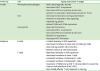
The process of aging is closely related to significant alterations in both innate and adaptive immune systems (Table 1). In terms of innate immunity, aging can result in both quantitative and qualitative changes, including decreased number of circulating monocytic and dendritic cells (DCs), reduced phagocytic activities of macrophages or migratory neutrophils, and impaired Ag presentation abilities by DCs. With respect to T cells, aging can result in the reduction of TCR repertoire, due to the thymic involution at puberty, and the accumulation of senescent or exhausted T cells that are functionally inert or dormant. Several factors, including chronic viral infection and the release of damage-associated molecular patterns (DAMPs), can contribute to age-dependent immune dysregulation that drives age-associated diseases, such as atherosclerosis, Alzheimer's diseases, and infectious diseases. Furthermore, vaccine efficacy in aging people is influenced by age-related alterations, ranging from decreased number of circulating naïve B and T cells, limited diversity in the BCR repertoires, and defective Ab response to new Ags (678).
Table 1
Summary of the immune changes associated with aging
Multiple studies have shown how aging induces immunological changes during viral infections and vaccine responsiveness. For instance, immunosenescence in the elderly contributes to reduced vaccine efficacy and increased susceptibility to infectious diseases. The elderly population is the most vulnerable to viral infections with Influenza virus (IFV) and Varicella Zoster virus (VZV), which are among the pathogens causing the most common infectious diseases worldwide. Vaccines for both influenza and shingles have been available for a long time; however, the low vaccine efficacy and effectiveness in the elderly suggest an aging-associated decline in the immunogenicity induced by vaccination.
This review will highlight recent advances and perspectives on the modifications of cellular and molecular characteristics of both innate and adaptive immune responses during aging. Furthermore, we discuss how aging cells or organisms respond to viral infections and vaccines, as well as the recent implications of next generation strategies for vaccine development for the elderly.
AGING-ASSOCIATED CHANGES IN INNATE IMMUNE CELLS
Receptors/sensors
TLRs are highly conserved receptors that can recognize a variety of stimuli, including pathogen-associated molecular patterns such as bacterial lipoproteins, lipopolysaccharides, and bacterial or viral DNA/RNA. TLRs play a key role in the innate immune system as regulators of the innate immunity against microbial infections. Recent studies have elucidated the consequences of aging on TLR function in human cohorts, adding to the existing findings that have been observed in animal models. TLR expression and function in monocytes (9), DCs (10), and neutrophils (11) decline with advancing age. Similar to humans, old mice demonstrate lower levels of TLR expression in splenic and peritoneal macrophages (12). While macrophages from young mice upregulate expression of most TLRs in response to specific ligands, TLR expression in macrophages of old mice is poorly upregulated, and TLR3 expression, in particular, is poorly detected (12).
Interestingly, not all TLR expressions were attenuated in the aged groups. For example, surface expression of TLR1 decreases significantly with age while surface TLR2 expression is unchanged by aging (9). Low surface expression level of TLR1 and defects in TLR1/2 signaling in aged groups led to an attenuated MAPK signaling, and further decreased the production of TNF-α and IL-6 in human monocytes. Similar to this finding, macrophages from aged mice secreted lower levels of IL-6 and TNF-α after stimulation with TLR ligands, especially TLR1/2, TLR2/6, TLR3, TLR4, TLR5, and TLR9 (12).
Furthermore, in monocytes from old individuals, elevated level of TLR5 expression resulted in an increase in TLR5-induced production of cytokines (13). Lim et al. (14) reported that TLR5 signaling is well maintained throughout the course of aging, and in vitro overexpression of caveolin-1 enhanced TLR5 mRNA through the MAPK pathway and prolonged the half-life of TLR5 through direct interaction. Overall, expression of TLRs, except for TLR5, decreases with advancing age, and the impaired localization of TLRs can induce alterations in cytokine and chemokine production that ultimately affect the immune response.
In addition to TLRs, the inflammasome—a multi-protein complex containing NACHT, LRR and PYD domains-containing protein 3 (NLRP3), apoptosis-associated speck-like protein containing a CARD, and caspase 1, which is activated by DAMPs, including microbial genome, endotoxin, extracellular ATP, β-amyloid and intracellular uric acid—has been suggested as an important modulator of age-associated inflammatory changes (15). Furthermore, inflammaging has been suggested to be associated with the canonical NLRP3 inflammasome (16). Aging can induce changes in NLRP3 expression levels in age-related disease model, as evident by the higher NLRP3 gene expression in the old subjects relative to the young subjects (1718). In addition, studies using macrophages isolated from aged mice have demonstrated how the aging-associated increase in ROS and endoplasmic reticulum stress, mainly due to unfolded proteins, downregulated the activity of caspase 1 and normal activation of NLRP3 during Streptococcus pneumoniae infection (19). Moreover, impaired NLRP3 function was observed in aged mice during the IFV infection (20). Youm et al. (21) has also highlighted the importance of NLRP3 in aging, during which NLRP3 deficiency in mice not only improved glycemic control, but also attenuated bone loss and thymic demise. Notably, NLRP3 inflammasome-dependent IL-1 inhibition can improve cognitive function and motor performance in aged mice, suggesting that the abrogation of NLRP3 inflammasome can be an innovative therapeutic target for multiple age-related neurological disorders.
Monocytes and macrophages
Despite the lack of significant differences in the number of total monocyte subsets between the young and older, global analysis of circulating monocytes in various age groups shows dramatic age-associated changes in humans (22). As an example, non-classical CD14+CD16+ monocytes significantly increased with age, but displayed reduced HLA-DR and CX(3)CR1 surface expression in the elderly. On the contrary, classical CD14+CD16- monocyte counts did not vary with age, although concentrations of serum MCP-1, but not MIP-1α, MIP-1β, or fractalkine (CX3CL1) increased with age (23). In response to TLR agonists, human monocyte subsets were found to have different transcriptional or functional levels according to age, and this difference induced alterations in surface molecule expression and reduced production of interferons and cytokines like IL-1β (24). Interestingly, monocytes from older individuals exhibit impaired phagocytosis but contain shortened telomeres and significantly higher intracellular levels of TNF-α both at the basal level and following TLR4 stimulation, suggesting dysfunctional monocytes in the aged (25).
In addition to changes in monocytic function, aging can also affect macrophage function. As previously described, the expression of TLR on macrophages is reduced in humans and mice of advanced age (1226). Decreased TNF-α and IL-6 and increased IL-10 production levels following stimulation with TLR ligands in the aged mice are well described by Chelvarajan et al. (27). Also, aged macrophages have reduced number of CD14 and TLR4 expressing cells, and this led to the reduction of cytokines such as IL-6, TNF-α, IL-1β and IL-12 (27). Additionally, LPS stimulation, TLR activation, and IFN-γ stimulation are less effective on the expression of MHC class II molecules in aged macrophages (28). Very recently, van Beek et al. (29) proposed that inflammaging can lead to the accumulation of alternatively activated (M2-like) macrophages, which remain pro-inflammatory in tissues, and express senescence markers. These findings, therefore, demonstrate that aging in macrophages influences many processes including TLR signaling, polarization, phagocytosis, and wound repair.
DCs
A number of studies show that aging does not alter the number of myeloid DCs (mDCs), but reduce the number and function of plasmacytoid DCs (303132). Of note, specialized DCs like the Langerhans cells present in the epidermis and mucosal tissues are reduced in number with aging (3334). Functionally, DCs from elderly individuals displayed a significantly reduced ability to phagocytose Ags. Furthermore, changes in the Ag presentation and migratory capacity of DCs can cause malfunctions in adaptive immunity, during which T cells and DCs are involved in immune tolerance, and lead to autoimmune diseases (353637). According to Agrawal et al. (3638), in comparison to young mDCs, old mDCs induce increased levels of IL-6 and TNF-α in response to LPS, ssRNA, and self-DNA. This induction is attributed to age-associated alterations in signaling pathways leading to PI3K, NF-κB, or type I IFN response. In DCs from old individuals, increased basal activity level of p65 during NF-κB pathway, in addition to increased IFN regulatory factor 3 activation and IFN-α secretion levels in response to self-DNA, has been observed. Overall, age-associated changes in signaling pathways in DCs can impact their function and result in outcomes such as dysfunctional cytokine secretion in response to pathogens or self-DNA and reduced phagocytosis and migration abilities.
Neutrophils
Neutrophils are major phagocytic cells that are specialized in early defense against invading pathogens (39). While the number of neutrophils remain unchanged with increasing age, neutrophils in the elderly tend to exhibit dysfunctional phagocytic and chemotactic abilities (40). Although high level of energy is required for neutrophils to carryout phagocytosis, aging inhibits hexose transport and increases intracellular calcium level in order to inhibit energy uptake and ultimately phagocytosis. Furthermore, increased activity of phosphorylated PI3K, which controls phagocytosis, degranulation, and chemotaxis, in the older induces inaccurate migration of neutrophils and damages normal tissue instead of abnormal tissues at the site of inflammation or infection (41). Neutrophils from older adults, compared to those from younger adults, showed impaired phagocytosis of opsonized Escherichia coli and S. pneumoniae (4243).
Aging also affects the recruitment of neutrophils, which are the first cells to migrate to the infection site, as demonstrated by studies that have observed impaired neutrophil-mediated chemotaxis in relation to advanced age (4044). In addition, neutrophils of the elderly produce fewer neutrophil extracellular traps (NET) that are comprised of nuclear components and granule proteins, and are able to bind and trap extracellular pathogens to defend against infections (4546). Consequently, age-related reduction in NET production can lead to delayed wound healing and high level of susceptibility to invasive methicillin-resistant Staphylococcus aureus (45). Given the recent findings that highlight the phenotypic diversity of neutrophils, it will be important for researchers to characterize aged neutrophils using global transcriptomic and proteomic analysis tools.
AGING-ASSOCIATED CHANGES IN ADAPTIVE IMMUNE CELLS
B cells
Diversity in B cell repertoire is essential for an effective immune response, given that B cells have to provide a variety of specific Abs to recognize a wide range of challenging Ags. Many elderly individuals are known to have limited diversity in B cell repertoire, potentially contributing to the older more prone to infectious diseases, less able to response well to vaccination and more likely to have autoreactive Abs. A previous report indicates that aging may cause significant changes in the selection process during affinity maturation of B cells (47), and also that the B cell repertoire is often less diverse in old age with evidence of non-pathogenic clonal expansions (8). This loss of diversity is likely to be correlated with poor vaccine responses against many pathogens (6). In terms of age-associated changes in B cell number, aging in mice reduces the number of naïve B cells and plasma cells while increasing the population of CD27+ memory B cells (48). Also, human peripheral B cell percentages and numbers significantly decrease with age, and although B lymphopoiesis is active throughout life, there is a decline in B cell production in the bone marrow in aged groups (495051).
Changes in the B cell population are also associated with Ab responses, during which age decreases the ability of B cells to mount an appropriate Ab response against new or known Ags, and the Ab response consists of defective isotype switching and short period of activation (52). Activation-induced cytidine deaminase (AID) is the principal regulator of class switch recombination (CSR) and somatic hypermutation (SHM), both of which are cellular processes that generate diverse Abs. Alteration in MAPK signaling, along with reduced mRNA stability and DNA binding affinity of E2A-encoded transcriptional factor E47, further contributes to the suppressed AID gene expression in the older. Subsequently, these intrinsic changes in B cells ultimately decrease Ab diversity and recently, Frasca et al. (53) has suggested miRNA-155 and miRNA-16 as contributing factors to these molecular alterations associated with B cell aging via downregulation of AID and E47. Aging-related downregulation of E2A result in defects in CSR of IgM memory B cells, and lower AID gene expression level in B cells has been shown to induce deterioration of CSR in the older (5455). Overall, it is highly likely that lower levels of AID and E47 expressions in the older also reduce the number and size of germinal centers, where Ab affinity maturation processes, including SHM occurs, and in turn decrease Ab affinity maturation and the number of circulating Ab from plasma cells (56).
Aging can also affect the quality of Abs from B cells and result in defects such as increased number of self-recognition Abs and skewed variable gene usage (5758). Furthermore, Han et al. (59) demonstrated a substantially higher level of Ab-forming cells in the spleens of aged mice than in those of younger controls. On the other hand, a significant decrease in the number of high-affinity, class-switched Ab-forming cells was observed in the spleen of aged mice. The lack of a considerable reduction in the total number of IgG1 splenic plasma cells suggests that a deficiency in isotope switching was not the cause of the accumulation of low-affinity IgM Ab-forming cells in the spleens of aged animals. Remarkably, both low and high affinity plasma cells were significantly diminished in the bone marrow of aged mice. Additionally, age-associated exposure to chronic cytomegalovirus (CMV) infections can lead to altered B cell function. Wang et al. (60) examines the repertoire of genes encoding the immunoglobulin heavy chain in young and elderly adults to determine the effects of aging and CMV infection on B cell populations. Age did not affect the use of variable, diversity, or joining segments; however, the loss of selection against longer complementarity-determining region 3 segments in the older indicate an age-associated difference between the tolerance mechanisms utilized by different age groups.
T cells
T cells undergo profound and complex changes with aging, including epigenetic and metabolic modifications, affecting a range of subsets including naïve, memory, and effector T cells (6162). Although aging does not change the level of IL-7, a key maintenance factor of T cell homeostasis, it generally decreases the number of naïve T cells and increases the number of senescent T cells (52). Aging T cells undergo distinct changes, which limit Agic specificity and decrease the expression of TCR, that lead to age-associated alteration of TCR-inducible gene expression in human CD4+ T cells (6364). Analysis using high-throughput Illumina sequencing platform revealed age-associated reduction in TCR diversity, indicating a significant reduction in the number of naïve T cells and TCR-beta diversity by the age of 40 (65). Additionally, thymus involution and changes in the expression level of transcriptional factors result in defective T cells that induce inflammaging and increased susceptibility to infection by decreasing vaccine efficacy.
Given that cytokines are key regulatory molecules of T cell-mediated immune response, it has been noted that aging-related T cell defects may originate from alterations of cytokine production. In particular, a shift in the cytokine profiling indicates that aged T cells predominantly show Th2-like phenotype (66). Th17 cells defend the host against extracellular pathogens and are associated with the development of autoimmune diseases and chronic inflammatory diseases in humans (67). The ratio of Th17 to Treg appears to increase with age and Schmitt et al. (68) suggest that this variation in the ratio may explain the increased frequency of autoimmune disease and diminished response to infections in the elderly. In particular, age-dependent increase in the ratio of Th17 cells to Treg cells in the elderly may trigger a shift in the basal levels of pro-inflammatory cells and contribute to autoimmune diseases and reduced immune response to infection.
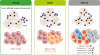
Fig. 2 shows the impact of immunosenescence on T cells involved in chronic viral infections. As an example, aging results in a decreased number of naïve CD8+ T cells, reduced diversity of the TCR repertoire, and elevated senescent, exhausted, or inflationary T cells, which are CMV-specific CD8+ T cells. Recently, Tahir et al. (69) has defined senescence-associated T (SA-T) cell phenotype as PD-1+/CD153+ memory phenotype CD4+ T cells. SA-T cells exhibit features of cellular senescence, which is characterized by defective TCR-mediated proliferation and T cell cytokine production. In particular, these cells secrete abundant atypical pro-inflammatory cytokines, potentially accumulating and causing persistent inflammation in tissues under metabolic stress or tumors (70).
Figure 2
The impact of immunosenescence on persistent viral infection and immunity. Aging leads to numerous changes in major components of both the innate and adaptive immune systems. In response to a viral infection, innate immune cells can trigger the activation of IFN pathways to clear the virus-infected cells. Age-associated defects in innate immune cells can lead to reduced IFN production. Persistent viral infection, such as CMV persistence, can have a profound effect on alterations in adaptive immunity, in particular, T cell composition and function. In the elderly, there are decreased numbers of naïve T cells, but increased numbers of senescent, inflationary, or exhausted T cells that are functionally inert or dormant.
In addition to increased senescent T cells, progressive and prolonged expansion of inflationary T cells also increases in the aged groups (71). Inflationary T cells show limited TCR repertoire, low levels of lymph node homing markers that induce accumulation in non-lymphoid peripheral organs and co-stimulatory receptors, and high levels of inhibitory receptors (7273). Furthermore, inflationary T cells from old mice with CMV infection express lower levels of CD62L and CD127, which are associated with central memory T cells (74). Similar to inflationary T cells, senescent T cells from both human and mice display loss of CD28 and gain of CD57 expression (7576). The elderly also exhibit greater T cell exhaustion, which is a state of T cell dysfunction that arises during chronic viral infections or cancers. In particular, higher expression levels of CTLA-4, PD-1 and TIM-3 by T cells were observed in the elderly (777879). Given the increasing evidence that highlight the important role of T cell immunosenescence in diverse age-related chronic disorders and cancer, targeted elimination of SA-T cells represents a promising strategy for controlling chronic inflammatory disorders and possibly cancer.
IMPACT OF AGING ON VIRAL INFECTION AND IMMUNITY
Multiple viruses establish a persistent infection by evolving evasion mechanisms of the host immune system. Certain viruses can establish latency at low levels of viral replication and also be reactivated to cause devastating symptoms in the absence of appropriate immunity. Infections, particularly those of the respiratory tract, can cause complications that can result in high morbidity and mortality rate among the elderly. Even though the advancement of biotechnology has increased vaccine efficacy while minimizing the side effects, the level of vaccine-induced immune response remains low in older individuals. The detailed mechanism and role of senescence underlying the increased susceptibility to viral infection have not been well elucidated, and this lack of knowledge highlights the need for further studies.
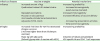
Table 2 shows current updates on the efforts in vaccine development strategies for the elderly. One key strategy to enhance vaccine immunogenicity is to use adjuvants. For example, oil-in-water emulsions MF59 and AS03 have been licensed as seasonal or pandemic influenza vaccines and have proven to increase vaccine immunogenicity and efficacy in the elderly (8081). Furthermore, AS01, a liposome-based adjuvant, has recently been approved as a recombinant protein vaccine against VZV infection in the elderly and demonstrated a high protection rate against herpes zoster in vaccine trials (8283). A more thorough understanding of the basic immunological changes that occur with age and the mode of action of novel adjuvants is a prerequisite for the development of formulations that specifically aim to overcome the limitations of the immune system associated with aging.
Table 2
Strategies towards more effective vaccines for the elderly
IFV
Influenza is an important contributor to morbidity and mortality worldwide as infections with IFV result in frequent hospitalization and deaths among the elderly. Greater cases of complications and hospitalization due to seasonal influenza are observed among people ≥65 years of age relative to younger individuals, and up to 90% of influenza-related deaths occur in the elderly groups (84). Therefore, World Health Organization recommends annual immunization against seasonal influenza for people ≥65 years of age (World Health Organization, 2018). However, vaccine effectiveness in the elderly does not mirror that observed in younger populations vaccinated with non-adjuvanted trivalent inactivated influenza vaccines (85). The efficacy and effectiveness of influenza vaccines decrease with age due to the detrimental impact of aging on the immune system's ability to function (6). Recently, Henry et al. (86) compared how B cells and Abs from elderly and younger adults respond to vaccination with different IFV strains. Interestingly, B cells from younger subjects showed a continuous accumulation of mutations, whereas the elderly appeared to have an essentially fixed B cell repertoire, lacking recent adaptations that would allow the evolution of B cells to divergent IFV strains. Additionally, in terms of T cells, old individuals who received influenza vaccination display defects, which result in poor immune response to vaccines against IFV, in the production of granzyme B and IFN-γ from CD8 T+ cells (8788).
Currently, the following approaches have been taken to tailor influenza vaccines to the elderly population: increased amounts of Ag, mucosal or intradermal route of vaccine delivery, and use of vaccine adjuvants (89). To fully understand the underlying role of aging in increased susceptibility to influenza infection and decreased vaccine efficacy, it will be necessary to use more translational approach such as systems vaccinology to evaluate new technologies suitable for vaccine development.
VZV
Shingles is caused by the reactivation of VZV that has persisted in latency within the dorsal root ganglia following an earlier episode of chickenpox, characterized by a painful rash and affects a significant proportion of the elderly population. Aging can contribute to the waning of VZV-mediated immunity, and the limited efficacy and duration of zoster vaccines suggest that aging-associated decline in immunogenicity can be improved by vaccination (90). The underlying mechanisms involved in VZV reactivation and susceptibility among the elderly or immunocompromised populations are currently unclear, although recent studies suggest aging cells as the cause of the inefficient clearance of virus-infected cells (9192). Few markers that correlate with age-related severity in shingles or poor response to vaccination have also been identified (939495). In particular, elderly residents in long-term care experience elevated incidence of shingles and post-herpetic neuralgia due to the high degree of immunosenescence, malnutrition, and existing chronic conditions. C-reactive protein level was suggested to be inversely correlated to vaccine responsiveness in the elderly (95). Meanwhile, the absolute numbers of CD3+, CD4+, and CD8+ T cells in herpes zoster patients were significantly lower compared to those in the control (94). Given these findings, it will be important to further characterize age-associated changes in immune cell compartments in shingles patients on a larger scale.
CMV
CMV is a herpesvirus that is prevalent worldwide. Although primary CMV infection induces innate and adaptive immune responses, CMV has developed various immune evasion strategies to modulate host immune activation during lytic and latent infections. CMV persistence has a profound effect on alterations in adaptive immunity, particularly T cell composition and function. The reduction in naïve T cell levels, specifically, was detected in the elderly infected with CMV (96). One hallmark of latent CMV infection is called “memory inflation,” which results in extensive expansion of CMV-specific memory CD8+ T cells over time, whereas CMV-specific memory CD4+ T cells accumulate to a lesser extent (97). In fact, previous evidences suggest that CMV-specific memory T cells gradually increase in numbers in the elderly, and in fact, 50% of the entire memory CD8+ T cell population is occupied by CMV-specific cells (719899).
Several studies have focused on the relationships between CMV serostatus and efficacy of vaccines, especially against the IFV and CMV has been associated with poor humoral response to influenza vaccination in the elderly (100101). In influenza-specific CD4+ T cell response, CMV seropositive elderly exhibit a lower response level compared to CMV-negative elderly. The late-differentiated (CD45RA+CCR7−CD27−CD28−) CD4+ T cells, but not CD8+ T cells were associated with poorer vaccine response. Thus, latent CMV infection, which has a deleterious effect on influenza Ab responses in the elderly, may be mediated by CD4+ T cells lacking CCR7, CD27, and CD28, but re-expressing CD45RA (102).
CONCLUSION AND FUTURE PERSPECTIVES
Immunosenescence is a major cause of increased incidence and severity of viral infections in the elderly, and contributes to impaired immunogenicity and efficacy of vaccines. Given the rapidly increasing elderly population in the world, improved vaccine efficacy has become a priority for the maintenance of global public health. Understanding the biological basis for age-associated alterations in viral immunity and vaccine immunogenicity is a challenge with substantial clinical importance. Subsequently, the use of systems biology approaches in combination with computational model systems will be crucial to understand the complexity of age-associated changes in the immune system by identifying molecular networks that orchestrate immunity to vaccinations in humans and potentially define correlates of protection.
Going forward, it will be important to better understand how environmental factors, such as diet, physical activity, co-morbidities, and pharmacological treatments, delay or contribute to the decline of the capability of the aging immune system to appropriately respond to infectious diseases and vaccination. Given the plasticity nature of aging and rapidly growing field of systems biology, molecular profiling of the aging-related changes is increasingly being examined at a single cell level by high-throughput omics technologies, including genomics, metagenomics, transcriptomics, and metabolomics (103). Specially, aging of the immune cells is affected by changes in homeostasis via cytokine levels, and by modifications in the metabolic pathways (104). Caloric restrictions (CR) affected a marked improvement in the maintenance and/or production of naïve T cells and the consequent preservation of TCR repertoire diversity. Furthermore, CR also improved T cell function and reduced production of inflammatory cytokines by memory T cells, suggesting that CR can delay T cell senescence and potentially contribute to extended lifespan by reducing susceptibility to infectious diseases (105).
A key area for future exploration in the immunosenescence field is the role of the secondary lymphoid organs as a critical partner in the development and function of the aging human immune system. Although most human studies focus on changes in lymphocytes collected from blood, but it will be important to analyze age-related changes in secondary lymphoid organs, lymph nodes and spleen, given the aging-associated decrease in the size of lymph nodes (52). Lymph nodes not only serve as the key initiating region of the immune response, but they also play an important role in maintaining naive lymphocytes. Richner et al. (106) reports detailed immunological and microscopic analyses of the defects in germinal center development in the draining lymph node and impaired migratory capacity of naïve CD4+ T cells within days of West Nile virus infection. Moreover, an increase in the suppressive T follicular regulatory cells combined with impaired function of aged T follicular helper cells in lymph nodes reduce T cell-dependent Ab responses in aged mice (107). These observations emphasize the importance of further analysis of the cellular and molecular characteristics of aging lymph nodes in the future.
Next, investigation of how extracellular vesicles (EVs) are linked to aging could be a promising area of interest. EVs are membrane-bound vesicles released by multiple cell types that include immune cells (108). In addition to SASPs, some senescent cells are reported to show increased secretion of EVs that are able to change protein composition and exert pro-proliferative function in some cancer cell lines (109). Evidence from cellular models suggests that exosomes released by macrophages from older are more pro-inflammatory than those released by macrophage from younger. In particular, mRNA levels of IL-6 and IL-12, but not TNF-α, in macrophage-derived exosomes were significantly higher in serums of older subjects (110). Given that EVs play an important role in immune cell network and cellular senescence, the profiles of secretome and the function of senescent immune cells will soon be revealed as the EV research field progresses.
Finally, the field of immunology has experienced a rapid increase in the use of single-cell sequencing approaches to characterize immune cells in the recent years (111). With the wide range of technologies available, researchers are able to use single cell transcriptomics and mass spectrometry to quantify changes in cellular activity states of various immune cell types and tissue proteome from young and old human samples.
Furthermore, massive efforts have been dedicated to the development of senolytic drugs, which selectively induce apoptosis of senescent cells. For example, UBX0101, a potent senolytic small molecule inhibitor of the MDM2/p53 protein interaction, is under evaluation for the treatment of musculoskeletal disease with an initial focus on osteoarthritis of the knee (112) and it is now awaiting clinical trial II. Hence, discovery of senolytic drugs that alleviate multiple senescence-related phenotypes in pre-clinical models can reduce the burden of aging-associated diseases and help to develop effective elderly-targeted vaccines and immunotherapies.
 XML Download