Journal List > Immune Netw > v.10(4) > 1033251

Kim, Han, and Kim: Heterogeneity of IL-22-producing Lymphoid Tissue Inducer-like Cells in Human and Mouse

Abstract

Lymphoid tissue inducer (LTi) cells have been characterized in mouse as a key cell when secondary lymphoid tissues are organized during development and memory T cells are formed after birth. In addition to their involvement in adaptive immune responses, recent studies show that they contribute to innate immune responses by producing large amount of interleukin (IL)-22 against microbial attack. Here, we compare IL-22-producing LTi and LTi-like cells in human and mouse and discuss their heterogeneity in different tissues.

INTRODUCTION

Since a lymphoid tissue inducer (LTi) cell was found in mouse lymphoid tissues (1), many groups have studied on its functions. Firstly, it has a key role in secondary lymphoid tissue development through the lymphotoxin-α1β2 expression during ontogeny (1-5), and provides survival signals to memory CD4 T cells through the OX40-ligand (OX40L) expression in adulthood (6-8). Secondly, they are also involved in ectopic lymphoid tissue development where lymphocytes infiltrate and organize lymphoid tissue like structure (9-11). In addition, thymic LTi cells promote the expression of AIRE (for autoimmune regulator), which is a transcription factor regulating the expression of self-tissue-restricted antigens on thymic medullary epithelial cells (12). Although LTi cells were actively studied in mouse during the last decade, the human counterpart was just recently identified in fetal mesentery and postnatal tonsils and reported to express large amount of IL-22 (13-15). Coincidently IL-22-producing LTi-like cells were reported in mouse gut (16,17), and referred as 'LTi-like' not 'LTi' cells because these cells express both a natural killer (NK) marker, NKp46 and a LTi characteristic marker, transcription factor retinoid-related orphan receptor (ROR) γt whose expression is critical for their development (18,19).
IL-22 is a Th17 cytokine and IL-22-producing cells are involved in innate immune responses against microbial attack (20,21). Here, the similarity and difference of mouse splenic and mucosal IL-22-expressing LTi and LTi-like cells and human IL-22-expressing LTi and LTi-like cells are discussed based on studies published within two years. In addition, the gene expression patterns of human LTi cells isolated from different secondary lymphoid tissues are compared with mouse splenic LTi cells.

SIMILARITY AND DIVERSITY OF IL-22-PRODUCING LTi-LIKE AND NK CELLS IN HUMAN AND MOUSE

'Conventional' LTi cells found in mouse lymph nodes and spleen are lineage-CD4+CD127+CD117+ (3,22,23) and OX40L+ in adulthood (23,24). They do not express molecules related to cytotoxicity such as interferon (IFN)-γ and granzyme, and do depend on RORγt expression for their development (18,22,23). These characteristics are distinct from those of NK cells (13). In addition, NK subsets are heterogeneous, and develop through four stages according to their expression of CD56, CD34, CD117 and CD94 (25,26). The phenotype of immature NK cells in stage 3 are similar to that of LTi cells; they are CD56-CD127+CD117+CD94- (13).
Takatori et al. showed that adult mouse splenic LTi-like cells express IL-22 and IL-17 after injection of zymosan in vivo (27), and Luci et al. showed that NKp46+RORγt+CD3- cells in mouse gut constitutively express IL-22 transcripts and produce IL-22 rapidly after stimulation (17). In addition, Sanos et al. showed that RORγt+NKp46+NK1.1int cells in mouse gut produce IL-22 and their emergence is dependent on RORγt expression (16). Because IL-22-producing cells described by Luci et al. and Sanos et al. express NKp46, they assumed that the cells could be derived from LTi cells. Recently, Buonocore et al. showed that Thy1+SCA1+RORC+ IL-23R+CD4-CD117- cells in mouse colon enhance IL-22 expression after IL-23 stimulation and the cell number is increased about 10 times more than other leukocytes by intestinal inflammation (28). The difference between these cells and conventional LTi cells is CD117 expression.
Several groups reported IL-22-expressing cells in human mucosa-associated lymphatic tissues. Cella et al. reported NK-22 cells which are IL-22-expressing NKp44+ cells found in human tonsil and Peyer's patches (29) and redefined them as CD56+NKp44+CCR6+CD103- (30). NK-22 cells stimulated by IL-23 and IL-1β increase not only IL-22 but also IFN-γ expression. They also identified NK-22 cells in mouse gut; ~40% of CD3-CD19-NKp46+ NK1.1- cells express IL-22 after IL-23 stimulation (30). Crellin et al. showed that CD56+ CD127+ cells are similar to NK-22 cells and CD56-CD127+ cells are LTi cells, and both express IL-22 and proliferate in the presence of IL-15 (15). Our group reported that CD3- CD117+CD56-OX40L+ cells in tonsil are very similar to LTi cells (31), and OX40L+ cells in this population express large amount of IL-22 (submitted).
In summary, several IL-22 expressing subsets have been reported in human and mouse. In human NK-22 cells by Cella et al. were identified in NK subsets, and the IL-22-producing cells by the others were studied after excluding NK cells. Cella et al. supposed that NK-22 may be derived from LTi cells. Many groups studying on IL-22 expression in mouse gut showed that IL-22-expressing cells are NKp46+ (16,17,29,30). The common factor of all IL-22-producing cells is RORγt expression which is critical for LTi development (18,32) and not expressed in conventional NK cells leading to the IL-22-producing cells are related to LTi cells.

HETEROGENEITY OF HUMAN LTi CELLS IN DIFFERENT TISSUES

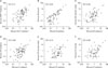
As mentioned above, not only LTi cells but also other IL-22-producing cells are heterogeneous. Lane's group previously reported that mouse LTi cells are heterogeneous according to their chemokine receptor expression and exist as CD4+ and CD4- cells indicating different functions in particular organs (22). In comparison, most human LTi cells which have been reported are CD4- (13,14). We therefore speculated that LTi cells in different tissues may express different molecules. To answer the question, we isolated CD3-CD117+OX40L+ cells in human spleen, lymph node, and tonsil, and compared 90-immune related gene expression patterns to mouse LTi cells isolated from spleen (Fig. 1). The gene expression was normalized to β-actin signals.
Firstly, the correlation between cells of different types in human and mouse was compared. Correlation coefficient (CC) between human CD4 memory T cells and mouse Th1 primed cells showed 0.71, between human and mouse B cells was 0.65, and between human and mouse dendritic cells (DCs) was 0.65 (Fig. 1A-C). Next, gene expression patterns of mouse splenic LTi cells were compared with human LTi cells isolated from different tissues; spleen (CC=0.80), lymph node (CC=0.74) and tonsil (CC=0.69) (Fig. 1D-F). Human splenic LTi cells showed the strongest correlation as mouse LTi cells were isolated from spleen. We further analyzed genes which are expressed over 10 times more in each tissue than genes expressed in mouse LTi cells; human splenic and lymph node LTi cells expressed higher levels of mRNA for BAFF (for B cell activating factor belonging to the tumor necrosis factor family) and Toll-like receptor 9, and tonsilar LTi cells expressed higher levels of mRNA for CD70, OX40, and GITR (glucocorticoid induced tumor necrosis factor gamily related gene). This indicates that LTi cells in specific tissues have different functions. This finding encourages us to investigate other functions of LTi cells in different tissues and diverse immune circumstances. In particular, their involvement in diseases such as cancer and autoimmunity has not been studied yet and may provide new insights for understanding human immune system.

CONCLUSION

LTi cells, LTi-like cells producing IL-22 in spleen and mucosa-associated lymphatic tissue and NK-22 cells produce large amount of IL-22 by IL-23 stimulation in bacterial infection. In comparison of LTi cells which do not express NK markers, LTi-like and NK-22 cells express NK cell marker(s). However, all of the cells are dependent on RORγt expression for their development indicating they may share a certain developmental stage with LTi cells. In addition, several differences of gene expression patterns by LTi cells in different tissues suggest their heterogeneity and various functions.

Figures and Tables

Figure 1
Correlation between the gene expression patterns of human and mouse cell populations. (A) Mouse splenic Th1 primed cells in vitro for 6 days in the presence of IL-4 and anti-IL-12 and human tonsilar CD45RO+ memory CD4 T cells. (B) Mouse splenic B cells and human tonsilar B cells. (C) Mouse splenic CD11c+ dendritic cells (DC) and human lymph node CD11c+ DC. (D-F) Mouse splenic LTi cells and human LTi cells isolated from spleen (D), lymph node (E), and tonsil (F). Over 0.01% expression of individual mRNAs of β-actin signals were plotted. X- and Y- axes show mRNA expression levels relative to the β-actin signal (β-actin signal=100%, log scale). Each plot compares two cell types, one on each axis (as labeled). CC=correlation coefficient. Analyzed 90 genes are: LT-α, TNF-α, LT-β, OX40L, CD40L, FASL, CD70, CD30L, 4-1BBL, TRANCE, TWEAK, APRIL, BAFF, LIGHT, TL1, TNFR1, TNFR2, LTβR, OX40, CD40, FAS, CD30, 4-1BB, RANK, TWEAKR, BAFFR, HVEM, GITR, DR3, CCR7, CXCR3, CXCR5, Bcl-2, Bcl-6, Bcl-XL, ROR-γ, GATA3, Foxp3, Myd-88, TLR2, TLR3, TLR4, TLR5, TLR7, TLR9, IL-1α, IL-1 β, IL-2, IL-3, IL-4, IL-5, IL-6, IL-7, TSLP, IL-9, IL-10, IL-12p35, IL-12p40, IL-13, IL-15, IL-18, IFN-α1, IFN-β, IFN-γ, TGF-β1, IL-2Rα, IL-2Rβ, IL-2Rγ, IL-4Rα, IL-7Rα, IL-10Rα, IL-10Rβ, IL-12Rβ1, IL-12Rβ2, IFNγR1, IFNγR2, CD80, CD86, DC-SIGN, CathepsinS, integrin alpha x, CTLA4, ICOS, ICOSL, β-2m, T-bet, CD4, CD74, perforin, and granzymeB.
in-10-115-g001

ACKNOWLEDGEMENTS

This work was supported by the National Research Foundation of Korea (NRF) grant funded by the Korean Government (MEST) (NRF-2009-0073480).

Notes

The authors have no financial conflict of interest.

References

1. Mebius RE, Rennert P, Weissman IL. Developing lymph nodes collect CD4+CD3- LTbeta+ cells that can differentiate to APC, NK cells, and follicular cells but not T or B cells. Immunity. 1997. 7:493–504.
crossref
2. Fu YX, Molina H, Matsumoto M, Huang G, Min J, Chaplin DD. Lymphotoxin-alpha (LTalpha) supports development of splenic follicular structure that is required for IgG responses. J Exp Med. 1997. 185:2111–2120.
crossref
3. Mebius RE. Organogenesis of lymphoid tissues. Nat Rev Immunol. 2003. 3:292–303.
crossref
4. Körner H, Cook M, Riminton DS, Lemckert FA, Hoek RM, Ledermann B, Köontgen F, Fazekas de St Groth B, Sedgwick JD. Distinct roles for lymphotoxin-alpha and tumor necrosis factor in organogenesis and spatial organization of lymphoid tissue. Eur J Immunol. 1997. 27:2600–2609.
crossref
5. Fütterer A, Mink K, Luz A, Kosco-Vilbois MH, Pfeffer K. The lymphotoxin beta receptor controls organogenesis and affinity maturation in peripheral lymphoid tissues. Immunity. 1998. 9:59–70.
crossref
6. Gaspal FM, Kim MY, McConnell FM, Raykundalia C, Bekiaris V, Lane PJ. Mice deficient in OX40 and CD30 signals lack memory antibody responses because of deficient CD4 T cell memory. J Immunol. 2005. 174:3891–3896.
crossref
7. Kim MY, Gaspal FM, Wiggett HE, McConnell FM, Gulbranson-Judge A, Raykundalia C, Walker LS, Goodall MD, Lane PJ. CD4(+)CD3(-) accessory cells costimulate primed CD4 T cells through OX40 and CD30 at sites where T cells collaborate with B cells. Immunity. 2003. 18:643–654.
crossref
8. Lane PJ, Gaspal FM, Kim MY. Two sides of a cellular coin: CD4(+)CD3- cells regulate memory responses and lymphnode organization. Nat Rev Immunol. 2005. 5:655–660.
crossref
9. Cupedo T, Jansen W, Kraal G, Mebius RE. Induction of secondary and tertiary lymphoid structures in the skin. Immunity. 2004. 21:655–667.
crossref
10. Meier D, Bornmann C, Chappaz S, Schmutz S, Otten LA, Ceredig R, Acha-Orbea H, Finke D. Ectopic lymphoid-organ development occurs through interleukin 7-mediated enhanced survival of lymphoid-tissue-inducer cells. Immunity. 2007. 26:643–654.
crossref
11. Drayton DL, Liao S, Mounzer RH, Ruddle NH. Lymphoid organ development: from ontogeny to neogenesis. Nat Immunol. 2006. 7:344–353.
crossref
12. Rossi SW, Kim MY, Leibbrandt A, Parnell SM, Jenkinson WE, Glanville SH, McConnell FM, Scott HS, Penninger JM, Jenkinson EJ, Lane PJ, Anderson G. RANK signals from CD4+3- inducer cells regulate development of Aire-expressing epithelial cells in the thymic medulla. J Exp Med. 2007. 204:1267–1272.
crossref
13. Kim MY, Kim KS, McConnell F, Lane P. Lymphoid tissue inducer cells: architects of CD4 immune responses in mice and men. Clin Exp Immunol. 2009. 157:20–26.
crossref
14. Cupedo T, Crellin NK, Papazian N, Rombouts EJ, Weijer K, Grogan JL, Fibbe WE, Cornelissen JJ, Spits H. Human fetal lymphoid tissue-inducer cells are interleukin 17-producing precursors to RORC+ CD127+ natural killer-like cells. Nat Immunol. 2009. 10:66–74.
crossref
15. Crellin NK, Trifari S, Kaplan CD, Cupedo T, Spits H. Human NKp44+IL-22+ cells and LTi-like cells constitute a stable RORC+ lineage distinct from conventional natural killer cells. J Exp Med. 2010. 207:281–290.
crossref
16. Sanos SL, Bui VL, Mortha A, Oberle K, Heners C, Johner C, Diefenbach A. RORgammat and commensal microflora are required for the differentiation of mucosal interleukin 22-producing NKp46+ cells. Nat Immunol. 2009. 10:83–91.
crossref
17. Luci C, Reynders A, Ivanov II, Cognet C, Chiche L, Chasson L, Hardwigsen J, Anguiano E, Banchereau J, Chaussabel D, Dalod M, Littman DR, Vivier E, Tomasello E. Influence of the transcription factor RORgammat on the development of NKp46+ cell populations in gut and skin. Nat Immunol. 2009. 10:75–82.
crossref
18. Eberl G, Marmon S, Sunshine MJ, Rennert PD, Choi Y, Littman DR. An essential function for the nuclear receptor RORgamma(t) in the generation of fetal lymphoid tissue inducer cells. Nat Immunol. 2004. 5:64–73.
crossref
19. Sun Z, Unutmaz D, Zou YR, Sunshine MJ, Pierani A, Brenner-Morton S, Mebius RE, Littman DR. Requirement for RORgamma in thymocyte survival and lymphoid organ development. Science. 2000. 288:2369–2373.
crossref
20. Zheng Y, Danilenko DM, Valdez P, Kasman I, Eastham-Anderson J, Wu J, Ouyang W. Interleukin-22, a T(H)17 cytokine, mediates IL-23-induced dermal inflammation and acanthosis. Nature. 2007. 445:648–651.
crossref
21. Liang SC, Tan XY, Luxenberg DP, Karim R, Dunussi-Joannopoulos K, Collins M, Fouser LA. Interleukin (IL)-22 and IL-17 are coexpressed by Th17 cells and cooperatively enhance expression of antimicrobial peptides. J Exp Med. 2006. 203:2271–2279.
crossref
22. Kim MY, Rossi S, Withers D, McConnell F, Toellner KM, Gaspal F, Jenkinson E, Anderson G, Lane PJ. Heterogeneity of lymphoid tissue inducer cell populations present in embryonic and adult mouse lymphoid tissues. Immunology. 2008. 124:166–174.
crossref
23. Kim MY, McConnell FM, Gaspal FM, White A, Glanville SH, Bekiaris V, Walker LS, Caamano J, Jenkinson E, Anderson G, Lane PJ. Function of CD4+CD3- cells in relation to B- and T-zone stroma in spleen. Blood. 2007. 109:1602–1610.
crossref
24. Kim MY, Toellner KM, White A, McConnell FM, Gaspal FM, Parnell SM, Jenkinson E, Anderson G, Lane PJ. Neonatal and adult CD4+ CD3- cells share similar gene expression profile, and neonatal cells up-regulate OX40 ligand in response to TL1A (TNFSF15). J Immunol. 2006. 177:3074–3081.
crossref
25. Freud AG, Caligiuri MA. Human natural killer cell development. Immunol Rev. 2006. 214:56–72.
crossref
26. Freud AG, Yokohama A, Becknell B, Lee MT, Mao HC, Ferketich AK, Caligiuri MA. Evidence for discrete stages of human natural killer cell differentiation in vivo. J Exp Med. 2006. 203:1033–1043.
crossref
27. Takatori H, Kanno Y, Watford WT, Tato CM, Weiss G, Ivanov II, Littman DR, O'Shea JJ. Lymphoid tissue inducer-like cells are an innate source of IL-17 and IL-22. J Exp Med. 2009. 206:35–41.
crossref
28. Buonocore S, Ahern PP, Uhlig HH, Ivanov II, Littman DR, Maloy KJ, Powrie F. Innate lymphoid cells drive interleukin-23-dependent innate intestinal pathology. Nature. 2010. 464:1371–1375.
crossref
29. Cella M, Fuchs A, Vermi W, Facchetti F, Otero K, Lennerz JK, Doherty JM, Mills JC, Colonna M. A human natural killer cell subset provides an innate source of IL-22 for mucosal immunity. Nature. 2009. 457:722–725.
crossref
30. Cella M, Otero K, Colonna M. Expansion of human NK-22 cells with IL-7, IL-2, and IL-1beta reveals intrinsic functional plasticity. Proc Natl Acad Sci USA. 2010. 107:10961–10966.
crossref
31. Kim S, Han S, Kim MY. Effects of interleukin-15 on human CD3(-)CD117(+)CD56(-)OX40L(+) cell differentiation. Hum Immunol. 2010. 71:745–750.
crossref
32. Eberl G, Littman DR. Thymic origin of intestinal alphabeta T cells revealed by fate mapping of RORgammat+ cells. Science. 2004. 305:248–251.
crossref
TOOLS
Similar articles