

 PDF
PDF ePub
ePub Citation
 Print
Print


A 25-year-old male patient was admitted to the cardiology outpatient clinic with atypical chest pain. Physical examination and electrocardiograms were normal.
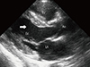
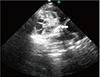
Chest radiography revealed irregular border of the cardiac silhouette, which was further evaluated by transthoracic echocardiography (TTE). Parasternal long axis view revealed mild local hypertrophy of the interventricular basal anterior septum (IBS) (Fig. 1) and by angulating the probe 60° clockwise (Fig. 2), a 6.5×5.5 cm, multilocular, thin walled strongly suggestive of echinococcus cyst type II appeared in the IBS.1)
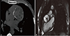
Thorax computed tomography and cardiac magnetic resonance imaging (CMRI) were performed to determine the exact location of the cyst. A 7×5.4 cm, multilocular, and thin walled cyst with rim like calcification was clearly demarcated by contrast enhanced computed tomography (CT) (Fig. 3A). CMRI further revealed the characteristics of the cyst (Fig. 3B). CT imaging of the brain, abdomen, and thorax were normal.
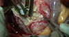
The patient underwent surgery for cystotomy with cyst enucleation (Fig. 4). Pathologic examination confirmed the diagnosis of echinococcus cyst and treatment involving albendazole (800 mg) administration was continued for 6 months.
Early diagnosis of cardiac hydatid cyst is of utmost importance for prevention of catastrophic complications. Because the contractions prevent settling of embryos to the heart isolated cardiac involvement of hydatid cyst is very rare with the incidence of 0.02-2%;2) furthermore, involvement of IBS is even more rare (5-9%).2)3) A lobular mass in the IBS seen by TTE should be considered as a hydatid cyst. In such cases, contribution of CMRI for diagnosis has been well defined.4)
 XML Download