

 PDF
PDF ePub
ePub Citation
 Print
Print


INTRODUCTION
For the treatment of symptomatic paroxysmal atrial fibrillation (PAF), catheter ablation is considered the first treatment strategy,1) and pulmonary vein isolation (PVI) is an essential and standard approach for both paroxysmal and persistent atrial fibrillation (AF).2)3)
Radiofrequency catheter ablation (RFCA) has been a mainstay in arrhythmia treatment including PVI for AF. Cryoablation for arrhythmias has been used for cardiac surgery for decades,4) and transvenous catheter cryoablation for arrhythmias has been used since the 2000s.
Cryoablation gained distinguished attention when it was used for AF therapy. The introduction of cryoballoon ablation (CBA) to isolate pulmonary veins (PVs) was considered breakthrough technology because of its single energy application for encircling lesions at the antral level of PVs, whereas conventional RFCA was characterized by point-by-point multiple energy applications for isolating PVs. The FIRE AND ICE trial has shown that the cryoballoon is not inferior to irrigated-tip radiofrequency ablation (RFA) in terms of efficacy and safety,5) and that CBA is becoming one of the most widely used ablation technologies in the field of AF treatment.
In this article, the basic biophysics of the cryoballoon, tissue changes due to cryothermal energy and practical applications are described. To determine the current efficacy and safety of CBA, a systematic review was performed by reviewing 12 studies comparing the second-generation cryoballoon (CB-2) and RFA.
BASIC BIOPHYSICS OF THE CRYOBALLOON
Tissue changes due to cryothermal energy were characterized by intracellular and extracellular ice crystal formation, osmotic cellular damage, microvascular injury resulting in hemorrhage, and inflammation, ischemia, fibrosis, and apoptosis of the surrounding tissue. In contrast to RFA lesions, cryoablation lesions are characterized by the preservation of the tissue architecture, including fibrocytes and collagen with significantly fewer large vascular structures or endocardium.6)
Based on this biophysical characteristic of preserving the tissue architecture, cryoablation was preferred to reduce the risk of causing destruction to the normal structure during specific arrhythmias such as atrioventricular nodal reentrant tachycardia or tachycardia originating near the His bundle region. These characteristics explain the lower risk of cardiac perforation or thrombogenicity compared to RFA.7)
Cryothermal energy is produced during refrigerant injected through a fine tube. The refrigerant vaporizes at the tip of a cryoablation catheter and can freeze the adjacent tissue. While freezing, the catheter tip adheres to the affected tissue, which enables the application of stable energy. The cryoballoon has the same overall structural mechanism of cryoenergy production, except the tip is composed of balloons.
The first-generation Arctic Front™ cryoballoon system (Medtronic, Minneapolis, MN, USA) has been commercially available since 2010. The cryoballoon contains inner and outer balloons, a central lumen for a guide wire and contrast, a thermocouple for inner balloon temperature monitoring, and lumens for the cryorefrigerant (an intake lumen and an exhaust lumen). Pressurized cryorefrigerant (nitrous oxide [N2O]) is delivered to the distal aspect of the inner balloon to allow the temperature to decrease to a maximum of −80°C. Heat is absorbed from the surrounding tissue. Then, the cryorefrigerant is returned to the console through a lumen under a vacuum.8)
The first-generation cryoballoon (CB-1) has an equatorial cooling region that could cause ineffective encircling of PVs (Figure 1). To compensate for this disadvantage, the CB-2, the Arctic Front Advance™ (Medtronic), was developed. It has similar basic structures but features a modified refrigerant injection system that renders more homogeneous cooling of the complete distal hemisphere, including the distal tip.9) With this development, the 1-year success rate was improved and complication rates decreased from those of CB-1.10)11)
Figure 1
Compared to the first-generation cryoballoon (A), the second-generation cryoballoon has homogeneous cooling system in distal hemisphere (B) (Courtesy; Medtronic, Minneapolis, MN, USA).

There are 2 size options for the cryoballoon, 23 and 28 mm. The 28-mm cryoballoon is preferable because it avoids common complications that can occur when the balloon is deeply seated in PVs. The size can be chosen according to the anatomical characteristics of PVs.
ABLATION PROCEDURES
A single interatrial septal puncture is enough for CBA because the spiral Achieve™ (Medtronic) catheter for the PV electrogram recording is inserted through the central guide wire lumen of the cryoballoon. An Achieve™ mapping catheter is installed before the cryoballoon is inserted in the patient's body.
The septal puncture technique is the same as that for RFA using a standard transseptal sheath, but a low anterior septal puncture is recommended to allow more space for the cryoballoon to move and provide better support to the PVs, especially the right inferior PV. Intracardiac echocardiography (ICE) is strongly recommended during septal puncture to ensure safety.
The inserted standard transseptal sheath is replaced by a steerable transseptal sheath (FlexCathAdvance™; Medtronic) with a 12-Fr inner diameter and 15-Fr outer diameter over a long stiff guidewire. This steerable sheath is stiffer than usual transseptal sheaths and is designed to steer and support the cryoballoon throughout the procedure. A cryoballoon and an Achieve™ mapping catheter can be inserted through the sheath. While the cryoballoon is being moved inside the left atrium, the Achieve™ catheter should lead the cryoballoon outside the sheath because the distal tips of both the cryoballoon and sheath are stiff to avoid cardiac damage.
When the cryoballoon is located near a PV, the next most important task is complete PV occlusion with the distal hemisphere of the cryoballoon. A cryoballoon should be inflated outside the PV and advanced to the PV antrum to be fitted. Because of low pressure, the inflated cryoballoon rarely causes cardiac damage, but it is better to avoid inflating the balloon inside the PV. With 1–2 mL of contrast injection, PV angiography should be performed to assess the complete sealing of the PV by the balloon. If the contrast leakage is seen, then the balloon can be repositioned. When the balloon fits the PV without any contrast leakage, instead of ablation, the balloon can be withdrawn slightly to see some leakage. This technique will show whether the balloon is at the appropriate antral position. Sometimes, the balloon could be inside the PV, which causes more complications to surrounding structures such as phrenic nerves (phrenic nerve injury [PNI]).
After the balloon is positioned appropriately, the Achieve™ mapping catheter could be adjusted to obtain the best possible PV recordings. Electrical isolation of the PVs is detectable, and the time required to achieve electrical PV isolation could be used as an indicator of long-term success of PV isolation.
When cryoenergy is applied, compliance of the balloon can be changed and the cooled cryoballoon adheres to the surrounding tissue. Therefore, to avoid cardiac damage, the cryoballoon should not be moved while cryoenergy is being applied. The 180-second initial ablation with a CB-2 is recommended by experts, but the optimal duration of cryoenergy application has not been determined.
To prevent complications, esophageal temperature and diaphragmatic motion during phrenic nerve stimulation should be monitored.
EFFICACY AND SAFETY: A SYSTEMATIC REVIEW
Since CB-1 and the more developed CB-2 were released in 2010 and 2012, respectively, many studies showed comparable efficacy of CBA to that of RFA, until now. The FIRE AND ICE trial results were released in 2016. It was a large, randomized, controlled study including both CB-1 and CB-2 that provided important information indicating that CBA is not inferior to RFA with respect to efficacy and overall safety.5) Because the cryoballoon has been improved from the CB-1 to the CB-2,12)13) and because RFA has been improved from a simple irrigated-tip catheter to a force-sensing catheter,14) it is reasonable to compare the efficacy and safety of CB-2 and the contact-force RFA catheter, even though there are only a few studies regarding this.
We systematically reviewed studies comparing the CB-2 and/or the CB-1 with RFA for AF ablation. We searched the studies comparing CBA with RFA in PubMed, EBSCO, Web of Science, and the Cochrane Library, and we excluded studies using the CB-1 alone or mostly CB-1 for the CBA group. We included studies in the analysis only when recurrence of atrial tachyarrhythmia (ATA) was evaluated during follow-up. A total of 12 studies involving 4,228 non-overlapped patients were included.5)15)16)17)18)19)20)21)22)23)24)25)
Of the 12 studies, 7 included only CB-2 for the cryoablation group and 5 included CB-2 for a median of 41% of patients (range, 30.7–80.4%). Six studies used only the force-sensing RFA catheter, and the other studies had no particular description of which type of RFA catheter was used (Table 1). Three hundred eighty-three of 4,228 patients (9%) had persistent AF.
Table 1
Baseline characteristics of included studies
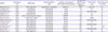
| Author | Publication year | Study type | Atrial fibrillation type | Number of patients of CB-2 ablation/CB ablation | Number of patients of RF ablation | RF catheter type | Mean follow-up (months) |
|---|---|---|---|---|---|---|---|
| Aryana et al.15) | 2015 | Retrospective | Paroxysmal, persistent | 773/773 | 423 | Non-CF | 12 |
| Jourda et al.16) | 2014 | Prospective | Paroxysmal | 75/75 | 75 | CF | 12 |
| Miyazaki et al.17) | 2016 | Prospective | Paroxysmal | 41/41 | 41 | Not described | 3 |
| Nagy et al.18) | 2016 | Retrospective | Paroxysmal | 38/38 | 58 | CF | 12 |
| Squara et al.19) | 2015 | Pros- & Retrospective cohort | Paroxysmal | 178/178 | 198 | CF | 12 |
| Straube et al.20) | 2016 | Prospective cohort | Paroxysmal | 86/107 | 99 | Mixed | 12 |
| Wasserlauf et al.21) | 2015 | Retrospective | Paroxysmal | 31/101 | 100 | Not described | 12 |
| Ciconte et al.22) | 2015 | Retrospective | Persistent | 50/50 | 50 | CF | 12 |
| Juliá et al.23) | 2015 | Retrospective | Paroxysmal | 41/100 | 186 | Not described | 12 |
| Khoueiry et al.24) | 2016 | Retrospective | Paroxysmal | 103/311 | 376 | CF | 14±8 |
| Kuck et al.5) | 2016 | Prospective RCT | Paroxysmal | 279/374 | 376 | Not described | 18 |
| Kardos et al.25) | 2016 | Retrospective | Paroxysmal | 40/40 | 58 | CF | 12 |
Acute procedural success
Electrical PVI was investigated using the index procedure in 8 studies. The acute procedural success rate ranged from 98.4% to 100% in the CB-2 group and from 97.4% to 100% in the RFA group; the median success rate was 99% in both groups. There was no significant difference between the 2 groups.
Recurrence of ATA during follow-up
Studies were included only when the patients were followed-up for at least 3 months. The follow-up period was 12 months in 9 studies; other studies included a follow-up period of 3–18 months.
Recurrence of ATA after a single procedure was similar between the 2 groups (12 studies; 4,228 patients; 25.2% CB-2 vs. 29.5% RF; odds ratio [OR], 0.81; 95% confidence interval [CI], 0.62–1.05; p=0.11) (Figure 2A).
Figure 2
Recurrence of atrial tachyarrhythmias. (A) The second-generation cryoballoon (CB-2) vs. radiofrequency (RF) ablation. (B) CB-2 vs. force-sensing RF catheter ablation (CF-RF).
CI = confidence interval.
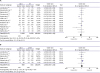
When the radiofrequency (RF) group was confined to force-sensing RFA, there was no difference between the CB-2 and force-sensing RF groups (6 studies; 1,507 patients; 19.5% CB-2 vs. 18.3%; OR, 1.08; 95% CI, 0.83–1.40; p=0.58) (Figure 2B).
Procedural and fluoroscopy time
For the CB-2 group, an electroanatomical mapping system was used for some patients. In contrast, in the RFA group, most RFA was performed with the assistance of an electroanatomical mapping system. However, the exact numbers of those using an electroanatomical mapping system were not provided.
Procedural time ranged from 73.5±16 minutes to 192.9±44 minutes in the CB-2 group and from 118.5±15 minutes to 283.7±78.0 minutes in the RF group. The procedure time was much shorter in the CB-2 group (mean difference [MD], −37.59 minutes; 95% CI, −56.78, −18.4; p<0.001) (Figure 3A).
Figure 3
Comparison of procedure time and fluoroscopy time between the second-generation cryoballoon (CB-2) vs. radiofrequency (RF) ablation group. (A) Procedure time. (B) Fluoroscopy time.
SD = standard deviation.
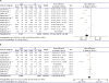
The fluoroscopy time was from 13.8±4 minutes to 39.8±14.9 minutes in the CB-2 group and from 15.8±6 minutes to 73.0±30.1 minutes in the RF group. The procedure time was shorter in the CB-2 group, but the difference was small (MD, −3.1 minutes; 95% CI, −6.93, −0.73; p=0.11) (Figure 3B).
COMPLICATIONS
PNI
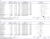
Transient PNI was evaluated in 11 studies, and permanent PNI was evaluated in 9 studies. Transient PNI is relatively common in CBA. Of 11 studies that included 2,088 patients, transient phrenic injury occurred in a median of 5% (range, 0.99–17.3%). Permanent PNI occurred in only 4 studies, with a median incidence rate of 1% (range, 0.26–2.5%). On the contrary, in the RFA group, only 2 cases of transient PNI occurred in 1,854 patients (0.1%) and permanent PNI never occurred (transient PNI was higher in the CB-2 group; OR, 9.69; 95% CI, 4.18–22.49; p<0.001) (Figure 4A).
To prevent PNI, monitoring of the phrenic nerve function is important, especially while the right superior PV (RSPV) is being ablated, because the risk of PNI is highest in the RSPV. Right phrenic nerve stimulation with palpation of the strength of diaphragmatic contractions or monitoring of the diaphragmatic compound motor action potential recordings can be used.26) With the assistance of these methods, the PNI rate can be decreased to less than 1.5%.27)
Other serious complications
Serious complications such as cardiac tamponade, gastrointestinal bleeding, or documented esophageal ulcer and thromboembolic events such as stroke occurred much less often in the CB-2 group (0.78% in the CB-2 group vs. 2.53% in the RF group; OR, 0.39; 95% CI, 0.23–0.68; p<0.001).
To avoid esophageal injury during CBA, esophageal temperature monitoring is highly recommended.28)29) Limiting the esophageal temperature to stop cryoenergy application has not been confirmed by large controlled trials, but discontinuation of cryoablation should be considered when the esophageal temperature decreases to 21–25°C.
Summary
Cryoenergy is safe and effective for the treatment of cardiac arrhythmia. The cryoballoon allowed faster and more convenient AF ablation procedures and showed clinical outcomes comparable to those of conventional and the more developed force-sensing RFCA. Even though CBA is safe; serious complications such as cardiac tamponade and significant bleeding occur less frequently with RFA, extra care should be taken to avoid PNI.
 XML Download