Journal List > Korean Circ J > v.48(4) > 1093995

Miura, Shirai, Hayashi, Jinnouchi, Isotani, Kakumoto, Arai, and Ando: Atypical Annulus Rupture after Transcatheter Aortic Valve Implantation
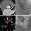
An 86-year-old female underwent transcatheter aortic valve implantation (TAVI) for severe aortic stenosis (AS) via a transfemoral approach. The aortic valve was a severely calcified bicuspid valve with fusion of the right and non-coronary cusps (Figure 1A). Therefore, a SAPIEN XT 26-mm valve (Edwards Lifesciences, Irvine, CA, USA) was implanted using a 1-mL-under balloon volume (Figure 1B). Transthoracic echocardiography (TTE) showed no problem one day after TAVI, but predischarge TTE at 1 week revealed a continuous shunt from the aorta to right ventricle (Figure 1C, Supplementary Video 1) and the pulmonary blood flow (Qp)/systemic blood flow (Qs) was 1.1. Aortography showed blood flow from the aortic annulus to the right ventricle (Figure 1D, Supplementary Video 2). She had no symptoms and was discharged on foot at 19 days after TAVI. At 18 months after TAVI, she was doing well without heart failure.
The silent onset of annulus rupture is very rare. Most annulus ruptures are observed during the initial procedure, and the patient's hemodynamic status is extremely unstable.1) In our case, the patient was very stable and the complication was late-onset. The mechanism of this complication may be erosion between the implanted valve edge and the aortic wall.2) Especially when there is a bicuspid valve with fusion of the right and non-coronary cusp, we need to carefully think about the type of implanted valve and the method of implantation. In addition, we need to carefully observe the patient for iatrogenic shunt, and percutaneous or surgical closure may be needed if the condition of the patient gets worse.

Figures and Tables

Figure 1
(A) Multislice computed tomography showing a severely calcified aortic valve with fusion of the right and non-coronary cusps. (B) A 26-mm SAPIEN XT (Edwards Lifesciences, Irvine, CA, USA) deployed via a transfemoral approach. (C) Transthoracic echocardiography showing atypical annulus rupture and a continuous shunt from the Ao to RV (arrow). (D) Aortography showing blood flow from the aortic annulus to RV (arrow).
Ao = aorta; LA = left atrial; LV = left ventricle; RV = right ventricle.
kcj-48-332-g001

Notes

Conflict of Interest The authors have no financial conflicts of interest.

Author Contributions

  • Conceptualization: Miura M, Shirai S, Hayashi M, Jinnouchi H, Isotani A, Kakumoto S, Arai Y, Ando K.

  • Data curation: Miura M.

  • Investigation: Miura M.

  • Project administration: Miura M.

  • Supervision: Shirai S, Ando K.

  • Writing - original draft: Miura M.

References

1. Hayashida K, Bouvier E, Lefèvre T, et al. Potential mechanism of annulus rupture during transcatheter aortic valve implantation. Catheter Cardiovasc Interv. 2013; 82:E742–E746.
crossref
2. Hagiwara K, Saito N, Yamazaki K, Kimura T. Aorto-right ventricular fistula following transcatheter aortic valve implantation using a 29 mm SAPIEN XT valve. BMJ Case Rep. 2017; 2017:bcr-2017-219247.
crossref

SUPPLEMENTARY MATERIALS

Supplementary Video 1

Transthoracic echocardiography showing atypical annulus rupture and a continuous shunt from the aorta to right ventricle.

Supplementary Video 2

Aortography showing blood flow from the aortic annulus to right ventricle.
TOOLS
ORCID iDs

Mizuki Miura
https://orcid.org/0000-0003-3252-3861

Similar articles