

 PDF
PDF ePub
ePub Citation
 Print
Print


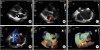
A 42-year-old woman presented with a 2-year history of shortness of breath. She was evaluated via transthoracic echocardiography at another institution and diagnosed with ostium primum atrial septal defect and advised to undergo surgery. Transthoracic echocardiography revealed a mildly enlarged right ventricle and mild tricuspid regurgitation with an estimated systolic pulmonary artery pressure of 40 mmHg. However, unroofed coronary sinus (UCS) was suspected when detailed reexamination was performed. Two- and three-dimensional (3D) transesophageal echocardiography (TEE) confirmed the diagnosis. The defect was repaired surgically. Enhanced temporal and spatial resolution 3D imaging enabled us to delineate the complex anatomy of an UCS without additional imaging modalities.
A suspicious drop in the roof of the coronary sinus was observed on transthoracic parasternal long axis view (arrow; Figure 1A, Supplementary Video 1). Color Doppler revealed significant flow through the coronary sinus (Figure 1B, Supplementary Video 2). However, when a probe was angulated, observations suggested the presence of a dilated coronary sinus ostium. Moreover, a silhouette of the coronary sinus was not observed. TEE revealed the UCS (arrow; Figure 1C). Color Doppler demonstrated significant shunting through the UCS and a small shunt through the foramen ovale (Figure 1D). 3D TEE depicted the en face view of the defect from the left atrium (Figure 1E, Supplementary Videos 3 and 4).The unroofed portion of the coronary sinus was visualized in detail (white arrow: coronary sinus, red arrow:unroofed portion; Figure 1F, Supplementary Video 5). Contrast echocardiography using agitated saline revealed the absence of a persistent left superior vena cava (Supplementary Video 6). Our case emphasizes the utility of 3D TEE for the evaluation of congenital heart defects.
 XML Download