Journal List > J Korean Orthop Assoc > v.54(1) > 1117037

송, 연, 이, 이, 엄, 권, Song, Yon, Lee, Lee, Um, and Kwon: Ponseti 방법으로 치료를 시작한 선천성 만곡족 환자에서 수술적 치료 여부를 예측할 수 있는 방사선적 지표

Abstract

목적

선천성 만곡족 환자에서 Ponseti 방법으로 치료한 환자들에서 잔여 혹은 재발 변형에 대한 수술적 치료의 필요성을 예측하고 치료 결과를 평가하는 데 있어 어떤 방사선적 기준선이 유용할지에 대하여 연구하고자 하였다.

대상 및 방법

2005년부터 2013년까지 Ponseti 방법으로 치료를 시작한 환자 102족(65명, 양측성 37명)을 연구대상으로 했다. 환자는 수술 여부에 따라 두 군(A군: Ponseti 방법의 결과가 성공적이어서 추가 수술을 시행하지 않은 군, B군: Ponseti 방법의 결과가 만족스럽지 않아 수술을 시행한 군)으로 나누었고, 두 군의 방사선적 족부 변형 상태 비교를 위해 전후면 및 측면 거종간 각, 전후면 거골-제1중족골간 각, 발목 완전 배굴 상태에서의 측면 경종간 각을 측정하였다. 각 방사선 사진은 관찰자 내 신뢰도를 평가하기 위해 한 정형외과 의사가 두 번씩 검토한 후 평균을 분석하였으며 관찰자 간 신뢰도를 평가하기 위해 난수표를 통해 20족을 선정하여 각도를 측정하였다.

결과

A군은 73족(71.6 %)이었고, B군은 29족(28.4%)이었다. 초기 방사선 소견상 측면 거종간 각 및 측면 경종간 각에서 두 군 간 의미 있는 차이가 있었고, 관찰자 간 및 관찰자 내 신뢰도의 편향은 관찰되지 않았다. 전후면 거골-제1중족골간 각 및 측면 경종간 각은 양군에서 모두 치료 전후로 유의한 차이가 있었다.

결론

선천성 만곡족의 치료에서 Ponseti 방법으로 치료한 후 70% 이상의 환자에서 만족스러운 결과를 보였다. 방사선적으로 초기 측면 거종간 각 및 측면 경종간 각을 통해 수술적 치료 여부를 예측할 수 있었고, 전후면 거골-제1중족골간 각 및 측면 경종간 각을 통해 치료 후 선천성 만곡족의 변화 정도를 평가할 수 있었다.

초록

Purpose

Several radiologic reference lines have been used to evaluate individuals with a clubfoot but there is no consensus as to which is most reliable. The aim of this study was to identify which radiologic parameters have relevance to the predictability of additional surgery after Ponseti casting on clubfoot and the effect of clubfoot treatments that contain Ponseti casting and additional surgery.

Materials and Methods

A total of 102 clubfeet (65 patients, 37 bilateral) were reviewed from 2005 to 2013. The patients were divided into two groups (Group A, those for whom the result of the Ponseti method was successful and did not require additional surgery; and Group B, those for whom the result of the Ponseti method was unsuccessful and required additional surgery), and the following parameters were measured on the plain radiographs: i) talo-calcaneal angle on the anteroposterior and lateral view, ii) talo-1st metatarsal angle on the anteroposterior view, and iii) Tibio-calcaneal angle on the lateral view with the ankle full-dorsiflexion state. Each radiograph was reviewed on two separate occasions by one orthopedic doctor to characterize the intra-observer reliability, and the averages were analyzed. Next, 20 cases were chosen using a random number table, and two orthopedic doctors measured the angle separately to characterize the inter-observer reliability.

Results

Groups A and B included 73 clubfeet (71.6%) and 29 clubfeet (28.4%), respectively. The initial talo-calcaneal angle and tibio-calcaneal angle in the lateral view were significantly different among the groups. In addition, inter- and intra-observer biases were not detected. The talo-1st metatarsal angle on the anteroposterior view and tibio-calcaneal angle on the lateral view were significantly different after treatment in both groups.

Conclusion

Congenital clubfeet treated with the Ponseti method showed successful results in more than 70% of patients. The initial talo-calcaneal angle and tibio-calcaneal angle on the lateral view were the radiologic parameters that could predict the need for additional surgical treatments. The talo-1st metatarsal angle on the anteroposterior view and tibio-calcaneal angle on the lateral view could effectively evaluate the changes in clubfoot after treatment.

서론

선천성 만곡족은 근골격계 질환 중 가장 흔한 족부 질환 중 하나이다. 이는 족관절의 첨족, 후족부 내반, 전족부 내전, 요족 등의 특징적 변형을 가지며 이 변형을 초기에 교정하는 것이 치료의 목적이다.1) 초기 치료는 Ponseti 방법을 이용하여 조기에 치료하는 것이 가장 선호되고 있다.2345)
선천성 만곡족의 교정 평가를 위하여 방사선적 평가가 많이 이용되고 있으나,67910) 어떠한 방사선적 기준선이 치료 결과의 예측 및 평가에 있어서 객관적인 의미를 가는지에 대해서는 아직 논란의 여지가 있다.11) 이에 본 저자들은 Ponseti 방법으로 치료한 환자들에 있어 수술적 치료의 필요성을 예측하는 데 방사선적 기준선들의 유용성을 알아보고 치료 효과를 평가할 수 있는 방사선적 기준선을 알아보고자 하였다.

대상 및 방법

2005년 2월부터 2013년 5월까지 계명대학교 동산의료원에서 선천성 만곡족으로 진단 받고 Ponseti 방법12)으로 교정 치료한 환자 중 2년 이상 추적 관찰이 가능하였던 65명(102족)의 환자를 대상으로 하였다. 37명은 양측성 만곡족을 보였고, 28명은 일측성 만곡족을 보였다. 1) Ponseti 방법 후 1주 이내에 수술적 치료를 받은 환자, 2) 2년 미만의 추시 관찰기간을 가지거나 추시 소실된 환자, 3) 선천성 만곡족 이외의 선천성 증후군 및 신경근육 이상을 보이는 환자는 연구 대상에서 제외하였다. 모든 환자에서 비수술적 방법인 Ponseti 방법12)을 이용한 석고붕대 교정방법으로 치료를 시작하였으며, 모든 술기는 소아 정형외과 경력이 20년 이상 되는 제1저자에 의하여 시행되었다. 본 연구는 계명대학교 동산의료원 e-IRB에서 승인 받아 진행되었다(IRB No. 2016-05-011-005).
Ponseti 방법12)은 전족부를 회외전시킨 후 제1 중족골두를 밀어 올리면서 요족을 교정하고자 하였고 족근동의 거골 경부측에 손가락을 대어 반대로 압력을 가하면서 전족부를 외전시켜 전족부 내전을 교정하였다. 이때 종골-입방골 관절 경첩이 되지 않도록 주의하면서 장하지 석고붕대 고정술을 시행하였다. 석고 고정은 1주일 간격으로 교체하였고 4–6회 시행 후 교정이 만족스럽게 이루어져 족배 굴곡이 20도 이상 가능한 환아에서 외전족배 보조기 착용을 시작하였으며 족배 굴곡이 20도 이하이거나 첨족 변형이 남아 있는 환아 39명(63족)에서는 국소마취하에 아킬레스건 절단술을 시행하여 족관절을 20도 족배 굴곡시키고 70도 외회전시킨 상태로 2주간 장하지 석고붕대 고정술을 시행하였다.
Ponseti 교정방법의 초기 성패는 추가적인 수술 여부로 결정하였으며 수술 시행 여부로 두 군(A군: 초기 치료가 성공하여 수술을 시행하지 않은 군, B군: 재발 혹은 잔존 변형으로 인하여 수술을 시행한 군)으로 나누었다. Ponseti 방법에 의해 초기 치료가 완료된 상태에서 결과가 만족스럽지 못하거나 결과가 만족스러워도 추시 기간 중 재발한 환아의 경우 수술적 치료를 시행하였다. 수술적 치료는 평균 생후 24개월(8–56개월)에 시행하였다. 초기 치료 후 결과가 만족스럽지 못하거나 추시 기간 중 재발한 환아, 경골 염전이 동반되어 있으며 내족지 보행이 있는 환아의 경우 추가적으로 수술적 치료를 시행하였다.
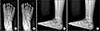
모든 대상 환아에 대하여 전후면 족부 및 최대 족배 굴곡 상태에서 족관절을 포함하는 측면 족부 단순 방사선 촬영을 시행하였다. 전후면 족부 방사선 촬영은 정형외과 의사가 환아의 발을 잡은 상태에서 X선 방사 각도가 발등에 수직이 될 수 있게 하여 촬영하였고, 측면 족부 방사선 사진은 정형외과 의사가 블록을 이용하여 환아의 발바닥 전체를 접촉시킨 상태에서 최대 족배 굴곡을 시킨 상태에서 촬영하였다. 두 군의 방사선적인 족부 변형 상태 비교는 전후면 거종간 각(talo-calcaneal angle on anteroposterior view, TCA), 측면 거종간 각(talo-calcaneal angle on lateral view, LTCA), TCA 및 LTCA의 합(sum of TCA and LTCA, TCA anteroposterior+laterior [AP+Lat]), 전후면 거골-제1중족골간 각(talo-1st metatarsal angle on anteroposterior view, TFMA), 측면 경종간 각(tibio-calcaneal angle on lateral view at ankle full-dorsiflexion state, TicalA)을 지표로 정하여 측정하였다(Fig. 1). 모든 기준선은 해당 뼈의 장축을 기준으로 선을 그렸고 그 사이의 각을 측정하여 각 도를 기록하였다. 각 방사선 사진은 관찰자 내 신뢰도를 평가하기 위해 한 정형외과 의사가 2회씩 검토한 후 평균을 분석하였으며, 관찰자 간 신뢰도를 평가하기 위해 난수표를 통해 20족을 선정하여 각도를 측정하여 급내 상관 변수(intra-class correlation)를 통하여 분석하였다.
두 그룹 간의 초기 족부 변형 상태를 비교하고자 최초 방문 시 각 방사선적 지표를 측정하여 통계적으로 비교하였다. Ponseti 방법을 시행 후 족부 변형의 호전 정도의 차이를 확인하기 위해 A군에서는 최초 방문 시와 Ponseti 방법으로 치료 후 최종 추시 시 방사선적 지표들을 측정하였으며 B군에서는 최초 방문 시와 수술적 치료 직전, 최종 추시 시의 방사선적 지표들을 측정하여 각 시기별로 호전 정도를 평가하였다.
모든 결과는 IBM SPSS package ver. 21.0.1 (IBM Co., Armonk, NY, USA)을 이용하여 분석하였다. 정규성 가정을 만족하는 항목에 대해서는 paired t-test를 시행하였고, 정규성 가정을 만족하지 못하는 항목에 대해서는 Mann-Whitney 및 chi square 검사, Wilcoxon 부호순위 검정을 시행하였다. 95%의 신뢰 구간을 기준으로 분석하였으며, p-value 0.05 미만에서 통계적 의의를 지닌다고 해석하였다.

결과

Ponseti 방법으로 치료를 시작할 당시의 환아들의 평균 연령은 생후 1.63주(A군: 1.45주, B군: 2.07주)였고, 최종 추시 시의 평균 연령은 6.67세(A군: 6.16세, B군: 7.86세)였다. 환자들의 추시 기간은 평균 3.95년(2.0-8.3년)이었다. 73족(71.6%: A군)에서 Ponseti 방법으로 치료 후 추가적인 수술 없이 성공적인 결과를 얻었고, 29족(28.4%: B군)에서 Ponseti 방법으로 치료 후 결과가 만족스럽지 못하여 추가적인 수술을 요하였다. 재발하는 족부 변형에 대하여 pie-crusting 술식을 이용한 경골-종골간 연장술(tibio-calcaneal lengthening) 9예, 입방골 설상 절골술을 포함한 거골-주상골 관절 유리술(tibio-navicular release+cuboid wedge osteotomy) 2예, 후경골근 유리술과 입방골 설상 절골술을 포함 혹은 포함하지 않은 거골-주상골 관절 유리술(posterior tibialis release+tibionavicular release±cuboid wedge osteotomy) 14예를 시행하였으며, Ponseti 교정에도 불구하고 잔존하는 족부 변형에 대하여 중족골 교정 절골술(metatarsal corrective osteotomy) 1예 및 경골 회전 절골술(tibia derotational osteotomy) 3예를 시행하였다. 추가적인 수술은 평균 나이 25.8개월(3–64개월)에 시행하였다. 추후 18족(17.6%)에 대해서는 추가적 2차 수술을 시행하였다(Table 1). Ponseti 치료 시 아킬레스건 절단술은 63족(61.8%)에서 시행하였고, A군에서 39족(53.4%), B군에서 24족(82.8%)에서 시행하였고, 아킬레스건 시행 여부에 따른 통계적 유의한 차이는 없었다.
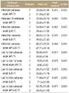
두 명의 정형외과 의사가 무작위로 선택한 20족의 방사선 사진을 통해 각각 각도를 측정하여 관찰자 간 신뢰도를 평가하였으며, 신뢰성 점수는 모든 각도에서 85% 이상이었다. 모든 각도는 한 명의 정형외과 의사가 두 차례에 걸쳐 검토하였으며, 관찰자 내 신뢰도 점수는 모든 각도에서 95% 이상이었다. 치료 전후 및 초기 변형 각도를 평가하기 위해 각 측정값의 평균값을 구하여 통계적 분석을 시행하였다(Table 2, 3).
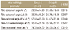
초기 방사선 소견상 LTCA 및 TicalA에서 두 군 간에 의미 있는 차이가 있었다. LTCA는 A군에서 33.99°±15.24°, B군에서 24.78°±15.25°, TicalA는 A군에서 79.03°±28.72°, B군에서 94.83°±24.13°로 초기 변형이 심할수록 수술 비율이 높게 나타났다(p=0.007, 0.010)(Table 4).
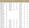
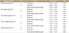
A군에서 평균 13.93주간 Ponseti 방법으로 치료 후 TCA가 평균 7.31°, TCA와 TCA의 합(TCA AP+Lat)이 평균 9.59°, TFMA가 평균 21.12°, TicalA이 평균 15.75°만큼의 호전(p=0.000)을 보였으며, LTCA에서는 통계적으로 의미 있는 변화가 없었다(p=0.179).
B군에서 평균 22.17주간 Ponseti 방법으로 치료 후 TFMA가 평균 11.77°만큼 호전(p=0.014)을 보였으며, TicalA이 평균 12.71°만큼의 호전(p=0.012)을 보였다. 그 외 방사선적 지표에서는 통계적으로 의미 있는 변화가 없었다(TCA, p=0.770; LTCA, p=0.973; TCA AP+Lat, p=0.865). TFMA, TicalA은 A군과 B군 모두에서 초기 평가와 비교하였을 때, 최종 추시상 의미 있는 호전이 있었다(Table 5).

고찰

지금까지 유·소아의 족부 변형에 있어서 단순 방사선 사진을 이용한 방사선적 측정을 통한 평가는 완전히 신뢰하기는 어려운 것으로 회자되어 왔다.13141516) Simons17)는 1978년 유·소아에서는 단순 방사선 사진상 골화 중심이 없거나 매우 작아서 선천성 만곡족의 평가에 있어서 방사선적 측정을 사용하는 것은 논란의 여지가 있을 수 있다고 기술하였다. 하지만 동시에 유·소아의 족부 위치의 오류로 인한 각도의 변화량은 매우 미미하다고도 기술하고 있다.
방사선적 평가의 질을 높이기 위해서는 첫째, 단순 방사선 사진 촬영 시의 올바른 발의 위치와 모양이 중요하며 둘째, 골화 중심의 축을 해부학적인 축과 최대한 일치하게 하여 오차를 줄이는 데 있다. 특히 후자의 경우 대상 환아의 나이가 어릴수록 골화 중심이 둥글기 때문에 각도를 잴 때 더 큰 오차가 생길 수 있으므로 주의해야 한다. 실제로 본 연구에서는 A군과 B군의 초기 방사선적 지표를 비교 분석했고 A군과 B군에서 각각 Ponseti cast를 통한 초기 치료 결과에 대해 분석했으며 B군에서는 초기 치료 후 술적 치료까지 마친 후의 치료 결과도 분석하였다. 본 연구의 결과에 따르면 LTCA 및 TicalA가 수술적 치료의 필요성을 예측할 수 있는 중요한 지표가 될 수 있을 것이다.
또한 TFMA, TicalA는 선천성 만곡족의 치료 후 치료 효과를 평가하는 데 있어 유용한 것으로 평가되었다. 이에서 종골과 거골이 모두 재태 26주경에 골화 중심 형성을 시작하나 종골의 경우 거골에 비해 장축의 형성이 빠르기 때문에1819) 경골과 종골의 장축을 비교하는 경종간 각 및 제1중족골의 장축을 측정하는 데 오차가 제일 적었을 것임을 짐작할 수 있다
본 연구의 제한점으로는 첫째, 본 연구는 후향적으로 이루어진 연구이며 두 그룹 간 치료 전 초기 족부 변형상태를 Diméglio score20) 등을 통하여 임상적으로 평가하지 못하여 방사선적 족부 상태의 평가와 임상적인 환아의 족부 상태의 평가가 실제로 연관성을 가지는지 확인하지 못하였다는 점이다. 둘째, 많은 연구에서 보조기 착용 순응도가 예후에 큰 영향을 끼친다는 보고21)가 있었음에도 불구하고 보조기 착용 순응도를 나타내는 객관적인 기준이 모호22)하여 본 연구에 포함하지 못하였다. 마지막으로 측정각들의 표준 편차가 커서 임상적으로 수술적 치료 여부를 결정하는 데 적용할 수 있는 기준치를 정하지 못하였다. 이들 제한점은 추후 환자군에 대한 지속적인 추시 및 추가적인 환자군의 포함을 통하여 파악할 수 있을 것으로 생각한다.

결론

선천성 만곡족의 치료에서 방사선적으로 초기 LTCA 및 TicalA를 통해서 수술적 치료 여부를 예측할 수 있었으며, 초기 LTCA가 작을수록, 초기 TicalA가 클수록 수술적 치료를 요하는 경우가 더 많았다. TFMA 및 TicalA가 선천성 만곡족의 치료에서 전족부 내전 및 요족 변형의 호전 정도를 평가하는 데 가장 객관적인 지표로 확인되었다.

Figures and Tables

Figure 1

(A) Talo-calcaneal angle on the anteroposterior view. (B) Talo-1st metatarsal angle on the anteroposterior view. (C) Talo-calcaneal angle on the lateral view. (D) Tibio-calcaneal angle on the lateral view.

jkoa-54-59-g001
Table 1

Surgical Treatments after the Ponseti Method

jkoa-54-59-i001

*Supramalleolar derotational osteotomy 1 case, subtalar posterior capsular release 2 cases, tibialis anterior tendon partial transposition 1 case.

Table 2

Inter-Observer Reliability of the Radiologic Parameter Evaluation in Randomized 20 Cases

jkoa-54-59-i002

SD, standard deviation; AP, on anteroposterior view; Lat, on lateral view with ankle full-dorsiflexion; Talo-calcaneal angle (AP+Lat), sum of talo-calcaneal angle AP and talo-calcaneal angle Lat; f/u, follow-up.

Table 3

Intra-Observer Reliability of a Radiologic Parameter Evaluation

jkoa-54-59-i003

Group A, those for whom the result of the Ponseti method was successful and did not require additional surgery; Group B, those for whom the result of the Ponseti method was unsuccessful and required additional surgery; SD, standard deviation; AP, on anteroposterior view; Lat, on lateral view with ankle full-dorsiflexion; Talo-calcaneal angle (AP+Lat), sum of talo-calcaneal angle AP and TALO-calcaneal angle Lat; Pre-op, preoperative; f/u, follow-up.

Table 4

Initial Radiologic Findings of Groups A and B

jkoa-54-59-i004

Values are presented as mean±standard deviation. Group A, those for whom the result of the Ponseti method was successful and did not require additional surgery; Group B, those for whom the result of the Ponseti method was unsuccessful and required additional surgery; AP, on anteroposterior view; Lat, on lateral view with ankle full-dorsiflexion; Talo-calcaneal angle AP+Lat, sum of talo-calcaneal angle AP and talo-calcaneal angle Lat.

Table 5

Statistical Value of the Change in Radiologic Findings after the Treatment of Clubfoot

jkoa-54-59-i005

Group A, those for whom the result of the Ponseti method was successful and did not require additional surgery; Group B, those for whom the result of the Ponseti method was unsuccessful and required additional surgery; AP, on anteroposterior view; Lat, on lateral view with ankle full-dorsiflexion; Talo-calcaneal angle AP+Lat, sum of talo-calcaneal angle AP and talo-calcaneal angle Lat.

Notes

CONFLICTS OF INTEREST The authors have nothing to disclose.

References

1. Dobbs MB, Rudzki JR, Purcell DB, Walton T, Porter KR, Gurnett CA. Factors predictive of outcome after use of the Ponseti method for the treatment of idiopathic clubfeet. J Bone Joint Surg Am. 2004; 86:22–27.
crossref pmid
2. Herzenberg JE, Radler C, Bor N. Ponseti versus traditional methods of casting for idiopathic clubfoot. J Pediatr Orthop. 2002; 22:517–521.
crossref pmid
3. Cooper DM, Dietz FR. Treatment of idiopathic clubfoot. A thirty-year follow-up note. J Bone Joint Surg Am. 1995; 77:1477–1489.
crossref pmid
4. Laaveg SJ, Ponseti IV. Long-term results of treatment of congenital club foot. J Bone Joint Surg Am. 1980; 62:23–31.
crossref
5. Ponseti IV, Smoley EN. The classic: congenital club foot: the results of treatment. 1963. Clin Orthop Relat Res. 2009; 467:1133–1145.
pmid pmc
6. Radler C, Manner HM, Suda R, et al. Radiographic evaluation of idiopathic clubfeet undergoing Ponseti treatment. J Bone Joint Surg Am. 2007; 89:1177–1183.
crossref pmid
7. Kite JH. Principles involved in the treatment of congenital club-foot. 1939. J Bone Joint Surg Am. 2003; 85:1847. discussion 1847.
pmid
8. Kite JH. The clubfoot. New York: Grune & Stratton;1964. p. 232.
9. Kite JH. Conservative treatment of the resistant recurrent clubfoot. Clin Orthop Relat Res. 1970; 70:93–110.
crossref pmid
10. Kite JH. Nonoperative treatment of congenital clubfoot. Clin Orthop Relat Res. 1972; 84:29–38.
pmid
11. Park SS, Kim SW, Jung BS, Lee HS, Kim JS. Selective soft-tissue release for recurrent or residual deformity after conservative treatment of idiopathic clubfoot. J Bone Joint Surg Br. 2009; 91:1526–1530.
crossref pmid
12. Ponseti IV. Congenital clubfoot: fundamentals of treatment. Oxford: Oxford University Press;1996. p. 140.
13. Simons GW. Analytical radiography of club feet. J Bone Joint Surg Br. 1977; 59:485–489.
crossref pmid
14. Colburn M, Williams M. Evaluation of the treatment of idiopathic clubfoot by using the Ponseti method. J Foot Ankle Surg. 2003; 42:259–267.
crossref pmid
15. Frick SL. The Ponseti method of treatment for congenital clubfoot: importance of maximal forefoot supination in initial casting. Orthopedics. 2005; 28:63–65.
crossref pmid
16. Hamel J, Becker W. Sonographic assessment of clubfoot deformity in young children. J Pediatr Orthop B. 1996; 5:279–286.
crossref pmid
17. Simons GW. A standardized method for the radiographic evaluation of clubfeet. Clin Orthop Relat Res. 1978; 107–118.
crossref
18. Shapiro F, Glimcher MJ. Gross and histological abnormalities of the talus in congenital club foot. J Bone Joint Surg Am. 1979; 61:522–530.
crossref pmid
19. Fritsch H, Eggers R. Ossification of the calcaneus in the normal fetal foot and in clubfoot. J Pediatr Orthop. 1999; 19:22–26.
crossref pmid
20. Diméglio A, Bonnet F, Mazeau P, De Rosa V. Orthopaedic treatment and passive motion machine: consequences for the surgical treatment of clubfoot. J Pediatr Orthop B. 1996; 5:173–180.
pmid
21. Thacker MM, Scher DM, Sala DA, van Bosse HJ, Feldman DS, Lehman WB. Use of the foot abduction orthosis following Ponseti casts: is it essential? J Pediatr Orthop. 2005; 25:225–228.
pmid
22. Shim JS, Seo SW, Kim CY, Jung C. Congenital clubfoot treated with the Ponseti method. J Korean Orthop Assoc. 2009; 44:634–641.
crossref
TOOLS
ORCID iDs

Chang Jin Yon
https://orcid.org/0000-0003-3580-4175

Similar articles