

 PDF
PDF ePub
ePub Citation
 Print
Print


Abstract
Purpose
An experimental animal study was performed to compare the bone fusion capacity of an allograft and porous hydroxyapatite.
Materials and Methods
Three milliliters of allograft or porous hydroxyapatite particles were inserted between the 4th and 5th lumbar transverse processes of New Zealand white rabbits weighing 3-3.5 kg. The total number of rabbits was 30, which were divided randomly into 2 groups. The bone formation and fusion capacity were evaluated 12 weeks after surgery through the gross findings and manual palpation, as well as radiological, biomechanical, and histological studies. Six rabbits in the allograft group died during breeding but the autopsy finding did not show any evidence suggesting an infection or graft rejection. The allograft was harvested from the iliac crest of the rabbits of the same species aseptically and was preserved at -80℃ for at least 7 days before implantation.
Results
The fusion rates were 55.6% (5/9) and 66.7% (10/15) in the allograft and porous hydroxyapatite groups, respectively. The mean values of the tensile strengths were 140.7 N in the allograft group and 189.6 N in the porous hydroxyapatite group. Histological analysis of 2 specimens from each group revealed theporous hydroxyapatite group to show a slightly better osteoconduction capacity.
Figures and Tables
Fig. 2
Serial posteroanterior radiographs. In both groups, implanted allograft and hydroxyapatite granules showed marginal blurring at 6 weeks, however, the radioopacities of the centers of the granules was sustained for up to 12 weeks.
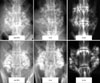
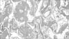
Fig. 3
Light microscopic findings of the fusion masses in the allograft group (H&E stain, ×100, decalcified). (A) Case 1. The allograft was transformed to mature bone including the osteoid formation with osteoblastic rim, newly formed bone marrow and intervening fibrous tissue. (B) Case 2. Newly formed bone and fibrous tissue are intermingled.

Fig. 4
Light microscopic findings of the fusion masses in the porous HA group (H&E stain, ×100, decalcified). (A) Case 1. Remodeling of new bone containing osteoid, bone marrow, and the surrounding new vessels can be observed within the pores. (B) Case 2. It shows a mixture of new vessels, osteoid, and bone marrow within the pore structure along the remodeled bone suggesting excellent osteoconduction.

References
1. Bauer TW, Muschler GF. Bone graft materials. An overview of the basic science. Clin Orthop Relat Res. 2000. 371:10–27.
2. Betz RR. Limitations of autograft and allograft: new synthetic solutions. Orthopedics. 2002. 25:Suppl 5. S561–S570.

3. Chang BS, Hong KS, Yeom JS, et al. Effects of pore size on osteoconduction at the porous hydroxyapatite. J Korean Orthop Assoc. 1999. 34:37–44.
4. Chang BS, Lee CK, Hong KS, et al. Osteoconduction at porous hydroxyapatite with various pore configurations. Biomaterials. 2000. 21:1291–1298.
5. Cheng JC, Guo X, Law LP, Lee KM, Chow DH, Rosier R. How does recombinant human bone morphogenetic protein-4 enhance posterior spinal fusion? Spine. 2002. 27:467–474.
6. Chung SS, Hong KS, Youn HJ, et al. Difference of bonding behavior between four different kinds of hydroxyapatite plate and rabbit\'s bone. J Korean Orthop Assoc. 1998. 33:158–167.
7. Ehrler DM, Vaccaro AR. The use of allograft bone in lumbar spine surgery. Clin Orthop Relat Res. 2000. 371:38–45.
8. Emery SE, Fuller DA, Stevenson S. Ceramic anterior spinal fusion. Biologic and biomechanical comparison in a canine model. Spine. 1996. 21:2713–2719.
9. Enneking WF, Campanacci DA. Retrieved human allografts: a clinicopathological study. J Bone Joint Surg Am. 2001. 83:971–986.
10. Friedlaender GE, Strong DM, Tomford WW, Mankin HJ. Long-term follow-up of patients with osteochondral allografts. A correlation between inmunologic responses and clinical outcome. Orthop Clin North Am. 1999. 30:583–588.
11. Hall EE, Meffert RM, Hermann JS, Mellonig JT, Cochran DL. Comparison of bioactive glass to demineralized freeze-dried bone allograft in the treatment of intrabony defects around implants in the canine mandible. J Periodontol. 1999. 70:526–535.
12. Hamson KR, Toth JM, Stiehl JB, Lynch KL. Preliminary experience with a novel model assessing in vivo mechanical strength of bone grafts and substitute materials. Calcif Tissue Int. 1995. 57:64–68.
13. Le Huec JC, Lesprit E, Delavigne C, Clement D, Chauveaux D, Le Rebeller A. Tri-calcium phosphate ceramics and allografts as bone substitutes for spinal fusion in idiopathic scoliosis: comparative clinical results at four years. Acta Orthop Belg. 1997. 63:202–211.
14. Lee DH, Ryu HS, Lee SL, et al. Comparison of Osteosynthesis in various types of porous calcium phosphate compounds: an experimental study by posterolateral fusion of rabbit's lumbar vertebrae. J Korean Soc Spine Surg. 2001. 8:455–467.
15. Lee JH, Ha JH, Lee DH, et al. Evaluation of biodegradation and osteosynthesis in CaO-SiO2-B2O3 glass-ceramics by posterolateral fusion of rabbit lumbar vertebrae. J Korean Orthop Assoc. 2003. 38:612–618.
16. Lee JH, Lee DH, Ha JH, et al. Porous beta-calcium pyrophosphate as a bone graft substitute in a canine bone defect model. J Korean Orthop Assoc. 2003. 38:384–392.
17. Marchesi DG. Spinal fusions: bone and bone substitutes. Eur Spine J. 2000. 9:372–378.
18. Strong DM, Friedlaender GE, Tomford WW, et al. SImmunologic response in human recipients of osseous and osteochondral allografts. Clin Orthop Relat Res. 1996. 326:107–114.
19. Tanaka J, Asaka M, Imamura M. T-cell co-signalling molecules in graft-versus-host disease. Ann Hematol. 2000. 79:283–290.
 XML Download