

 PDF
PDF ePub
ePub Citation
 Print
Print


INTRODUCTION
The major serum metabolite of vitamin D (Vit D) is 25-hydroxyvitamin D (25[OH]D), which has been providing new insights into breast cancer. Compelling meta analyses have suggested that serum 25(OH)D concentrations are inversely associated with breast cancer development and increased risks of recurrence and death in patients with early-stage cancer [12]. A recent prospective cohort study also demonstrated that serum 25(OH)D levels were an independent prognostic factor in women with breast cancer [3]. However, contradictory results have also been reported [45]. Furthermore, no effect of serum Vit D levels on the pathologic complete response (pCR) has been demonstrated in neoadjuvant settings [67].
Vit D is closely linked to various disease conditions, including malignancy and skeletal health; Vit D deficiency is highly prevalent worldwide and an important threat to human health [89]. During treatment for breast cancer, serum 25(OH)D levels have been reported to dramatically change, with the main effect being decreased 25(OH)D concentrations, especially during chemotherapy [1011]. Although dietary intake and ultraviolet B exposure are important factors influencing 25(OH)D levels, Vit D supplements have been investigated for the purpose of cancer prevention and active forms are considered as adjuvants to chemotherapy for malignancies [812]. Therefore, it is worthwhile to explore the emerging roles of Vit D in patients with breast cancer.
Herein, to address the association between serum 25(OH)D concentrations and the outcomes of patients with breast cancer treated with neoadjuvant chemotherapy (NCT), we examined the sequential changes in serum 25(OH)D levels prior to and after receiving NCT and exploratively analyzed the associations with pCR and survival in patients with breast cancer.
METHODS
Demographics and serum 25(OH)D levels
A total of 374 consecutive patients who received NCT and subsequently underwent definitive surgery of the breast and axilla between January 2010 and December 2013 were retrospectively selected. All patients in the study cohort were examined for their serum 25(OH)D levels both prior to and after receiving NCT. The serum 25(OH)D levels of the patients at baseline and after NCT were evaluated according to the manufacturer's protocol at the Department of Nuclear Medicine, Severance Hospital, Seoul, Republic of Korea. A gamma counter (1470 Wizard; Perkin-Elmer, Turku, Finland) with a radioimmunoassay (25-Hydroxyvitamin D 125I RIA Kit; DiaSorin, Stillwater, USA) was used to measure serum 25(OH)D concentrations. Using a cutoff of ≥20 ng/mL for sufficient 25(OH)D levels [13], the patients were categorized into the “both deficient” group, wherein patients had deficient Vit D levels at baseline and after NCT, or the “either sufficient” group, wherein patients had sufficient Vit D levels either at baseline or after NCT.
The NCT regimen mainly comprised four cycles of anthracycline plus cyclophosphamide (AC) followed by four cycles of taxane±titanium silicate-1 in 342 patients (91.4%). Fourteen patients (3.7%) received AC alone or a cyclophosphamide, methotrexate, and fluorouracil regimen. Of the remaining 18 patients (4.8%), 10 were treated with a taxane, carboplatin, and bevacizumab regimen; four with anthracycline plus taxane or a taxane, anthracycline, and cyclophosphamide regimen; and four with taxane plus trastuzumab. All patients received radiation therapy postoperatively, and endocrine therapy was initiated according to their hormone receptor status. Among the 16 administrative districts of the Republic of Korea, the capital city (Seoul), surrounding metropolitan area (Gyeonggi), and six other metropolitan cities (Busan, Daegu, Incheon, Gwangju, Daejeon, and Ulsan) were categorized as urban. The remaining regions (Gangwon, Chungbuk, Chungnam, Jeonbuk, Jeonnam, Gyeongbuk, Gyeongnam, and Jeju) were categorized as rural. This study was approved by the Institutional Review Board of Severance Hospital, Yonsei University Health System, Seoul, Korea (IRB number: 4-2016-0367), and the need for written informed consent was waived.
Pathologic examination
The absence of in situ or invasive carcinomas or residual in situ carcinoma alone without invasive disease in the breast and no evidence of metastatic tumors in the axillary lymph nodes were considered an achievement of pCR post-NCT. Expression of biomarkers, including estrogen receptor (ER) and progesterone receptor (PR), was reviewed through pathology reports. Positivity of hormone receptors was defined as tumors with ≥1% nuclear-stained cells on immunohistochemistry assessment of biopsy specimens according to the American Society of Clinical Oncology/College of American Pathologists (ASCO/CAP) guidelines [14]. Human epidermal growth factor receptor 2 (HER2) immunostaining was scored from 0 to 3+ and in situ hybridization was performed in cases with HER2-equivocal results. Criteria for positivity of HER2 followed the ASCO/CAP guidelines of HER2 testing [15]. The Ki-67 indexes were scored by counting the number of positively stained nuclei and were expressed as the percentage of total tumor cells. Ki-67 >15% was used as a cutoff for high proliferative indexes.
Based on the ER, PR, and HER2 expressions and Ki-67 indexes, the molecular phenotypes were categorized into the following four subgroups: luminal A-like (ER- and/or PR-positive, HER2-negative, and Ki-67 ≤15%), luminal B-like (ER- and/or PR-positive, HER2-negative, and Ki-67 >15%; or ER- and/or PR-positive and HER2-positive), HER2-enriched (ER-negative, PR-negative, and HER2-positive), and triple-negative breast cancer (TNBC; ER-negative, PR-negative, and HER2-negative). In patients with unavailable Ki-67 results, histologic grade III was considered as high proliferation.
Statistical analysis
Differences between the groups according to clinicopathological parameters were evaluated using the chi-square test, and Fisher exact test was applied when appropriate. The independent t-test and one-way analysis of variance (ANOVA) with Bonferroni correction were used to compare the means of continuous numerical data. Disease-free survival (DFS) was measured from the date of curative surgery to the date of first locoregional or distant recurrence or death before any type of relapse. Overall survival (OS) was calculated from the date of first surgery to the date of last follow-up or death from any cause. Univariable associations between predefined events and parameters were assessed using the Kaplan-Meier method; the groups were compared using the log-rank test. The Cox proportional hazard model was used to identify variables independently associated with survival. All statistical tests were two-sided and p-values <0.05 were considered statistically significant. SPSS version 23.0 (IBM Inc., Armonk, USA) was used for all analyses.
RESULTS
Changes in serum 25(OH)D levels and patient characteristics
The mean age of all patients was 48.7±9.7 years and the mean follow-up duration was 52.3±16.5 months. The overall pCR rate was 25.9%. The median 25(OH)D levels were 12.94 ng/mL (range, 3.57–46.28 ng/mL) at baseline and 10.52 ng/mL (range, 2.54–39.57 ng/mL) after NCT. The mean time interval of 25(OH)D examination between baseline and completion of NCT was 163±19.6 days. At baseline, 63 patients (16.8%) showed sufficient 25(OH)D levels, and after the completion of NCT, 41 (11.0%) showed sufficient levels. Compared to baseline 25(OH)D levels, 246 patients (65.8%) showed decreased 25(OH)D levels after NCT (median difference before vs. after NCT, −2.69 ng/mL; range, −24.20 to 25.57 ng/mL). The “either sufficient” group comprised 89 patients (23.8%). In patients who achieved pCR, the mean serum 25(OH)D levels at baseline and after NCT were 14.60 ng/mL and 12.68 ng/mL, respectively. The mean 25(OH)D levels before and after NCT were 14.39 ng/mL and 11.87 ng/mL, respectively, in patients who did not achieve pCR. There were no differences in 25(OH)D levels according to pCR (p=0.795 at baseline and p=0.314 after NCT).
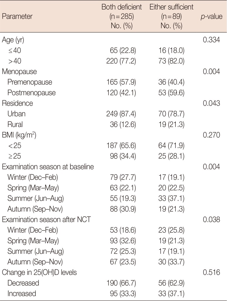
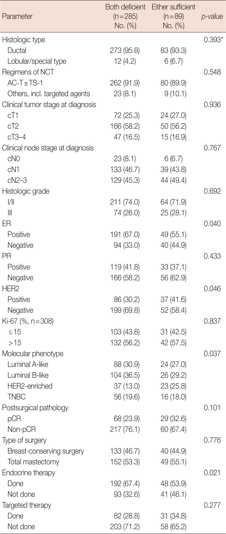
Table 1 shows the patient characteristics according to the 25(OH)D levels prior to and after receiving NCT. The “either sufficient” group demonstrated higher proportions of postmenopausal status, residence in rural areas, and baseline summer examinations. At completion of NCT, the “either sufficient” group frequently underwent examinations of 25(OH)D levels in the autumn and winter seasons. The histopathological features and treatment modalities are presented in Table 2. ER-negative and HER2-positive tumors were more common in the “either sufficient” than in the “both deficient” group. Therefore, the HER2-enriched subtype was significantly more frequent in the “either sufficient” group and endocrine therapy was more frequently performed in the “both deficient” group. Other tumor-associated characteristics, including achievement of pCR or the Ki-67 proliferation index, did not differ. When the clinicopathological parameters were compared according to 25(OH)D levels at baseline and after NCT, postmenopausal status, rural residence, examination in the summer season, and ER-negative and HER2-positive tumors were more frequent in patients with sufficient baseline 25(OH)D concentrations. However, only the positive association between ER-negativity and sufficient 25(OH)D concentrations after NCT was maintained when the clinicopathological characteristics were compared according to the Vit D status after NCT.
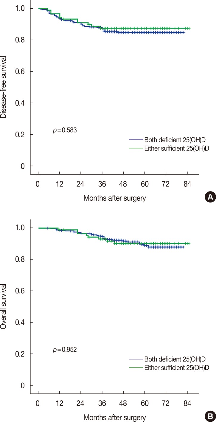
Survival analyses
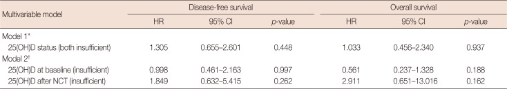
DFS and OS curves according to 25(OH)D status are presented in Figure 1 and showed no statistical significance. When each 25(OH)D status before and after NCT was analyzed, no association with survival outcomes was determined. Achievement of pCR and the luminal A-like molecular phenotype were significant factors for improved survival (Supplementary Figure 1, available online). Multivariable analysis demonstrated no significance of each or combined 25(OH)D statuses for survival (Table 3). Advanced stage III at diagnosis, high grade, non-pCR, and the molecular phenotype were independently associated with increased risks for poor DFS and OS (data not shown).
Subgroup analyses stratified by molecular phenotype
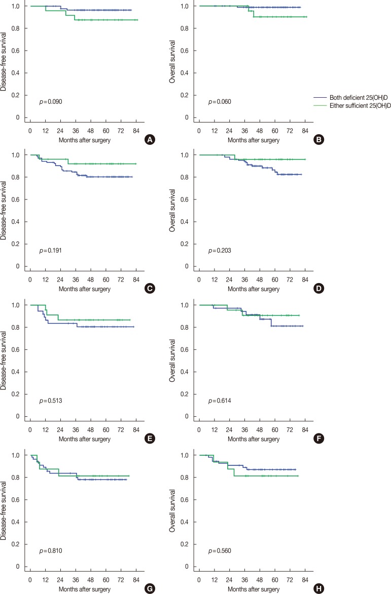
Finally, the association between changes in 25(OH)D levels and survival was explored according to the stratification by molecular phenotype. Figure 2 shows the changes in 25(OH) D levels during NCT according to the molecular phenotype and achievement of pCR. The mean baseline 25(OH)D levels were significantly higher in patients with the HER2-enriched subtype (17.67 ng/mL) than those in patients with the luminal A-like (13.51 ng/mL, p=0.001), luminal B-like (13.89 ng/mL, p=0.003), and TNBC (14.19 ng/mL, p=0.022) subtypes. However, 25(OH)D concentrations highly decreased after NCT in patients with the HER2-enriched subtype and no difference in 25(OH)D levels after NCT was determined among the molecular phenotypes (luminal A-like, 11.56 ng/mL; luminal B-like, 11.22 ng/mL; HER2-enriched, 13.78 ng/mL; and TNBC subtypes, 13.04 ng/mL; ANOVA with Bonferroni correction, p>0.05). There was no statistical difference in 25(OH)D levels between patients who achieved pCR and those who did not achieve pCR, irrespective of the molecular phenotype. The mean levels of 25(OH)D decreased in patients after NCT compared to those in patients at baseline, irrespective of the responsiveness to NCT or molecular phenotypes. Figure 3 demonstrates no differences in DFS and OS according to 25(OH)D status stratified by molecular phenotype.
DISCUSSION
In this study, 83.2% of patients (311/374) with breast cancer were determined to have Vit D deficiency at diagnosis and only 12 patients (3.2%) showed 25(OH)D levels of ≥30 ng/mL. After completion of NCT, 91.6% (285/311) still had deficient Vit D levels and only 23.8% (15/63) maintained sufficient 25(OH)D concentrations. Asian countries, including China, India, and Mongolia, have high prevalence rates of Vit D deficiency, and in Korea, 63.8% of women with a self-reported history of cancer and 73.4% of women without a cancer history show deficient Vit D status [916]. Although the sample size in the NEOZOTAC trial was small, the prevalence of Vit D deficiency increased from 38.3% before initiating NCT to 55.9% at the end of NCT, with a change in the Vit D level of −16.0 nmol/L (−6.4 ng/mL) after NCT [7]. Similarly, adjuvant chemotherapy is associated with decreased 25(OH)D concentrations of −5.52 ng/mL at 6 months and −1.24 ng/mL at 12 months [11]. However, daily supplements of Vit D3 (400 IU) for 1 year in patients with premenopausal status modestly increased 25(OH)D levels and partially prevented a decrease in serum concentrations by chemotherapy [17].
It has not been clearly determined whether chemotherapy directly affects changes in serum 25(OH)D levels, plays indirect roles through gastrointestinal side effects and behavioral changes toward avoiding sunlight exposure, or whether 25(OH)D concentrations are not significantly affected by chemotherapy [71017]. Miyoshi et al. [18] suggested that docetaxel could upregulate the cytochrome P450 3A4 enzyme, which might convert active forms of Vit D to inactive metabolites. The administration of corticosteroids as antiemetics during chemotherapy or estrogen deprivation by chemotherapy-induced amenorrhea might partially affect Vit D metabolism and serum concentrations [11].
In addition to dietary intake, supplementation, and sunlight, serum 25(OH)D levels are influenced by many genetic, environmental, and lifestyle factors, including genetic polymorphisms, race, ethnicity, age, sex, pregnancy, season, body mass index, pregnancy, skin pigmentation, and hereditary and acquired disorders, such as liver failure or chronic renal disease [1920]. The anticancer actions of Vit D signaling are exerted via antiproliferation, anti-inflammation, anti-invasion and metastasis, antiangiogenesis, and induction of apoptosis and differentiation [2122]. Calcitriol, an active Vit D form, has various functions through endocrine, paracrine, or autocrine modes. After binding to Vit D or retinoid X receptors, calcitriol plays a role via both genomic and nongenomic actions [23]. The net effects of altered gene expression are beneficial antitumor effects [2]. ER pathways may be influenced by calcitriol, like the suppression of aromatase in adipose tissues and the suppression of ER or estrogen-mediated signaling in cancer cells [24]. In patients with breast cancer, Vit D deficiency is associated with aggressive prognostic features, including advanced tumor stage, high grade, high Ki-67 indexes, or negative ER expression [32526]. Consistently, our study demonstrated that the “either sufficient” group was associated with postmenopausal status, rural residence, summer examinations, and molecular phenotypes. However, the body mass index, stage, grade, and Ki-67 index were not different between the groups. More comprehensive studies are necessary to determine the associations between serum 25(OH)D levels and clinicopathological characteristics in breast cancer.
Recent studies of the impact of Vit D on the responsiveness to NCT showed no significant association of serum 25(OH)D levels with pCR, similarly to that determined in our study [67]. When stratified by molecular phenotype, changes in Vit D status are not a predictive factor for pCR. Although the baseline patient characteristics differed from those in our study, patients with a favorable response of >90% decrease in tumor cells showed increased serum Vit D levels at the end of NCT [7]. Based on in vitro cell line and animal experiments, calcitriol has been shown to increase chemotherapy-induced cell death; however, in the in vivo milieu, the tumoricidal effects exerted by chemotherapeutic agents might be more complex [6]. As we could not evaluate Vit D receptor expression in the cancer tissues, further studies are required to conclude the association between serum Vit D concentrations and pCR in neoadjuvant settings.
Additionally, the present study demonstrated no clinical association between 25(OH)D status and survival in patients with breast cancer treated with NCT. Irrespective of the molecular phenotype, changes in 25(OH)D levels were not a prognostic factor. Although the follow-up duration of our cohort was relatively short, subgroup analysis from the I-SPY trial similarly determined no evidence of Vit D levels as a prognostic factor [6]. However, previous meta analyses showed that high 25(OH)D levels were significantly associated with lower risks of breast cancer mortality and overall death in adjuvant settings [2728]. On the contrary, an adjuvant clinical trial showed no evidence of the association between Vit D blood levels and relapse-free survival, breast cancer-specific survival, or OS [5]. More research should be conducted to determine the implications of 25(OH)D levels on survival outcomes in patients with breast cancer receiving NCT.
A strength of our study is the relatively large sample size with paired evaluation of serum 25(OH)D concentrations before initiation and after completion of NCT. We also analyzed the prognostic power of Vit D levels according to molecular phenotype. However, the retrospective nature and relatively short follow-up period are important limitations. Further, we could not assess several confounding factors for serum 25(OH)D concentrations, including dietary intake, supplements, application of sunblock, or physical activity. Finally, for the definition of molecular phenotype, Ki-67 indexes were substituted with histologic grade in 17.6% of cases.
In conclusion, Vit D3 deficiency was highly prevalent at the time of diagnosis in Korean patients with breast cancer and a significant decrease in serum 25(OH)D levels was demonstrated after completion of NCT. Sufficient 25(OH)D status either before or after NCT was not significantly associated with a favorable response to NCT or improved survival outcomes. Therefore, correction or maintenance of appropriate serum Vit D3 levels as comprehensive management of patients receiving NCT should be urgently focused on for skeletal health, but not for oncological outcomes until more evidence is accumulated. Additionally, the possible oncological aspects of Vit D3 should be further explored and researched comprehensively considering breast cancer subtypes.

 XML Download