

 PDF
PDF ePub
ePub Citation
 Print
Print


INTRODUCTION
Introduction Dental prostheses require physiologic stability, biocompatibility, strength sufficient for the occlusal load, an esthetic similar to the shape and the color of real tooth, and, lastly but most importantly, accurate fit to the abutment.12 Advances in computer-aided design and manufacturing (CAD/CAM) techniques in the last three decades have dramatically improved prosthetic devices machined directly in the dental clinic or laboratory.34 The excellent esthetic and biocompatible properties of zirconia restoration make it an attractive metal-free alternative for durable prosthetic reconstructions in high demand by patients.5 The introduction of zirconia-based ceramic materials is one of the most significant advances in the field of restorative dentistry. Once only used in engineering, this zirconia material combines several properties critical for dental applications, including high esthetics, excellent biocompatibility, low plaque accumulation, low thermal conductivity, and high strength.5
Current zirconia-based ceramics contain yttria-stabilized tetragonal zirconia polycrystals (Y-TZP). This material can efficiently arrest crack propagation.67 Two types of CAD/CAM systems are commonly used today, which employ either subtractive milling or an additive technique based on three-dimensional (3D) printing. However, the subtractive technique is more widely used because it is more economical than the 3D printing technique. Both layered zirconia with coping and monolithic zirconia are popular due to developments in the material properties of zirconia.
Precise fit of fixed prostheses reduces the prevalence of diseases associated with abutment teeth and increases long-term survival of prostheses.89 The first requirement of CAD/CAM systems should be their ability to produce prosthetic components with similar fit accuracy to that of conventional manufacturing processes using heat-pressed or casting techniques.810
Previous studies on the fit of conventional prostheses have reported marginal openings less than 120 µm to be clinically acceptable.1112 However, a minimal space between the prosthesis and its abutment is necessary to ensure accurate insertion of the prosthetic component and to allow interposition of an even layer of luting cement with mean values from 25 to 50 µm.13
Holmes et al. first defined the perpendicular measurement between the internal surface of the casting and the axial wall of the preparation as the internal gap; the same measurement at the margin of the casting is defined as the marginal gap.14 Another important measurement, the absolute marginal discrepancy, is the angular combination of the marginal gap and extension error (over- or under-extension); in other words, the combination of the vertical and horizontal marginal discrepancies.15
Various methods for measuring and evaluating the marginal gap are described in the literature, all with individual pros and cons. Sorensen classified the available methods into four basic categories: direct view, cross-sectional view, impression technique, and use of explorer with visual examination (x-rays).16
The silicone weight technique is one popular method currently used to evaluate marginal and internal misfit. With this technique, marginal and internal misfits are easily identified using low-viscosity impression material. This technique is nondestructive, and measurements can be repeated for comparison of results.17 Another recently favored method for measuring the marginal and internal gap is the replica technique. A number of studies have evaluated the accuracy of crowns and fixed dental prosthesis in vivo as well as in vitro. This technique offers the advantage that neither restoration nor abutment are destroyed during the assessment.1819
While many CAD/CAM systems are currently available, the present study evaluated the fit accuracy of prostheses generated by two widely used systems. This study assessed the Ceramill (Amann Girrbach AG, Koblach, Austria) and Zirkonzahn (Zirkonzahn GmbH, Bruneck, Italy) systems. Partially sintered zirconia blocks were used in each system, and the press-over technique (IPS e.max ZirPress; Ivoclar Vivadent, Amherst, NY, USA) was used for porcelain veneering. Few studies have assessed the fit of monolithic crowns, coping, and layered zirconia crowns, and even fewer have investigated press-over techniques for porcelain veneering. 2021
The aims of this study are to evaluate the internal and marginal adaptation of two widely used CAD/CAM systems and to evaluate the effect of porcelain pressing on prosthesis adaptation. Two non-destructive analysis methods were used in this study. The first, the silicone weight technique, uses low-viscosity materials. The second is the replica technique. The null hypothesis was that there is no significant difference in the internal and marginal fit among restorations fabricated by the two different CAD/CAM systems. Another null hypothesis was that there is no significant difference in the internal and the marginal fit between zirconia coping with or without veneering porcelain.
MATERIALS AND METHODS
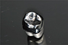
In master die preparation, the first molar of a lower jaw typodont resin model (KaVo Basic study model; KaVo Dental GmbH, Biberach, Germany) was prepared to accommodate an all-ceramic restoration. The model was prepared by adjusting for a 1.0 mm circumferential chamfer, an occlusal reduction of 2.0 mm, and a 5° convergence angle. The area of the resin model including the prepared tooth was then duplicated as an abrasion-resistant master model made of Ceramill sintron (Amann Girrbach AG, Koblach, Austria) using Ceramill system (Fig. 1). Based on 40 individual impressions of this master die (Aquasil Ultra XLV and Monophase, Dentsply, Milford, DE, USA), 40 stone dies in class IV stone (Fuji Rock, GC, Leuven, Belgium) were made and subsequently used to fabricate the Y-TZP restorations. The 40 stone dies were randomly assigned to four groups (n = 10). Each twenty stone die was used to fabricate copings and to manufacture monolithic crowns.
In the fabrication of monolithic crown and coping, copings and crowns were fabricated using Ceramill and Zirkonzahn CAD/CAM systems. As this study is designed to compare two different CAD/CAM systems, burs and zirconia blocks recommended by each system were used for the experiment. Designated cement space follows the guideline of each system. The Ceramill system was used to manufacture 10 copings and 10 monolithic crowns by CAD/CAM. After each stone die was scanned by the Ceramill map400 (Amann Girrbach AG, Koblach, Austria), a monolithic crown and coping were designed using the Ceramill mind design program (Amann Girrbach AG, Koblach, Austria). The design of the final prosthesis after porcelain veneering was also drawn while designing the coping. Ceramill Zolid FX (Amann Girrbach AG, Koblach, Austria), a partially sintered zirconia block, was processed using a Ceramill Motion 2 (Amann Girrbach AG, Koblach, Austria), a five-axis milling machine, to manufacture the prostheses. Three kinds of burs with diameters of 2.5, 1.0 and 0.6 mm were used in the milling machine in regular sequence. Finally, they were sintered at 1450℃ for 11 hours in a Ceramill Therm furnace (Amann Girrbach AG, Koblach).
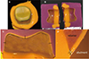
Another 10 copings and 10 monolithic crowns were manufactured using a Zirkonzahn system. In this system, a SCANNER S600 ARTI (Zirkonzahn GmbH, Bruneck, Italy) was used to scan the stone dies and a Zirkonzahn Modellier (Zirkonzahn GmbH, Bruneck, Italy) was used to design monolithic crowns and copings. As with the Ceramill system, the design of the final prosthesis after porcelain veneering was also drawn while designing the coping. Milling burs with diameters of 2.0, 1.0, and 0.5 mm were used in a MILLING UNIT M5 (Zirkonzahn GmbH, Bruneck, Italy), a five-axis milling machine. Partially sintered Prettau zirconia (Zirkonzahn GmbH, Bruneck, Italy) blocks were used and the prostheses were finally sintered at 1600℃ for 10 hours in a ZIRKONOFEN 600 furnace (Zirkonzahn GmbH, Bruneck, Italy) (Fig. 2).
In the analysis of fit accuracy, the copings and monolithic crown manufactured by both CAD/CAM systems fitted well to the master die without any adjustment by dental technicians. First, the weight technique was used to determine the overall fit accuracy: the inner side of the monolithic crowns and copings were filled with light-body silicone (Aquasil Ultra XLV; Dentsply, Milford, DE, USA), simulating the clinical application of a luting agent. Then, the full monolithic crowns and copings were placed onto the master die in order. The restorations were then seated on the master die using finger pressure. Following the removal of excess unpolymerized material at the margins, finger pressure was applied again for four minutes. After polymerization of the impression material, the crowns were carefully removed from the master die, and the weight of the additional silicone was measured using an analytical balance (Mettler AJ, San Francisco, CA, USA). A single operator performed all measurements. The same procedure was performed three times for each specimen.
Replica technique procedures were used to evaluate the internal and marginal fit of each specific part. The first steps of the replica technique were similar to those of the silicone weight technique. The master die was lubricated with separating fluid (Microfilm; Kerr Italia Srl, Salerno, Italy) before each prosthesis was adapted. First, monolithic crowns and copings were filled with light-body silicone (Aquasil Ultra XLV; Dentsply, Milford, DE, USA) and seated on the master die with finger pressure. After the light-body silicone had set, the restorations were removed from the master model while the thin silicone films representing the space between abutment teeth and retainers remained on the abutment teeth (Fig. 3A). The silicone films were then stabilized by contrasting heavy-body silicone (Aquasil Ultra Monophase; Dentsply, Milford, DE, USA) using a customized impression tray. The trays were designed to have generally equal space over the master die so that the light-body silicone is not deformed by the heavy-body silicone while making the replica. The replicas were then cut in the bucco-lingual direction in the center of the prosthesis using a No. 10 surgical blade to measure and photograph the internal and marginal gap with a stereoscopic microscope with surface illumination (MZ-16FA; Leica Microsystems, Wetzlar, Germany) using Leica microscope software (Fig. 3B).
The silicone film thickness was recorded using a stereoscopic microscope to measure five points for each replica. Marginal discrepancies, internal gap between the two points, and occlusal gap at one point of occlusal surface were measured (Fig. 3C), as well as three different points at the marginal area. The horizontal (x), vertical (y), and absolute marginal discrepancies (z) were evaluated. Horizontal marginal discrepancy is the horizontal distance between abutment margin and prosthetic margin, while vertical marginal discrepancy is the vertical distance between abutment margin and prosthetic margin. Absolute marginal discrepancy is the summation of the two previously-explained discrepancies (Fig. 3D).
For evaluating of porcelain veneering effect, copings were subsequently veneered with their respective ceramic material to produce the final restoration. In this study, the press-over technique was used in order to reduce dental technician errors and to produce uniform porcelain veneer thickness. The veneer form designed with the coping was duplicated using the castable milling wax produced by each company (Ceramill Wax; Amann Girrbach AG, Koblach, Austria and Zirkonzahn Wax; Zirkonzahn GmbH, Bruneck, Italy). The duplicated wax replicas and sprues were attached to each coping, which were then embeded. The wax was burnt out and the final prosthesis acquired by filling the space with IPS e.max ZirPress (Ivoclar Vivadent, Amherst, NY, USA), a pressable glass-ceramic ingot. Firing was performed in a proper ceramic furnace (Programat EP 5000, Ivoclar Vivadent, Amherst, NY, USA) at 910℃. After recovery of the restorations, they were finished according to the manufacturer's instructions and glazed at 750℃. After the veneering process, the previously described measurements were performed on every veneered restoration.
Statistical analysis was performed using software (SPSS Statistics for Windows, Version 20.0; IBM Corp., Armonk, New York, USA). The Shapiro-Wilk test was used to confirm the normal distributed of the silicone weight, internal gaps and marginal discrepancies. The mean values and standard deviations per group were calculated. One-way analysis of variance (ANOVA) was used to assess the influence of the CAD/CAM systems and the porcelain veneering process on the silicone weight, internal gaps and marginal discrepancies. Levene's tests were also performed to evaluate the equality of the variances. The level of significance was set at 0.05.
RESULTS
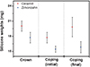
Table 1 shows the silicone weights for the cement space of the Ceramill (n = 10) and Zirkonzahn monolithic (n = 10) crowns measured using the weight technique. The average weights of silicone in the Ceramill and Zirkonzahn monolithic crown specimens were 20.02 ± 1.02 and 17.72 ± 1.05 mg, respectively (Fig. 4). This difference was statistically significant (Table 3). The initial silicone weight of the Ceramill coping specimen was 19.94 ± 2.39 mg; the silicone weight after porcelain veneering 19.82 ± 1.94 mg. In comparison, the initial silicone weight for the Zirkonzahn coping specimen was 15.43 ± 0.62 mg, which increased to 15.84 ± 1.11 mg after porcelain veneering (Table 1) (Fig. 4). The initial silicone weight and the silicone weight after porcelain veneer differed significantly between the Ceramill and Zirkonzahn systems. However, the silicone weight before and after porcelain veneering was not significantly different in both manufacturing system (Table 4, Table 5).
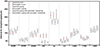
Analysis using the replica technique revealed internal gaps of the Ceramill monolithic crowns of 83.20 ± 10.0 µm at the bucco-axial surface, 213.40 ± 19.5 µm on occlusal surface, and 56.20 ± 8.2 µm at linguo-axial surface. The measured internal gaps of the Zirkonzahn crown were 43.70 ± 7.3 µm at the bucco-axial surface, 170.60 ± 14.2 µm on occlusal surface, and 42.90 ± 6.8 µm at linguo-axial surface (Table 2) (Fig. 5). The internal gaps measured at each position of the monolithic crowns differed significantly between the two manufacturing systems (Table 3).
The initial internal gaps of the Ceramill copings were 84.10 ± 13.48 µm at the bucco-axial surface, 206.00 ± 25.29 µm on the occlusal surface, and 60.20 ± 14.63 µm at the linguo-axial surface. After porcelain veneering, these gaps were 84.00 ± 11.57, 216.90 ± 29.35, and 64.00 ± 12.27 µm, respectively. In comparison, the initial internal gaps of the Zirkonzahn copings were 42.20 ± 13.23, 170.40 ± 16.21, and 47.60 ± 7.28 µm, respectively. After veneering, the gaps were 42.20 ± 4.08, 171.30 ± 13.10, and 54.80 ± 8.23 µm, respectively (Table 2) (Fig. 5). The internal gaps differed significantly before and after porcelain veneering; the coping also differed significantly between manufacturing systems (Table 4, Table 5).
The horizontal, vertical, and absolute marginal discrepancies were also measured. The discrepancies at the buccal margins of the Ceramill crowns were 89.91 ± 13.30, 55.40 ± 8.33, and 106.00 ± 12.48 µm, respectively, and 86.92 ± 15.52, 77.30 ± 10.44, and 117.00 ± 13.16 µm at the lingual margins. The horizontal, vertical, and absolute marginal discrepancies at the buccal margins of the Zirkonzahn crowns were 93.47 ± 16.06, 60.0 ± 8.82, and 111.60 ± 14.30 µm, and 93.48 ± 11.84, 66.80 ± 7.18, and 115.40 ± 7.95 µm, respectively, at the lingual margins (Table 2) (Fig. 5). Comparisons of the monolithic crowns revealed that only the linguo-vertical marginal discrepancies differed significantly between the two manufacturing systems (Table 4).
The horizontal, vertical, and absolute marginal discrepancies at the buccal margins of the Ceramill copings before porcelain veneering were 85.12 ± 10.62, 54.70 ± 6.68, and 101.30 ± 11.39 µm, respectively. The discrepancies at the lingual margins were 105.06 ± 21.03, 64.50 ± 10.15, and 124.10 ± 17.87, respectively. After veneering, the horizontal, vertical, and absolute marginal discrepancies at the buccal margins of the Ceramill copings were 90.09 ± 10.74, 64.70 ± 8.10, and 111.20 ± 10.49 µm, respectively, and 110.50 ± 13.25, 70.80 ± 11.85, and 131.40 ± 16.37 µm at the lingual margins. The horizontal, vertical, and absolute marginal discrepancies at the buccal margins of the Zirkonzahn copings before porcelain veneering were 105.72 ± 10.86, 55.40 ± 12.24, and 119.90 ± 11.13 µm, respectively, and 100.13 ± 12.90, 60.00 ± 3.97, and 116.90 ± 11.79 µm at the lingual margins. The horizontal, vertical, and absolute marginal discrepancies at the buccal margins of the Zirkonzahn copings after porcelain veneering were 115.64 ± 12.18, 60.50 ± 11.97, and 131.10 ± 10.99 µm, respectively. They were 106.87 ± 7.65, 72.50 ± 6.20, and 129.30 ± 7.17 µm, respectively, at the lingual margins (Table 2) (Fig. 5). There were significant differences in the marginal discrepancies of the copings between the two manufacturing systems. The marginal discrepancies before and after porcelain veneering also differed significantly in the bucco-vertical margins of the Ceramill system and the bucco-absolute, linguo-vertical, and linguo-absolute margins in the Zirkonzahn system (Table 4, Table 5).
DISCUSSION
Despite careful preparation of full-coverage restorations and precise cementation, small gaps will remain between the margins of the restorations and the finish lines of the prepared teeth, predisposing the teeth to caries and periodontal disease. The closer the margin of restoration to the finish line of the preparation, the smaller the marginal gap and thickness of the exposed cement layer at the margin.22 The clinically allowable marginal gaps of dental prostheses have been described in numerous studies. Some authors have reported marginal gaps under 120 µm to be clinically allowable for traditional fixed prostheses.13 Others have reported marginal gaps of 160 - 172 µm to be clinically acceptable for conventional crowns, with an acceptable marginal gap range of under 200 µm and occlusal gap range of under 250 - 300 µm.23242526 In this study, finger pressure was used to seat the crowns and copings to the master die to reproduce the clinical situations. Some previous literatures also used this method.27
While planning this research, micro-CT was also considered for measurement of the fit accuracy. However, it was excluded due to some limitations. First of all, it is not easy to find specific measuring point for internal gap and marginal discrepancy because the exact interface between materials with different radiation absorption coefficient is hardly defined in micro-CT. Furthermore, radiographic images could have artificial defects due to the reflection of radioactive rays.28
Kokubo et al.29 used a light-body silicone in place of luting cement to determine the relative marginal gaps in ceramic crowns. McLean and von Fraunhofe also used a light-body silicone to measure the thickness of the cement film, concluding that it is a convenient method to evaluate three-dimensional volume of the luting cement space.30 Nakamura et al.31 and May et al.32 both used a test-fit silicone paste to measure internal gaps.
In this study, the silicone weights of the cement spaces of crowns manufactured with the Ceramill system were significantly higher than those of the Zirkonzahn system. The Ceramill system recommends 50 µm of cement space, while the Zirkonzahn system recommends 35 µm. This gap might have contributed to the observed differences in silicone weights, as the prostheses were manufactured according to the recommendations for each system. However, higher silicone weight does not mean lower prosthesis fit accuracy. Excessively small cement space can disturb precise setting of the prosthesis. Therefore, the fit accuracy of prostheses cannot be determined only by the cement space. In this study, both replica and silicone weight techniques were used to more accurately measure the prosthesis fit accuracies.
Impression techniques with low-viscosity impression material (replica technique) are popular methods for evaluating marginal discrepancies between crowns and teeth. The replica technique is a methodology applicable to both in vitro and in vivo measurements of precision. The advantages of replica technique include the small probability of damaging the sample and abutment in the process, which makes it a non-destructive methodology. The majority of authors agree that, compared with other techniques, the replica technique offers more potential for verifiable and accurate results.3334 The replica technique can be used to measure the fit accuracy of prosthesis in each position and provide information about specific margins with critical impact on the fitness and prognosis of prostheses.
In this study, the marginal discrepancies of the monolithic crowns manufactured by both systems were less than 120 µm, well within the clinically acceptable range. The marginal discrepancies were relatively lower in the Ceramill system, and the internal gaps were smaller in the Zirkonzahn system.
The marginal discrepancies of copings made with the Ceramill and Zirkonzahn system were between 101 and 131 µm and 116 and 131 µm, respectively. Both were within the clinically allowable range, based on less strict criteria.3536 Similarly, analysis of the monolithic crowns showed that the Ceramill system had lower marginal discrepancies, while the Zirkonzahn system had smaller internal gaps. It can be assumed one of the factors contributed to the lower marginal discrepancies in the Ceramill system is that the prostheses are set to the correct position without any friction against the abutments because the Ceramill system requires 15 µm more cement space. Additional studies are necessary to investigate the optimal cement space required for lower marginal discrepancies with internal gaps within clinically acceptable ranges.
In this study, the press-over technique was used for porcelain veneering process. This technique resulted in porcelain veneers with equal shape and thickness, and also reduced the potential for dental technician errors.
Several studies have reported conflicting results on the effects of porcelain veneering on marginal fit. Balkata et al.37 compared the marginal fit of three all-ceramic systems. The results showed that porcelain firing significantly altered the marginal fit of the crowns. They concluded that the copings were not completely stable during the porcelain firing cycle and that the distortion might have been due to a nonuniform porcelain mass. Gemalmaz and Alkumru reported a small increase in the metal-ceramic crown's gap size after firing the body porcelain; this distortion was most evident during the first firing cycle.38
In a study comparing all-ceramic and porcelain-fused-to-metal (PFM) crowns, Castellani et al.39 reported a firing cycle to significantly influence the vertical discrepancy in both types of prosthesis. The results of the study showed the influence to be greater in all-ceramic crowns. Another study also reported that thermocycling changed the marginal discrepancies of all three crown types they tested.40
However, other studies argue that there are no changes in marginal discrepancies after veneering because the volumetric stabilities of the copings are maintained during the porcelain veneering procedure.4142
Torabi et al.43 measured changes in marginal fit of zirconia copings using three porcelain-veneering techniques, including layering, press-over, and CAD-on techniques. The results showed significant alterations in all three techniques; however, these alterations were in the range of clinically allowable marginal fit. The changes were smallest using the press-over technique, and significant compared with the conventional layering technique.
This difference may be explained by the fact that porcelain particles melt and gather to fill voids during the porcelain veneering procedure, and the resulting contraction of the porcelain mass causes a compressive force on the coping. The deformation of the coping under the stress of contracting porcelain is spread around the entire marginal circumference. Generally, the marginal openings of these crowns after porcelain veneering were within clinically acceptable standards; thus, the amount of distortion does not exclude their use in clinical applications.44
The results of this study revealed that the overall volume change before and after porcelain veneering was not statistically significant. Differences in marginal discrepancies before and after porcelain veneering were statistically significant only at some positions, and the effect of porcelain veneering was greater in the Zirkonzahn system than in the Ceramill system. Nevertheless, these differences do not exceed the acceptable limits in clinical settings.
The results of this study showed that full zirconia crowns and porcelain-fused zirconia coping crowns manufactured with Ceramill and Zirkonzahn systems are sufficient for use in clinical settings. The fit accuracy of the prostheses made by both systems compared favorably with that of conventional prostheses. Differences between the results of the present study and those of other studies may be related to differences in measurement methodologies, microscopes, microscope magnification, measurement location and number, or luting agents.
However, this experiment only compared marginal discrepancy values at two points of zirconia-based prostheses, it is hard to say that these values represent fit accuracy of the entire prostheses. More accurate and meaningful conclusions will be drawn if marginal discrepancy values are compared in more points.
CONCLUSION
The results of this study reject the null hypotheses that the internal and marginal fits of dental prostheses are not affected by different CAD/CAM systems or porcelain veneering process. There were significant differences in the internal crown and coping gaps between the two CAD/CAM systems. Porcelain press veneering did not significantly influence the internal gap, but did significantly affect the marginal fit at some position. And, the marginal discrepancies produced by two CAD/CAM systems were within the reported clinically acceptable ranges.


 XML Download