

 PDF
PDF ePub
ePub Citation
 Print
Print


INTRODUCTION
Implant-supported fixed dental prosthesis is a standardized restoration method that is often used when an aesthetically critical anterior single tooth is missing. It has similar 5-year survival rates as tooth-supported fixed dental prosthesis.12 Although the success rate of an all-ceramic crown was reported to be lower than that of a metal-ceramic crown, all-ceramic crowns displayed similar success rates in both tooth-supported fixed dental prosthesis and implant-supported fixed dental prosthesis.234 Titanium is a stable implant material, and titanium abutments have the advantage of supporting gingival health and preventing galvanic reaction between the fixture and abutment.56 However, when a titanium abutment is used in thin peri-implant mucosa in the anterior area, the metal part can be detected through the mucosa. For this reason, studies on alumina and zirconia, which are high-strength ceramics, have been conducted, and the study on implant abutments using zirconia has advanced, showing excellent material properties and biocompatibility. 78
The influence of the type of connection between a zirconia abutment and a titanium fixture were addressed previously. The types of implant-abutment connection can be divided into two major groups - internal connection and external connection. Sailer et al.910 reported that the internal implant-abutment connection type, including zirconia abutment with titanium insert, showed the highest strength, followed by external implant-abutment connection type and 1-piece internal type, when artificial aging was not conducted. However, in the above-mentioned experiment, only the implant-abutment fracture load was measured, without involvement of the crowns. Therefore, the fracture load was measured under non-physiological conditions, and intra-oral temperature changes and dynamic functional load were not applied. Truninger et al.11 investigated the fracture load of zirconia abutment with 5-year artificial aging. They showed that the fracture load was dependent on the types of connection, which was the same result as that of the former studies, while the strength was lower after artificial aging. However, that study faced the limitation that it cannot be applied to actual clinical situations, because the fracture load of the zirconia abutment was measured without crown restoration, and the fracture loads before and after artificial aging were not examined.
Due to the development of CAD/CAM systems (computer-aided design/computer-aided manufacturing system), the prosthesis using zirconia has replaced the conventional ceramic restoration and is now additionally being applied to implant abutments.1213 Park et al.14 investigated the fit of a customized zirconia abutment manufactured by the CAD/CAM system. They reported that while the fit was less precise than that of a prefabricated zirconia abutment, it was within the clinically acceptable range. The strength of the customized zirconia abutment was significantly higher than that of the prefabricated zirconia abutment.
When restoring all-ceramic implant crowns, shoulder or deep chamfer preparation of zirconia abutment was typically applied. Koutayas et al.15 measured the fracture loads of 1-piece internal full zirconia abutments with three preparation depths (0.5 mm, 0.7 mm, and 0.9 mm) that were restored using lithium disilicate. They reported that the fracture loads of all three specimens were higher than physiologic masticatory force, but preparation depths of over 0.7 mm were not recommended. Subsequent to that report, Mitsias et al.16 reported measurement of the fracture load of 1-piece internal full zirconia abutment with artificial aging. Preparation depths under 0.9 mm were found to have had no influence on the fracture load. Many literature reports have stated that the optimum preparation depth of zirconia abutment ranged from 0.5 mm to 1.0 mm.171819202122 However, further studies regarding standard indicators are still needed. Therefore, the purpose of this study was to investigate the change of fracture load of customized zirconia abutments with titanium insert according to the different preparation depths, with aging and chewing simulation. The null hypotheses were that the fracture loads of customized zirconia abutment with titanium inserts will not change depending on the various preparation depths, or as a result of aging and chewing simulation.
MATERIALS AND METHODS
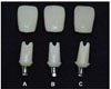
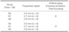
A commercial titanium fixture of 4.5 mm in diameter and 10 mm in length (Anyridge, Megagen, Gyeongsan, Korea) was used. The fixture was embedded in acrylic resin (Orthoresin Dentsply, Weybridge, Surrey, UK) according to the ISO-Normed protocol [ISO 14801].23 Prefabricated titanium inserts that fit into the titanium fixture (Prop abutment, Megagen, Gyeongsan, Korea) were used, and the zirconia supra-structures to be attached to the insert were fabricated with zirconia blocks (ZenostarZr translucent, Wieland, Germany) (Table 1). The preparation depths of the zirconia suprastructure were designed to 0.5 mm, 0.7 mm and 0.9 mm using the CAD system (3shape dental designer premium 2013, 3shape, Denmark), and the same-sized zirconia structures were manufactured using the CAM system (Zenotec T1, Wieland Dental + Technik GmbH, Pforzheim, Germany). The customized zirconia suprastructures and prefabricated titanium inserts were then bonded using dual cure self-adhesive resin cement (Rely X Unicem, 3M ESPE, St. Paul, MN, USA). Crowns fabricated to replace a maxillary right central incisor in Asian adult (8.6 mm in width and 11.9 mm in length) were made using lithium disilicate (IPS e.max press, Ivoclar-vivadent, Schaan, Liechtenstein) (Table 1).24 Crowns were designed to fit over the customized zirconia abutments using the CAD system (3shape dental designer premium 2013, 3shape, Denmark). Rapid prototype model of crowns with the same overall size and different preparation depths were fabricated by 3D printer (Digital Dental Printer, EnvisionTEC, Gladbeck, Germany). Lithium disilicate crowns were manufactured according to the previous conventional method (Fig. 1). Customized zirconia abutments with titanium insert were sandblasted for 30 seconds under the pressure of 0.5 bar using 50 µm Al2O3 particles (Cobra®, Renfert, Germany). After the surface processing, ultrasonic cleansing was performed on every specimen for 10 minutes using acetone and alcohol, after which the specimens were dried at room temperature. Next, zirconia primer (Metal/Zirconia primer, Ivoclar-vivadent, Schaan, Liechtenstein) was applied to the bonding surface. Following the manufacturer's recommendations, the inner surfaces of the lithium disilicate crowns were etched with hydrofluoric acid and silane finished (Monobond-S, Ivo c l a r-Vivadent, Schaan, Liechtenstein) for 1 minute. The fixture and abutment were then tightened with a torque of 30 Ncm, and the inside of the abutment was filled with cotton and temporary restorative material (Caviton, GC, Tokyo, Japan). The abutment and crown were then bonded using dual cure self-adhesive resin cement (Rely X Unicem, 3M ESPE, St. Paul, MN, USA), and finger pressure was applied for 5 minutes. After the bonding procedure, excessive cement was removed mechanically.
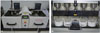
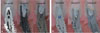
Half of the samples (n=18) were put into distilled water at 37℃ or 24 hours, after which thermocycling was conducted in water baths set to 5 ± 0.5℃ and 55 ± 0.5℃, according to the international standard ISO normed protocol (ISO 10477).25 Thermocycling were conducted to obtain 5-year artificial aging, based on previous reports (Fig. 2).2627 After thermocycling, the samples were placed at a 45 degree angle on a chewing simulator (Chewing simulator CS-4, SD Mechatronic GmbH, Germany) using a customized jig. Chewing simulation was performed 1,200,000 cycles at 1.67 Hz and 49 N, which corresponds to 5-year artificial aging.27 A cobalt-chrome steel indenter with a rounded tip (8 mm in diameter) was used as an antagonist. The fracture load of the abutment was measured using the universal testing machine (RB Model 301 Unitech M™, R and B, Korea). The samples were fixed with a custom-made jig. The maximum fracture load was determined by applying load on the palatal 2 mm of the incisal part of the abutment at the cross head speed of 0.5 mm/min. Following the a previously published method, 0.5 mm of aluminum foil was inserted between the sample and the testing machine for even distribution of the load.1128 Depending on the preparation depths and performance of artificial aging, the specimens were classified into 6 groups, including three non-artificial aging groups (N5, N7, N9) and three artificial aging groups (A5, A7, A9) (Table 2). To evaluate the fracture mode, all specimens were embedded in acrylic resin and sectioned in the middle of model. The fracture modes were then identified macroscopically (Fig. 3).
The fracture loads were processed statistically using the SPSS program (SPSS Version 21.0, SPSS Inc., Chicago, IL, USA). After confirmation of the equal variance assumption by Levene's test, two-way ANOVA for analysis and Tukey HSD post-hoc tests were conducted to determine the interactions between the changes in preparation depth and artificial aging on the fracture load of the customized zirconia abutment with titanium insert (P>.05). Under the equal variance assumption, one-way ANOVA was performed to analyze the fracture load depending on the preparation depths of the samples with artificial aging (P<.05), while the non-parametric Kruskal-Wallis test was used in groups without artificial aging because there was no assumption of equal variance (P<.017). The differences in fracture load between the artificial aging group and non-artificial aging group at each preparation depth were statistically analyzed using the independent t-test (P<.05). Multiple linear regression analysis was conducted to determine the relationships between variables and the interaction between artificial aging and changes in preparation depths or fracture load (P<.05).
RESULTS
One of the tested specimens showed crown fracture during the chewing simulation. All other specimens were stable, with no observable change in gross morphology.
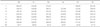
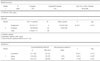
The means and standard deviations of the fracture loads of whole specimens are shown in Table 3. One sample in group A5 was broken during the chewing simulation, as it could not resist the masticatory force. Therefore, there were 35 valid samples, for which each fracture load was measured.
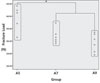
The mean fracture loads in groups N5, N7, and N9 (non-artificial aging groups) were 539.28 ± 63.11 N, 406.56 ± 28.94 N, and 366.66 ± 30.19 N, respectively. The fracture area of this group, without artificial aging, was found to be located in the implant-abutment titanium insert internal connection area, and all specimens showed the typical fracture or deformation. (Fig. 3A) The Kruskal-Wallis test indicated that the fracture load, depending on preparation depth, was higher in the N5 group than in the N7 and N9 groups (P<.017), while no significant difference in fracture load was observed between groups N7 and N9 (P>.017) (Fig. 4).
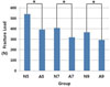
The fracture loads of groups A5, A7, and A9 (with artificial aging) were 392.616 ± 50.57 N, 317.94 ± 30.05 N, and 292.74 ± 37.15 N, respectively. Like the groups without artificial aging, the fracture areas of these groups were also located in the implant-abutment titanium insert internal connection area, and all specimens showed the typical fracture or deformation (Fig. 3B) According to one-way ANOVA, there were significant differences in the fracture load depending on the preparation depth (P<.05). Tukey HSD post hoc test indicated that the fracture load, depending on preparation depth, was significantly higher in the A5 group than the A7 and A9 groups, while no meaningful difference was found in the fracture load between groups A7 and A9 (P>.05) (Fig. 5).
The difference in fracture load was evaluated between each paired group (N5/A5, N7/A7, N9/A9), depending on whether artificial aging was performed or not. Significant differences in the fracture load were observed for every pair of preparation depth through independent t-test (P<.05) (Fig. 6).
Multiple linear regression analysis was conducted to predict the changes in fracture load depending on the three preparation depths and the performance of artificial aging, as well as to examine the influence of fracture load according to each variable. The regression model had a power of explanation of 73.4%, and was significant when assumed through analysis of variance (P<.05). Coefficient analysis revealed that fracture load decreased by 102.456 N in the case of artificial aging, and by about 68.56 N with increase in the preparation depth of 0.2 mm. Overall, changes in the preparation depth were found to have more influence on the fracture load than artificial aging (Table 4).
DISCUSSION
In the present study, the fracture loads of customized zirconia abutments with titanium inserts were investigated according to preparation depths and the performance of artificial aging (thermocycling & chewing simulations). Moreover, for representation of the 'crown-abutment-fixture complex', lithium disilicate crowns were fabricated, and the optimum preparation depth of the crown was also assessed. The results indicated that the fracture load was significantly influenced by the variance of preparation depth, as well as by artificial aging. In other words, both null hypotheses were rejected. The fracture load of preparation depth 0.5 mm (groups N5 and A5) was significantly higher than those of the other preparation groups (groups N7, N9, A7, and A9) regardless of artificial aging. After artificial aging and chewing simulations, all groups were significantly weakened.
The width and length ratio of the maxillary anterior crown is known to be an important factor for anterior esthetic restorations. Tsukiyama et al. reported that the average size of the central incisor in adult Asians was 8.6 mm in width and 11.9 mm in length.24 Culp and McLaran reported the use of lithium disilicate crowns with a variety of transparencies for maximization of esthetics.29 Many studies in the literature reported that the 6-degree angle of axial preparation offered adequate mechanical retention of the crowns and improved the adaptation of the crown margins. 30313233 In the present experiments, the crowns and abutments were fabricated based on the reported values. For restoration of all-ceramic implant crowns, zirconia abutments require shoulder or deep chamfer margins. Koutayas et al. selected three preparation depths which were thinner than the natural teeth.15 Although such specific limitation of the preparation depth resulted in a restricted space for the veneering materials, this disadvantage was outweighed by the favorable color of the underlying zirconia abutment. Koutayas et al. also stated that when static load was applied to the implant axis at 135 degrees, the thinnest portion of the abutment and fixture connection was fractured due to the levering effect within the internal connection of the 1-piece zirconia abutment.15 They thus named the thinnest point as the "Weakest link". Mitsias et al. also reported that all specimen of 1-piece zirconia abutment presented the typical fracture at the implant-abutment internal connection after dynamic loading.16 Substituting the link with metal materials may allow strengthening of the fractured joint.11 However, despite replacement of the link, all specimens in the present study presented fractures in the same area as the previous reports. It should be noted that fracture did not occurin all samples, as titanium insert and screw deformation appeared in some of the specimens. The physiological force during mastication and the swallowing of food ranges from 10-120 N, with the maximum masticatory force of between 108-299 N.3435 Herein, except for some specimens in the A7 and A9 groups, most specimens demonstrated tolerance of the physiological and maximum masticatory force.
Exposure of ceramic materials to mechanical stress and moisture results in low-temperature degradation,36 and artificial aging has an adverse impact on the mechanical properties of zirconia material. Moreover, the phase of zirconia could be transformed from tetragonal to monoclinic.37 Mitsias et al. reported that the fracture strength and durability of 1-piece full zirconia abutment was increased after dynamic loading in the chewing simulator. However, they stated that such results could not be supported by evidence-based scientific data.16 In the present study, the fracture loads of all preparation groups were decreased after artificial aging and chewing simulation. Therefore, unlike the results of the abovementioned study, the fracture load of zirconia abutment with titanium insert examined herein was reduced after the 5-year cyclic loading. The evidence-based scientific data also did not support the results. However, it could be expected that 5-year cyclic loading might weaken the physical properties of the zirconia abutment with titanium insert. Therefore, in order to determine the exact mechanisms involved, more advanced study will be needed. For the zirconia, phase aging and chewing simulation, as well as XRD (X-ray diffusion) analyses will be required.
In order to evaluate the correlation of the independent variables (preparation depth and artificial aging) on the dependent variable (fracture load), multiple linear regression analysis was performed. The results revealed that increase of the preparation depth by 0.2 mm tended to decrease the fracture load by 68 N, while the aging and chewing simulations tended to cause decreases of 102 N. Changes in the preparation depth of 0.2 mm had a higher effect than the aging and chewing simulations. Thus, appropriate setting of the preparation depth of zirconia abutment is critical to the survival rate, and hence, the clinical applicability.
CONCLUSION
Within the limitations of this in vitro study, the following conclusions could be drawn:
Regardless of chewing simulation, the circumferential preparation depth of 0.5 mm of zirconia abutments had a significantly higher fracture load than other groups.
Artificial aging caused a significant decrease of the fracture load for all groups of different preparation depths.
The change of preparation depth was more influential than the chewing simulation on the fracture load of the customized zirconia abutment with titanium insert.
Single implants restored with lithium disilicate crowns and zirconia abutments with titanium insert could with-stand maximum masticatory force in the incisive area when the preparation depth was 0.5 mm.

 XML Download