

 PDF
PDF ePub
ePub Citation
 Print
Print


Abstract
Respiratory bronchiolitis-associated interstitial lung disease is one of the smoking-related interstitial lung diseases. Histopathologically, it shows respiratory bronchiolitis, which is characterized by the accumulation of pigmented macrophages within the respiratory bronchioles, accompanying peribronchiolar inflammation. Clinically, it is presented with respiratory symptoms such as a cough, sputum and dyspnea on exertion. It is well known that the incidence of malignancy in interstitial lung disease is high, but in respiratory bronchiolitis-associated interstitial lung disease the report of accompanying malignancy is rare. Here we report a case of a 60-year-old male heavy smoker presented with a cough, sputum and clubbing finger. A chest computed tomography (CT) of the patient did not show any shadow suspected of malignancy, but adenocarcinoma was found on a transbronchial lung biopsy and on a surgical lung biopsy with respiratory bronchiolitis-associated interstitial lung disease.
Figures and Tables
Figure 1
Posteroanterior chest radiograph shows subpleural reticular opacity in both lower lung zones and small nodular lesion in left upper lung zone.
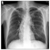
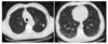
Figure 2
High resolution computed tomography shows peripheral ground-glass attenuation with multiple clustered air-filled cysts in the both lower lung zones and paraseptal emphysema in both upper lung zones. Well defined 10 mm sized calcified nodule was observed in left upper lobe.
References
1. Niewoehner DE, Kleinerman J, Rice DB. Pathologic changes in the peripheral airways of young cigarette smokers. N Engl J Med. 1974. 291:755–758.
2. Myers JL, Veal CF Jr, Shin MS, Katzenstein AL. Respiratory bronchiolitis causing interstitial lung disease. A clinicopathologic study of six cases. Am Rev Respir Dis. 1987. 135:880–884.
3. Rao RN, Goodman LR, Tomashefski JF Jr. Smoking-related interstitial lung disease. Ann Diagn Pathol. 2008. 12:445–457.
4. Patel RR, Ryu JH, Vassallo R. Cigarette smoking and diffuse lung disease. Drugs. 2008. 68:1511–1527.
5. Portnoy J, Veraldi KL, Schwarz MI, Cool CD, Curran-Everett D, Cherniack RM, et al. Respiratory bronchiolitis-interstitial lung disease: long-term outcome. Chest. 2007. 131:664–671.
6. Ozawa Y, Suda T, Naito T, Enomoto N, Hashimoto D, Fujisawa T, et al. Cumulative incidence of and predictive factors for lung cancer in IPF. Respirology. 2009. 14:723–728.
7. Izumi M, Takayama K, Yabuuchi H, Abe K, Nakanishi Y. Incidence of hypertrophic pulmonary osteoarthropathy associated with primary lung cancer. Respirology. 2010. 15:809–812.
8. Scheidl S, Kovacs G, Stacher E, Popper H, Olschewski H. A 55-year-old craftsman with dyspnea and clubbing: a case report. Cases J. 2009. 2:8579.
9. Tomita K, Caramori G, Lim S, Ito K, Hanazawa T, Oates T, et al. Increased p21(CIP1/WAF1) and B cell lymphoma leukemia-x(L) expression and reduced apoptosis in alveolar macrophages from smokers. Am J Respir Crit Care Med. 2002. 166:724–731.
10. Mavridou D, Laws D. Respiratory bronchiolitis associated interstitial lung disease (RB-ILD): a case of an acute presentation. Thorax. 2004. 59:910–911.
 XML Download