

 PDF
PDF ePub
ePub Citation
 Print
Print


Abstract
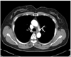
The middle mediastinum contains several important organs and pluripotent cells. It is difficult to make a definitive diagnosis in patients with middle mediastinal tumors due to a wide range of diseases. The likelihood of malignancy is influenced primarily by the following factors: patient age, size, tumor location, and the presence or absence of symptoms. We describe a case of a middle mediastinal tumor, which was suspected on chest x-ray; chest computed tomography revealed the eccentric mass of distal esophagus. This case emphasizes the diagnostic importance of the chest x-ray to the physicians. The possible differential diagnoses are reviewed.
Figures and Tables
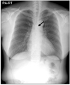
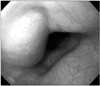
Figure 2
Endoscopic finding of esophageal submucosal tumor. There was a 1.7 cm-sized elevated lesion, which was covered with normal esophageal mucosa and showed positive rolling-sign, at 28 cm from incisor.
References
1. Lee HL, Kim SK, Kim HK, Chung KY, Lee DY, Kim SE, et al. Clinical study on primary mediastinal tumors and cysts: report of 344 cases. Tuberc Respir Dis. 1993. 40:575–583.
2. Davis RD Jr, Oldham HN Jr, Sabiston DC Jr. Primary cysts and neoplasms of the mediastinum: recent changes in clinical presentation, methods of diagnosis, management, and results. Ann Thorac Surg. 1987. 44:229–237.
3. Choong CK, Meyers BF. Benign esophageal tumors: introduction, incidence, classification, and clinical features. Semin Thorac Cardiovasc Surg. 2003. 15:3–8.
4. Choi SW, Park H, Lee SB, Chung JP, Lee SI, Hong SW. Three cases of secondary esophageal tuberculosis presenting as an esophageal submucosal tumor. Korean J Gastrointest Endosc. 2005. 30:80–85.
5. Huang YK, Wu YC, Liu YH, Liu HP. Esophageal tuberculosis mimicking submucosal tumor. Interact Cardiovasc Thorac Surg. 2004. 3:274–276.
6. Macchiarini P. Primary tracheal tumours. Lancet Oncol. 2006. 7:83–91.
7. Macchiarini P, Ostertag H. Uncommon primary mediastinal tumours. Lancet Oncol. 2004. 5:107–118.
 XML Download