

 PDF
PDF ePub
ePub Citation
 Print
Print


INTRODUCTION
The strength of the bond between the bracket and the enamel surface depends on 3 factors, namely, the retention mechanism of the bracket base, the adhesive material or bonding resin, and the preparation of the tooth surface.1 Commonly used adhesive systems employ an enamel conditioner, a primer solution, and an adhesive resin to bond the orthodontic brackets to the enamel surface. These adhesive systems generally contain 35 - 37% orthophosphoric acid, which conditions the enamel surface.
Zachrisson and Büyükyilmaz2 found that sandblasting improves the strength of bonds to gold, porcelain, and amalgam. Further, Faltermeier and Behr3 reported that the process of sandblasting improves the shear bond strength (SBS) of stainless steel brackets. Further, Chung et al.4 found that sandblasting is a more viable alternative to chemical etching techniques in terms of bond strength, while Berk et al.5 and Canay et al.6 reported that sandblasting the enamel surface does not provide adequate SBS for bracket bonding.
Laser energy enables localized melting and ablation of the enamel surface; it affects etching through a process of continuous vaporization and micro-explosions, which occur due to the vaporization of the water trapped within the hydroxyapatite matrix.7 Irrigation of the enamel by laser energy may be beneficial since it inhibits enamel demineralization and, thereby, caries formation.8
To date, studies on laser etching have addressed various issues, such as power output differences,9,10 application distance,11 and microleakage under orthodontic brackets.12 However, none of them compare all the known enamel-conditioning techniques.
The aim of this study was to compare the effects of different enamel conditioning techniques for bracket bonding in terms of the SBS, adhesive remnant index (ARI),13 and scanning electron microscope (SEM) findings.
MATERIALS AND METHODS
Ninety human premolars that had been extracted for orthodontic treatment were used in this study. After extraction, the teeth were stored at room temperature in distilled water containing thymol crystals (1% wt/vol) to inhibit bacterial growth. The teeth were cleaned and polished with a fluoride-free pumice slurry and rubber cups for 10 s and thoroughly washed and dried by exposure to oil-free air stream. They were then examined under a light stereomicroscope (SMZ460; Nikon, Osaka, Japan) at × 10 magnification to rule out caries and enamel cracks. Teeth with caries, restorations, and surface abnormalities were excluded from the study. The samples were randomly divided in 6 groups of 15 specimens each, by using a random numbers table. All samples were embedded vertically in cold-curing acrylic (Orthocryl; Dentaurum, Ispringen, Germany) by using metal ring moulds.
Phosphoric acid (35%) gel (Gel Etch; 3M Unitek, Monrovia, CA, USA) was used for etching the teeth in group 1 for 15 s. The teeth were then rinsed with water infused from a 3-in-1 syringe for 15 s and dried with an oil- and moisture-free source for 10 s.
The teeth in group 2 were treated with a self-etching primer (SEP; Transbond Plus; 3M Unitek, Monrovia, CA, USA), which was rubbed onto the enamel by gentle pressure for 5 s. Then, a gentle air burst was applied to dry the primer into a thin film.
In group 3, the teeth were sandblasted from a distance of 1 mm at 65 - 70 psi for 10 s with 50 µm aluminium oxide (Dynaflex Inc., St. Ann, MO, USA). To prevent unnecessary etching, a 4 × 5 mm aperture was made on a 0.040 inch thick thermoplastic retainer material (Dentsply Raintree Essix Inc., Sarasota, FL, USA), and the sandblaster device (Microetcher II; Danville Engineering, San Ramon, CA, USA) was directed perpendicular to the enamel surface through this aperture. After sandblasting, the specimens were thoroughly rinsed for 15 s and dried for 10 s.
In group 4, the samples were initially treated with the same procedure mentioned in group 3; thereafter, the enamel surface was etched by 35% phosphoric acid gel for 15 s. The teeth were then rinsed with water for 15 s and dried for 10 s.
The enamel surfaces of the teeth in group 5 were conditioned by Er:YAG laser (KaVo Key 3, hand-piece 2060TM; KaVo Dental GmbH, Biberach, Germany) administered at 350 mJ/pulse with a frequency of 4 Hz, from a distance of 1 mm. This frequency rate was chosen to ensure a homogenous ablation pattern by distributing 1 pulse per square millimeter to prevent excess ablation of the enamel. Since the bracket base area was determined to be 10.41 mm2, a 3 × 4 mm area was conditioned with 12 pulses. The tooth was prepared under water-spray cooling (7 ml/min), as per the recommendations of the manufacturer for hard tissue preparation. In order to standardize the procedure, an operation microscope (OPMI®Pico; Carl Zeiss Meditec, Munich, Germany) was used at 10 × magnification.
For group 6, in addition to the steps followed for group 5, the enamel surface was etched by 35% phosphoric acid gel for 15 s. The teeth were then rinsed with water for 15 s and dried for 10 s.
After the enamel-conditioning procedures of all the groups, the stainless steel premolar brackets (Mini Master Roth; American Orthodontics, Sheboygan, WI, USA) were bonded to the teeth with an orthodontic adhesive (Transbond XT; 3M Unitek). The brackets were pressed firmly onto the tooth surface, and excessive adhesive was removed using a sharp scaler. The adhesive was polymerized for 40 s by a halogen light source (Hilux Ultra Plus; 600 mW/cm2; Benlioglu Dental, Ankara, Turkey) placed at the mesial, distal, occlusal, and gingival aspects for 10 s each. The average base surface areas of the brackets were calculated as 10.41 mm2 by using a digital caliper (Absolute Digimatic; Mitutoyo, Miyazaki, Japan).
All specimens were stored in water at 37℃ for 24 h. The shear debonding test was performed using a universal testing machine (Instron Co., Canton, MA, USA). The specimens were subjected to stress from a vertical direction, at a crosshead speed of 1 mm/min. The maximum shear force necessary to debond each bracket was recorded in Newton and then converted into megapascal (MPa).
The debonded enamel surfaces were examined under a stereomicroscope (SMZ460; Nikon, Kyoto, Japan) at 20 × magnification to assess the residual adhesive remaining on the tooth surface by a blinded examiner (Ç.U). The ARI was used to quantify the amount of adhesive remaining on the tooth surface. The following scale was used to grade the amount of adhesive retained on the tooth surface: 0, indicating no adhesive; 1, less than half of the adhesive; 2, more than half of the adhesive; and 3, all the adhesive.
The mean SBS and standard deviations were calculated for each group by one-way analyses of variance (ANOVA) to determine whether there was any significant difference between the enamel conditioning systems. The Scheffé test was used for multiple comparison of the bonding forces. The ARI scores were evaluated by the chi-square test.
Six teeth, one from each group, were selected randomly for SEM evaluation. The coronal parts of the teeth were separated after debonding and prepared for observation under a SEM by serial dehydration of graded ethanol solutions (50 - 100%), mounted on aluminium stubs, and coated with platinum. The specimens were then observed under an SEM (JSM-7000F; JEOL, Tokyo, Japan) at an accelerating voltage of 15 kV to evaluate the intergroup difference in the surface quality.
RESULTS
The average bond strength forces; their standard deviations; and standard errors of the means, minimum, and maximum shear bond strengths are shown in Table 1. The groups subjected to laser and acid etching showed the highest mean SBS values (13.61 ± 1.14 MPa), while the group subjected to sandblasting yielded the lowest value (3.12 ± 0.61 MPa). One-way ANOVA test showed statistically significant differences among the 6 different surface-conditioning methods with respect to SBS (F: 228.709, p < 0.001; Table 2).
The results of the Scheffé post-hoc test showed that groups 3 and 6 were significantly different from other groups (Table 3). The intergroup differences and their levels of significance are shown in Table 3.
The chi-squared test revealed statistically significant differences in the ARI scores of the 6 groups (χ2 = 42.711, p < 0.001; Table 4).
DISCUSSION
Currently, research is on to develop time-conserving and tooth-friendly enamel conditioning systems for bracket bonding. In the present study, we evaluated the effects of all well-known enamel conditioning techniques for bracket bonding in terms of SBS, ARI, and SEM findings.
Phosphoric acid treatment is the most common technique used in the bonding procedure. However, acid etching has been implicated in decalcification and loss of enamel.14,15 Although the enamel-etching technique is a useful and accepted orthodontic procedure for bonding orthodontic brackets, it needs to be improved to establish clinically useful bond strengths while minimizing the amount of enamel loss.16
The bonding force in groups 3 and 6 were significantly different from those in all the other groups (Table 3). The bond strength values in all groups, except group 3, were consistent with the minimal bond strength values reported by Reynolds'17 as clinically acceptable (5.9 - 7.8 MPa; Table 1). Group 3 had a mean SBS value of 3.12 ± 0.61 MPa, which is not suitable for clinical usage. This result is consistent with those reported in studies evaluating the sandblasting technique for enamel conditioning.6,18,19 These findings suggest that as a form of macro-etching,19 only sandblasting the enamel may not be sufficient for orthodontic bonding. Another disadvantage of sandblasting is that the aluminium oxide-containing aerosol used may be swallowed or inhaled by the patient or doctor.
Laser irradiation results in an increase in the calcium to phosphorus ratio,20 thereby rendering the enamel more acid resistant and less susceptible to caries attack.8 Therefore, using laser for enamel conditioning may be beneficial for orthodontic bonding.
In the present study, group 5 had attained the bond strength of 9.45 ± 0.92 MPa, which was comparable to that achieved in the acid-etched group; however, group 6 had a mean SBS of 13.61 ± 1.14 MPa, which was much greater than that in the other groups, and some specimens in this group also showed fractured enamel surfaces (Table 1). This suggests that laser etching, both alone and with acid etching, provided enough SBS for bonding. These findings concur with the findings of some studies,11,21-23 but are contrary to those of others.7,9,24,25 This discrepancy may be attributed to differences between the studies in the power outputs, application distances, and laser types, but this study showed that treatment with Er:YAG laser at 350 mJ/pulse and a frequency of 4 Hz administered as 1 pulse/mm2, from a distance of 1 mm afforded sufficient SBS.
The use of the SEP in adhesive systems for enamel conditioning has become popular among orthodontists because it produces a gentler etch pattern compared to other methods26 and because the combination of the etchant and primer in this method simplifies the clinical procedure. In this study, group 2, which was subjected to SEP treatment, showed a mean SBS of 9.23 ± 0.91 MPa. Despite having the lowest SBS value, acceptable levels of bond strength were achieved in this group. Consistent with our results, the 4 SEP systems tested by Scougall Vilchis et al.26 afforded SBS of levels adequate for orthodontic bonding. Similarly, Özer et al.23 and Bishara et al.27 reported that SEPs provided SBS values adequate for orthodontic bonding.
A previous study reported that the amount of remnant adhesive tends to increase at high SBS.28 The ARI values in our study groups were significantly different, indicating discrepancies in the bond failure sites among the groups. Except for group 3, all the groups showed bond failures within the bracket base and the adhesive surface; in group 3, all the teeth had ARI scores of 0 since the bond failure occurred between the tooth surface and the adhesive. The teeth in groups 1 and 5 showed similar ARI scores but greater extent of adhesive on the tooth surface in group 6. Excessive residual adhesives result in increased chair time during debonding. Considering the high SBS value in group 6 and the acceptable SBS value in all the groups except group 3, we think that laser conditioning followed by acid etching may be unnecessary since it results in greater amount of remnant adhesive on the tooth.
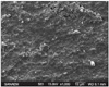
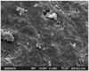
In this study, the SEM evaluation of the samples after debonding showed differences in the surface characteristics of the teeth in the 6 groups. For groups 1 (Figure 1) and 4 (Figure 4), the entire enamel surface was coated with resin, thereby indicating good enamel-resin bonding.
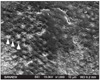
Photomicrography (Figure 2) revealed that primer bonding to the enamel surface was achieved in group 2. The enamel surface does not show a prismatic view with enamel rods, but spurs (arrowhead) can be observed on the tracings of rods. This appearance may be indicative of bond failure at the primer-resin interface.
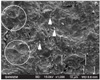
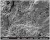
The photomicrographs obtained for group 3 showed physical roughness of the enamel surface, indicating that chemical demineralization did not occur with sandblasting (Figure 3). Fractured enamel surfaces (circles) due to sandblasting, sand particles (arrowheads), and a few remnant resins (brackets) can be observed.
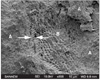
Group 5 exhibited the ablation of enamel surfaces and enamel rods (Figure 5). The ablated enamel surface may have fractured during debonding; this may have resulted in the separation of the enamel surface from the resin material and the formation of roughened area.
Teeth in group 6 (Figure 6) exhibited remnant adhesive sites (A), rough enamel rods (B), and fractured enamels (between arrows). The presence of fractured enamel sites reflects the high SBS between the enamel and resin. The irradiated and roughened enamel surface may have become more irregular with acid etching, and some of the residual resin may have been retained on the surface while some enamel fractures may have occurred during debonding.
CONCLUSION
Although laser conditioning afforded high SBS, the procedure resulted in considerable damage to teeth. Therefore, the acid-etching and self-etching techniques were found to be safer for orthodontic bracket bonding. Since the sandblasting method did not afford sufficient bonding strength alone, this technique must be accompanied by acid etching in order to achieve better results.


 XML Download