

 PDF
PDF ePub
ePub Citation
 Print
Print


Dear Editor,
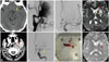
A 45-year-old man was transferred to our stroke unit due to acute right-sided weakness, right hemianopsia, and global aphasia at 390 min after symptom onset [National Institutes of Health Stroke Scale (NIHSS) score=20]. Computed tomography (CT) angiography (CTA) performed at another institution revealed occlusion of the left proximal middle cerebral artery (MCA) due a giant aneurysm of the ipsilateral cervical internal carotid artery (ICA). The second CT and CTA performed at our institution at 405 min after symptom onset showed two small hypodense areas within the left MCA territory [Alberta Stroke Program Early CT score (ASPECTS) score=8/10] as well a large extracranial aneurysm of the left ICA with arterial wall calcification and hypodense material within the lumen corresponding to an intraluminal thrombus (Fig. 1A and B). His neurological status deteriorated further (NIHSS score=23) at 410 minutes after symptom onset.
The patient did not fulfill two of the suggested inclusion criteria for mechanical thrombectomy (MT) in acute ischemic stroke (AIS) according to recent recommendations1 (pretreatment with intravenous thrombolysis and time window of ≤6 hours from symptom onset), and the pathology of the extracranial ICA was a contraindication in some of recent randomized controlled trials.2 However, we decided to pursue rescue MT at 415 minutes after symptom onset based on the high ASPECTS score in the second brain CT (8/10), good collaterals in CTA, and the presence of a severe neurological deficit at baseline with further neurological deterioration.
Digital-subtraction angiography revealed left proximal MCA occlusion and verified the presence of a giant, partially thrombosed aneurysm in the left ICA (Fig. 1C and D). MT was performed using a triaxial homocentric system approach with placement of a long sheath (Neuron-MAX-6F, Penumra, Alemeda, CA, USA) in the left ICA bulb and distal catheterization of the cavernous ICA through the extracranial aneurysm with a Penumbra 5Max-ACE device and a microcatheter (Rebar-18, Covidien, Dublin, Ireland). Left proximal MCA revascularization was successful with two passages of a Solitaire™-FR stent retriever (Covidien) that resulted in substantial thrombus removal (Fig. 1E and F). The procedure took 35 minutes under conscious sedation and resulted in complete reperfusion of the left MCA territory at 455 minutes after symptom onset (Thrombolysis In Cerebral Infarction grade=III). The patient exhibited significant improvement immediately after the procedure, with a NIHSS score of 14 at 460 min after symptom onset. Brain MRI performed after 24 hours (Fig. 1G and H) showed an acute left putaminal infarction with asymptomatic hemorrhagic transformation (hemorrhagic infarct grade=II), while transcranial Doppler ultrasonography showed sustained complete recanalization (TIBI grade=5). The patient experienced further neurological improvement during hospitalization, with mild residual expressive aphasia present at discharge 9 days after stroke onset (NIHSS score=3). Anticoagulation with subcutaneous enoxaparin (80 mg bid) was initiated at 72 hours and was recommended to him until the subsequent aneurysm repair of the aneurysm (scheduled for 3 months later). His diagnostic workup was unremarkable for other AIS etiopathogenic mechanisms, and he had a modified Rank Scale score of 1 at the 3-month follow-up.
This is the first case of MT in a proximal intracranial occlusion caused by a giant, extracranial aneurysm with an intraluminal thrombus. Even though simultaneous endovascular treatment of acute cervical ICA dissections (mostly with stenting) and distal thrombectomy of ipsilateral intracranial occlusions has been reported,3 this option was not feasible in our patient due to the size of the aneurysm and the limited time window to achieve reperfusion of ischemic brain tissue. We therefore decided to bypass the aneurysm and perform distal aspiration in order to minimize the risk of embolization. Aneurysmal rupture during thrombectomy is another potential complication of this therapeutic approach, but the risk of rupture is much lower in extracranial than in intracranial aneurysms. Zibold et al.4 reported recently on coincidental intracranial aneurysms in the target vessels of AIS patients with proximal intracranial occlusions treated with MT, and they attributed cerebral ischemia to the aneurysm in only one case.
The present case underlines the safety and efficacy of MT in AIS caused by distal embolization of an extracranial aneurysm with a concomitant intraluminal thrombus. The present findings support offering MT to patients with large-vessel occlusions and the possibility of good functional recovery, even if they do not meet the top-tier guidelines.5 We recommend in similar cases to bypass the extracranial aneurysm and perform distal aspiration in order to minimize the risk of embolization followed by MT using a stent retriever.
 XML Download