Journal List > J Clin Neurol > v.13(2) > 1048024

Hwang, Kim, Kang, Kim, and Liebeskind: Impact of Baseline Ischemia on Outcome in Older Patients Undergoing Endovascular Therapy for Acute Ischemic Stroke

Abstract

Background and Purpose

The outcome for older stroke patients who undergo endovascular revascularization remains unsatisfactory. We aimed to determine the effect of the extent of baseline ischemia on outcome according to age, testing the hypothesis that the restorative capacity for recovery is only marginal in older patients.

Methods

Two hundred and thirteen patients who underwent endovascular revascularization due to occlusion in the M1 segment of the middle cerebral artery (with or without internal carotid artery occlusions) were selected for analysis. Patients were categorized into three age groups: group A (<66 years), group B (66–75 years), and group C (>75 years). Using pretreatment diffusion-weighted imaging (DWI), the Alberta Stroke Program Early CT Score (ASPECTS) and lesion volume were independently measured and analyzed in relation to a favorable outcome.

Results

A favorable outcome was achieved in 111 of 213 patients overall: in 60 of the 94 (63.8%) patients in group A, in 36 of the 70 (51.4%) patients in group B, and in 15 of the 49 (30.6%) patients in group C (p=0.001). In older stroke patients (group C), a DWI ASPECTS ≥9 and lesion volume ≤5 mL were found to predict a favorable outcome, which was more restrictive than the cutoffs for their younger counterparts (groups A and B; DWI ASPECTS ≥8 and lesion volume ≤20 mL).

Conclusions

The age-adjusted pretreatment DWI lesion volume and ASPECTS may represent useful surrogate markers for functional outcome according to age. The use of more-restrictive inclusion criteria for older stroke patients could be warranted, although larger studies are necessary to confirm these findings.

INTRODUCTION

The effectiveness of endovascular revascularization for stroke patients with acute large-vessel occlusion in the anterior circulation has been clearly established in several recent randomized controlled trials.12345 However, recent increases in life expectancy mean that older stroke patients possibly constitute the population with the highest stroke incidence6 but are excluded from some of these landmark trials; this is probably due to the prognosis for this age group being expected to remain poor even after reperfusion. The rate of favorable functional outcome in older patients who undergo endovascular therapy (EVT) reportedly varies widely, from 0 to 28%, which might be at least partially due to the application of diverse patient selection criteria in previous cohort studies.7891011121314
Reperfusion is futile in almost 50% of older stroke patients; that is, the 3-month modified Rankin Scale (mRS) score remains ≥3 despite successful reperfusion, with this percentage being almost double that for younger patients.15 This may be partly attributed to the higher incidence of comorbidities and lower restorative ability of the brain in such patients, which results in poor functional recovery.16 It may therefore be necessary to define stricter inclusion criteria for EVT for older stroke patients. The low restorative ability of the brain in older patients might imply that the threshold of baseline ischemia for recovery–which is one of the most-reliable surrogate markers of stroke outcome–171819 could be lower than that in their younger counterparts.
We hypothesized that the threshold for baseline ischemia as a predictor of good functional outcome is lower in older patients. To test this hypothesis, which was not been explored previously in the clinical literature, we examined the relationship between the degree of baseline ischemia and clinical outcome according to age.

METHODS

Between May 2006 and December 2014, patients were retrospectively selected from a prospectively maintained acute stroke registry at our institution. The criteria for inclusion in this study were as follows: 1) EVT performed due to acute anterior circulation stroke with proximal vessel occlusion [T- or L-type internal carotid artery (ICA), M1 segment of the middle cerebral artery (MCA), or ICA/M1 tandem occlusion], 2) arrival at our emergency center within 6 hours of symptom onset, 3) evaluable pretreatment diffusion-weighted imaging (DWI), and 4) prestroke mRS score of 0–2. The study protocol was approved by the local Institutional Review Board.
Eligible patients underwent transfemoral cerebral angiography with contrast agent injected into both carotid arteries and the dominant vertebral artery during the late venous phase under local or general anesthesia, in order to define the angioarchitecture of the occluded vessel and assess the extent of collateral flow from all possible sources. If the treatable ICA or MCA occlusion persisted, EVT was initiated. Treatment strategies were selected on the basis of therapies available at the time of angiography, and they included intra-arterial thrombolytic infusion (urokinase or recombinant tissue plasminogen activator), mechanical clot disruption, mechanical thrombectomy with forced arterial suction thrombectomy or Solitaire thrombectomy, placement of rescue intra- or extracranial stenting, or a combination of these approaches.202122 The angiographic collateral flow grade, as observed during pretreatment angiography, was evaluated according to the American Society of Interventional and Therapeutic Neuroradiology/Society of Interventional Radiology Collateral Flow Grading System. This angiographic scale assigns patients to grades 0 to 4 according to the completeness and rapidity of retrograde collateral filling.23 The reperfusion status was measured using the Thrombolysis in Cerebral Infarction (TICI) scale.23
Information on demographic and clinical characteristics, medical history, admission blood pressure, and blood glucose were collected at baseline. The onset of stroke was defined as the time when the patient was last observed to be without any abnormal signs or symptoms. Before the initiation of treatment, baseline stroke severity was assessed using the National Institutes of Health Stroke Scale (NIHSS). All patients underwent a CT or MRI scan at 24–48 hours after the initiation of treatment. If evidence of hemorrhage was detected, the subtype of the stroke was classified as hemorrhagic infarction, parenchymal hematoma, subarachnoid hemorrhage, intraventricular hemorrhage, or mixed.24 Symptomatic intracranial hemorrhage was defined as any type of hemorrhage associated with an increase in the NIHSS score of 4 or more within 24 hours of treatment.25 The DWI findings at baseline were assessed using the Alberta Stroke Program Early CT Score (ASPECTS) for DWI by two raters (Y.W.K. and D.H.K.),26 with any disagreements resolved by consensus. The volume of the lesion on DWI was calculated using open-source image analysis software (OsiriX, Pixmeo, Geneva, Switzerland) by an experienced neurologist (Y.W.K.) who was blinded to the clinical status of the patient. Recovery of neurological function was assessed at 3 months using the mRS, with a favorable outcome being defined by an mRS score of 2 or less.2527
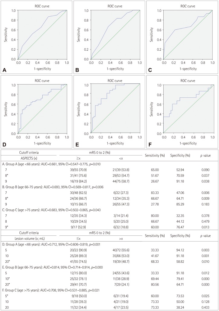
Statistical analysis was performed using the SPSS statistical package (version 20.0, SPSS Inc., Chicago, IL, USA). The statistical significance of intergroup differences was assessed using the chi-square test for categorical variables, one-way ANOVA for continuous variables, and the Kruskal-Wallis test for ordinal variables and continuous variables with skewed distributions. A receiver operator characteristic curve was plotted to calculate the sensitivity and specificity of the cutoff values for the DWI ASPECTS and lesion volume that predicted a favorable outcome. Multivariable regression analysis was performed to identify independent predictors of a favorable outcome relative to the prespecified cutoff ASPECTS and lesion volumes. The results were expressed as odds ratio (OR) estimates of the relative risk with a 95% confidence interval (CI). Probability values less than 0.05 were considered statistically significant.

RESULTS

Two hundred and fifty-seven patients who had proximal vessel occlusion in the anterior circulation and arrived within 6 hours of symptom onset were screened for inclusion in this study. Forty-four of these patients were excluded for the following reasons: nonavailability of pretreatment DWI (n=32), poor quality images (n=2), and prestroke mRS score of ≥3 (n=10). Thus, 213 patients were finally included in the analysis.

Clinical and imaging characteristics based on age groups

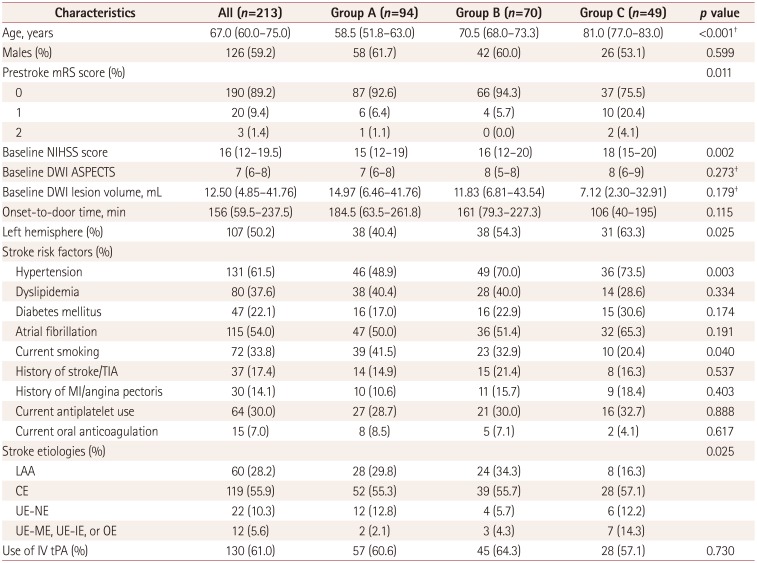
The following groups were defined based on the age at stroke onset to explore the trend between the degree of baseline ischemia and functional outcome relative to age: group A (n=94; <66 years), group B (n=70; 66–75 years), and group C (n=49; >75 years). At baseline and relative to the other two groups, group C had higher prestroke mRS and NIHSS scores, a different profile of stroke etiologies, a higher prevalence of left hemispheric involvement, a higher prevalence of underlying hypertension, and a lower prevalence of current smoking. The details of these baseline characteristics are provided in Table 1.
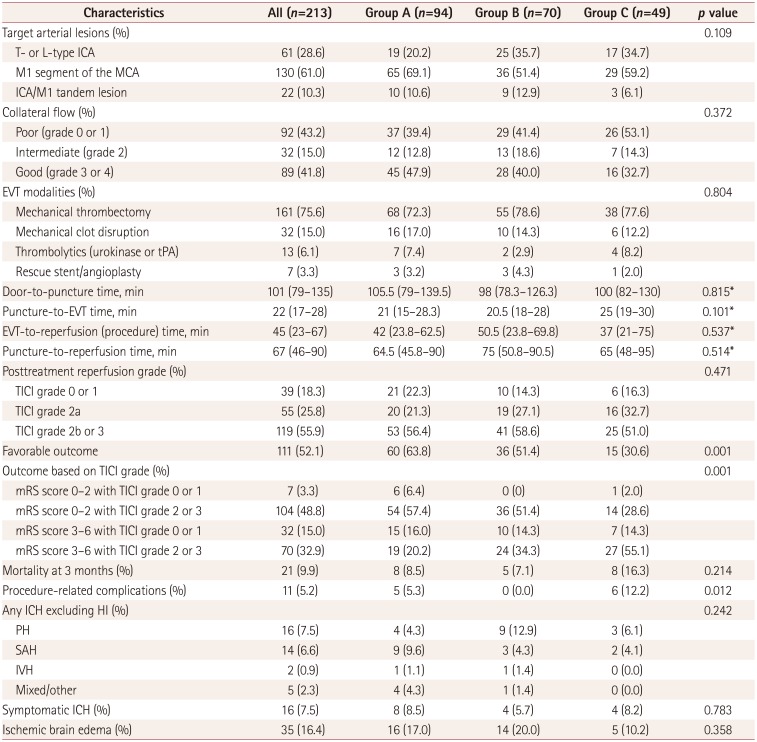
At baseline, the collateral-flow status tended to be worse in older patients [collateral flow grade 0 or 1: 26 of 49 patients (53.1%) in group C vs. 66 of 164 patients (40.2%) in groups A and B; p=0.112]. The duration from arterial puncture to the start of EVT tended to be longer in group C (p=0.101), which reflect the highest burden of vascular tortuosity and atherosclerosis in older patients during the preparation of EVT, but the total time spent on EVT (procedure time) was not different significantly in the groups, which may imply that the procedure itself was unrelated to the underlying vascular status. A favorable functional outcome was achieved in 111 of the 213 patients (52.1%) overall: in 60 of the 94 (63.8%) patients in group A, in 36 of the 70 (51.4%) patients in group B, and in 15 of the 49 (30.6%) patients in group C (p=0.001). The percentage of patients with TICI grade 2 or 3 reperfusion did not differ significantly in the three groups, but the percentage of patients with futile reperfusion was much higher in group C (55.1%, p=0.001). The rate of procedure-related complications was higher in older patients (12.2% in group C vs. 5.3 and 0% in groups A and B, respectively). The incidence rates of symptomatic intracranial hemorrhage and ischemic brain edema did not differ significantly in the groups. The details of angiographic and outcome characteristics are presented in Table 2.

Pretreatment DWI threshold for favorable outcome based on age groups

To determine the age-specific cutoffs for the pretreatment DWI ASPECTS and lesion volume, a receiver operating characteristic curve was constructed (Fig. 1). A DWI ASPECTS of 8 was found to predict a favorable outcome in groups A and B (sensitivity=51.67 and 66.67%, respectively; specificity=70.59 and 64.71%, respectively), while ASPECTS=9 was identified as favorable for group C (sensitivity=60.0%, specificity=76.47%). A DWI lesion volume of 20 mL was selected as the criterion for predicting a favorable outcome in groups A and B (sensitivity=68.33 and 80.56%, respectively; specificity=58.82 and 64.71%, respectively), while a volume of 5 mL was selected as for group C (sensitivity=60.0%, specificity=73.53%). After adjusting for age, prestroke mRS score, baseline NIHSS score, final TICI grade 2b or 3 reperfusion, and procedure time, age-specific DWI ASPECTS cutoff values remained statistically significant in predicting a favorable outcome (OR=2.387, 95% CI=1.219–4.676, p=0.011). Additionally, after adjusting for age, prestroke mRS score, baseline NIHSS score, final TICI grade 2b or 3 reperfusion, and procedure time, the age-specific DWI lesion volume cutoff values continued to remain statistically significant in predicting a favorable outcome (OR=3.595, 95% CI=1.789–7.223, p<0.001).

DISCUSSION

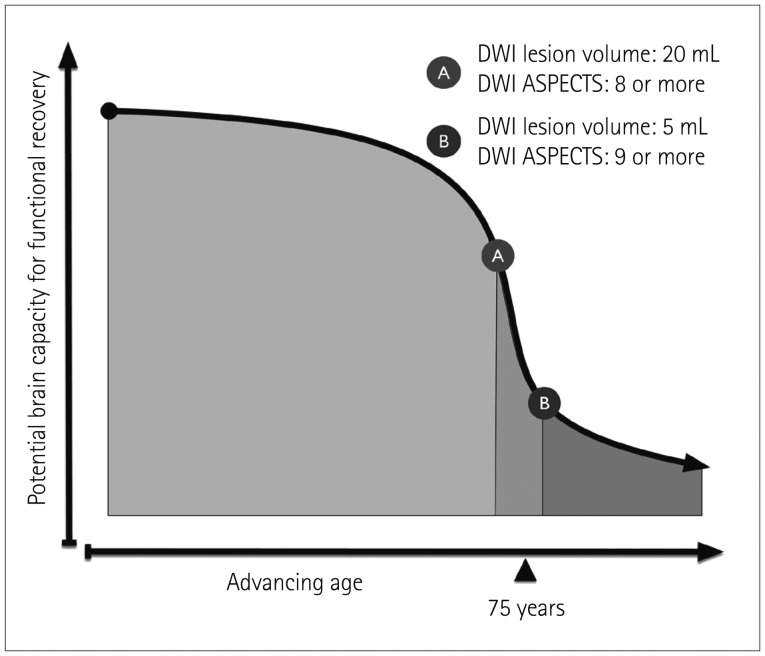
The novel finding in this study was that the baseline DWI ASPECTS and lesion volume cutoffs for a favorable outcome were much stricter in older stroke patients (≥9 and ≤5 mL, respectively) than in their younger counterparts (≥8 and ≤20 mL, respectively). Further, the rate of futile reperfusion exceeded 50% in group C (age >75 years). Both of these findings suggest that the restorative ability of the brain is weaker in older patients (Fig. 2).
In general, the risk of stroke is closely associated with age. A related finding is that the incidence of stroke is continuing to increase exponentially as an inevitable result of increased life expectancy. Older stroke patients have increased mortality and more-severe disability compared to their younger counterparts for a similar spectrum of stroke severity and pathology.28 This situation can result in physicians being reluctant to offer EVT to older stroke patients.
In our cohort, the rate of a favorable outcome was about 31% in older patients (>75 years), and the probability of them achieving a favorable outcome was much lower compared to their younger counterparts despite the similarity of their baseline characteristics and reperfusion rates. Previous studies have already shown that poor outcomes are more frequent in octogenarians.7891011121314 Poor outcomes in older patients have been attributed to higher levels of baseline disabilities, higher incidence rates of medical comorbidities and poststroke medical complications, less familial support, and withdrawal of care.29 Further, the poor restorative ability of the brain in older stroke patients (>75 years) can be a contributing factor.16 Also, a tendency for the collateral flow to be worse in older patients in our cohort could be a contributing factor to the endovascular outcome, with it being reported that the interaction between collateral adequacy and age could contribute to an adverse outcome.30
Our hypothesis that older stroke patients have a different ischemia cutoff for a favorable outcome can be investigated using baseline brain imaging. To accurately define the cutoff value for baseline ischemia, DWI is arguably superior to CT in predicting outcome.31 Ribo et al.19 examined follow-up CT images of patients obtained after 24–36 hours to determine the age-specific posttreatment infarct size predictive of a favorable outcome. They found that the target cutoff infarct volume that predicted a favorable outcome decreased with increasing age (49 mL for age <70 years, 32.5 mL for age 70–79 years, and 15.2 mL for age ≥80 years). However, that study did not examine the age-adjusted pretreatment infarct volumes. The present study examined the pretreatment lesion volume and ASPECTS, which were found to decrease and increase, respectively, with increasing age.
The limitations of this study include its retrospective design and the lack of control patients. Additionally, the type of acute treatment given or recommended to each individual patient varied considerably since it was based on the best available treatment at that time at our institute and the willingness of the patient's next of kin to proceed with the suggested active treatment approach. This means that the rate of TICI grade 2b or 3 reperfusion was lower than in recent endovascular trials, which may hinder the ability to define the optimal baseline ischemia threshold for clinical outcome despite TICI grades being adjusted in the statistical analyses. Another limitation is that the lower baseline infarct volume or higher DWI ASPECTS cutoffs for a favorable outcome might be related to a high prevalence of baseline intracranial atherosclerosis in our cohort. Finally, older patients were defined as those aged ≥76 years, which differs from many previous studies that analyzed patients aged ≥80 years.
In conclusion, our findings suggest that the age-adjusted pretreatment DWI lesion volume and ASPECTS are useful surrogate markers for functional outcome in older stroke patients. We further suggest that these results may help to define more-restrictive imaging criteria for older stroke patients in future trials. Further investigations of larger data sets from recent clinical trials would help to confirm our findings and justify their widespread clinical application.

Acknowledgements

This research was supported by Kyungpook National University Research Fund, 2014.
Dr. Hwang reports grants from Kyungpook National University, during the conduct of the study; Dr. Liebeskind reports grants from NIHNINDS, other from Medtronic, other from Stryker, outside the submitted work.
We thank Wade Martin of Emareye for his critical English revision.

Notes

Conflicts of Interest: The authors have no financial conflicts of interest.

References

1. Berkhemer OA, Fransen PS, Beumer D, van den Berg LA, Lingsma HF, Yoo AJ, et al. A randomized trial of intraarterial treatment for acute ischemic stroke. N Engl J Med. 2015; 372:11–20. PMID: 25517348.
2. Campbell BC, Mitchell PJ, Kleinig TJ, Dewey HM, Churilov L, Yassi N, et al. Endovascular therapy for ischemic stroke with perfusion-imaging selection. N Engl J Med. 2015; 372:1009–1018. PMID: 25671797.
crossref
3. Goyal M, Demchuk AM, Menon BK, Eesa M, Rempel JL, Thornton J, et al. Randomized assessment of rapid endovascular treatment of ischemic stroke. N Engl J Med. 2015; 372:1019–1030. PMID: 25671798.
4. Saver JL, Goyal M, Bonafe A, Diener HC, Levy EI, Pereira VM, et al. Stent-retriever thrombectomy after intravenous t-PA vs. t-PA alone in stroke. N Engl J Med. 2015; 372:2285–2295. PMID: 25882376.
crossref
5. Jovin TG, Chamorro A, Cobo E, de Miquel MA, Molina CA, Rovira A, et al. Thrombectomy within 8 hours after symptom onset in ischemic stroke. N Engl J Med. 2015; 372:2296–2306. PMID: 25882510.
crossref
6. Hong KS, Bang OY, Kang DW, Yu KH, Bae HJ, Lee JS, et al. Stroke statistics in Korea: part I. Epidemiology and risk factors: a report from the Korean Stroke Society and Clinical Research Center for Stroke. J Stroke. 2013; 15:2–20. PMID: 24324935.
crossref
7. Kim D, Ford GA, Kidwell CS, Starkman S, Vinuela F, Duckwiler GR, et al. Intra-arterial thrombolysis for acute stroke in patients 80 and older: a comparison of results in patients younger than 80 years. AJNR Am J Neuroradiol. 2007; 28:159–163. PMID: 17213448.
8. Qureshi AI, Suri MF, Georgiadis AL, Vazquez G, Janjua NA. Intra-arterial recanalization techniques for patients 80 years or older with acute ischemic stroke: pooled analysis from 4 prospective studies. AJNR Am J Neuroradiol. 2009; 30:1184–1189. PMID: 19342542.
crossref
9. Loh Y, Kim D, Shi ZS, Tateshima S, Vespa PM, Gonzalez NR, et al. Higher rates of mortality but not morbidity follow intracranial mechanical thrombectomy in the elderly. AJNR Am J Neuroradiol. 2010; 31:1181–1185. PMID: 20395387.
crossref
10. Mazighi M, Labreuche J, Meseguer E, Serfaty JM, Laissy JP, Lavallée PC, et al. Impact of a combined intravenous/intra-arterial approach in octogenarians. Cerebrovasc Dis. 2011; 31:559–565. PMID: 21487220.
11. Mono ML, Romagna L, Jung S, Arnold M, Galimanis A, Fischer U, et al. Intra-arterial thrombolysis for acute ischemic stroke in octogenarians. Cerebrovasc Dis. 2012; 33:116–122. PMID: 22179504.
crossref
12. Chandra RV, Leslie-Mazwi TM, Oh DC, Chaudhry ZA, Mehta BP, Rost NS, et al. Elderly patients are at higher risk for poor outcomes after intra-arterial therapy. Stroke. 2012; 43:2356–2361. PMID: 22744644.
crossref
13. Johnson JN, Haussen DC, Elhammady MS, Pao CL, Yavagal DR, Aziz-Sultan MA. Poor outcomes of elderly patients undergoing multimodality intra-arterial therapy for acute ischemic stroke. Clin Neurol Neurosurg. 2014; 123:136–141. PMID: 25012026.
crossref
14. Parrilla G, Carreón E, Zamarro J, Espinosa de Rueda M, García-Villalba B, Marín F, et al. Recanalization and mortality rates of thrombectomy with stent-retrievers in octogenarian patients with acute ischemic stroke. Cardiovasc Intervent Radiol. 2015; 38:288–294. PMID: 24923241.
crossref
15. Singer OC, Haring HP, Trenkler J, Nolte CH, Bohner G, Reich A, et al. Age dependency of successful recanalization in anterior circulation stroke: the ENDOSTROKE study. Cerebrovasc Dis. 2013; 36:437–445. PMID: 24281318.
crossref
16. Petcu EB, Sfredel V, Platt D, Herndon JG, Kessler C, Popa-Wagner A. Cellular and molecular events underlying the dysregulated response of the aged brain to stroke: a mini-review. Gerontology. 2008; 54:6–17. PMID: 18160818.
crossref
17. Inoue M, Olivot JM, Labreuche J, Mlynash M, Tai W, Albucher JF, et al. Impact of diffusion-weighted imaging Alberta stroke program early computed tomography score on the success of endovascular reperfusion therapy. Stroke. 2014; 45:1992–1998. PMID: 24923724.
crossref
18. Yoo AJ, Zaidat OO, Chaudhry ZA, Berkhemer OA, González RG, Goyal M, et al. Impact of pretreatment noncontrast CT Alberta Stroke Program Early CT Score on clinical outcome after intra-arterial stroke therapy. Stroke. 2014; 45:746–751. PMID: 24503670.
crossref
19. Ribo M, Flores A, Mansilla E, Rubiera M, Tomasello A, Coscojuela P, et al. Age-adjusted infarct volume threshold for good outcome after endovascular treatment. J Neurointerv Surg. 2014; 6:418–422. PMID: 23832414.
crossref
20. Hwang YH, Kang DH, Kim YW, Kim YS, Park SP, Suh CK. Outcome of forced-suction thrombectomy in acute intracranial internal carotid occlusion. J Neurointerv Surg. 2013; 5(Suppl 1):i81–i84. PMID: 22544821.
crossref
21. Kang DH, Hwang YH, Kim YS, Park J, Kwon O, Jung C. Direct thrombus retrieval using the reperfusion catheter of the penumbra system: forced-suction thrombectomy in acute ischemic stroke. AJNR Am J Neuroradiol. 2011; 32:283–287. PMID: 21087940.
crossref
22. Kang DH, Kim YW, Hwang YH, Park J, Hwang JH, Kim YS. Switching strategy for mechanical thrombectomy of acute large vessel occlusion in the anterior circulation. Stroke. 2013; 44:3577–3579. PMID: 24021683.
crossref
23. Higashida RT, Furlan AJ, Roberts H, Tomsick T, Connors B, Barr J, et al. Trial design and reporting standards for intra-arterial cerebral thrombolysis for acute ischemic stroke. Stroke. 2003; 34:e109–e137. PMID: 12869717.
crossref
24. Hacke W, Kaste M, Fieschi C, von Kummer R, Davalos A, Meier D, et al. Second European-Australasian Acute Stroke Study Investigators. Randomised double-blind placebo-controlled trial of thrombolytic therapy with intravenous alteplase in acute ischaemic stroke (ECASS II). Lancet. 1998; 352:1245–1251. PMID: 9788453.
crossref
25. Saver JL, Jahan R, Levy EI, Jovin TG, Baxter B, Nogueira RG, et al. Solitaire flow restoration device versus the Merci Retriever in patients with acute ischaemic stroke (SWIFT): a randomised, parallel-group, non-inferiority trial. Lancet. 2012; 380:1241–1249. PMID: 22932715.
crossref
26. Barber PA, Demchuk AM, Zhang J, Buchan AM. Validity and reliability of a quantitative computed tomography score in predicting outcome of hyperacute stroke before thrombolytic therapy. ASPECTS Study Group. Alberta Stroke Programme Early CT Score. Lancet. 2000; 355:1670–1674. PMID: 10905241.
27. van Swieten JC, Koudstaal PJ, Visser MC, Schouten HJ, van Gijn J. Interobserver agreement for the assessment of handicap in stroke patients. Stroke. 1988; 19:604–607. PMID: 3363593.
crossref
28. Sharma JC, Fletcher S, Vassallo M. Strokes in the elderly-higher acute and 3-month mortality-an explanation. Cerebrovasc Dis. 1999; 9:2–9.
29. Chandra RV, Leslie-Mazwi TM, Mehta BP, Yoo AJ, Simonsen CZ. Clinical outcome after intra-arterial stroke therapy in the very elderly: why is it so heterogeneous? Front Neurol. 2014; 5:60. PMID: 24808887.
crossref
30. Arsava EM, Vural A, Akpinar E, Gocmen R, Akcalar S, Oguz KK, et al. The detrimental effect of aging on leptomeningeal collaterals in ischemic stroke. J Stroke Cerebrovasc Dis. 2014; 23:421–426. PMID: 23583014.
crossref
31. McTaggart RA, Jovin TG, Lansberg MG, Mlynash M, Jayaraman MV, Choudhri OA, et al. Alberta stroke program early computed tomographic scoring performance in a series of patients undergoing computed tomography and MRI: reader agreement, modality agreement, and outcome prediction. Stroke. 2015; 46:407–412. PMID: 25538199.
Fig. 1

Receiver operating characteristic (ROC) curves for favorable outcome and the baseline diffusion-weighted imaging (DWI) Alberta Stroke Program Early CT Score (ASPECTS) and lesion volume according to the age groups. The tables display the test characteristics for the prespecified ASPECTS and lesion volume, including optimal values (*). AUC: area under the ROC curve, CI: confidence interval, mRS: modified Rankin scale.

jcn-13-162-g001
Fig. 2

Schematic of the hypothetical inverse sinusoidal relationships between advancing age and restorative brain capacity. Between points A and B, the restorative capacity decreases steeply for small age increase. ASPECTS: Alberta Stroke Program Early CT Score, DWI: diffusion-weighted imaging.

jcn-13-162-g002
Table 1

Baseline characteristics in different age groups*

jcn-13-162-i001
Characteristics All (n=213) Group A (n=94) Group B (n=70) Group C (n=49) p value
Age, years 67.0 (60.0–75.0) 58.5 (51.8–63.0) 70.5 (68.0–73.3) 81.0 (77.0–83.0) <0.001
Males (%) 126 (59.2) 58 (61.7) 42 (60.0) 26 (53.1) 0.599
Prestroke mRS score (%) 0.011
 0 190 (89.2) 87 (92.6) 66 (94.3) 37 (75.5)
 1 20 (9.4) 6 (6.4) 4 (5.7) 10 (20.4)
 2 3 (1.4) 1 (1.1) 0 (0.0) 2 (4.1)
Baseline NIHSS score 16 (12–19.5) 15 (12–19) 16 (12–20) 18 (15–20) 0.002
Baseline DWI ASPECTS 7 (6–8) 7 (6–8) 8 (5–8) 8 (6–9) 0.273
Baseline DWI lesion volume, mL 12.50 (4.85–41.76) 14.97 (6.46–41.76) 11.83 (6.81–43.54) 7.12 (2.30–32.91) 0.179
Onset-to-door time, min 156 (59.5–237.5) 184.5 (63.5–261.8) 161 (79.3–227.3) 106 (40–195) 0.115
Left hemisphere (%) 107 (50.2) 38 (40.4) 38 (54.3) 31 (63.3) 0.025
Stroke risk factors (%)
 Hypertension 131 (61.5) 46 (48.9) 49 (70.0) 36 (73.5) 0.003
 Dyslipidemia 80 (37.6) 38 (40.4) 28 (40.0) 14 (28.6) 0.334
 Diabetes mellitus 47 (22.1) 16 (17.0) 16 (22.9) 15 (30.6) 0.174
 Atrial fibrillation 115 (54.0) 47 (50.0) 36 (51.4) 32 (65.3) 0.191
 Current smoking 72 (33.8) 39 (41.5) 23 (32.9) 10 (20.4) 0.040
 History of stroke/TIA 37 (17.4) 14 (14.9) 15 (21.4) 8 (16.3) 0.537
 History of MI/angina pectoris 30 (14.1) 10 (10.6) 11 (15.7) 9 (18.4) 0.403
 Current antiplatelet use 64 (30.0) 27 (28.7) 21 (30.0) 16 (32.7) 0.888
 Current oral anticoagulation 15 (7.0) 8 (8.5) 5 (7.1) 2 (4.1) 0.617
Stroke etiologies (%) 0.025
 LAA 60 (28.2) 28 (29.8) 24 (34.3) 8 (16.3)
 CE 119 (55.9) 52 (55.3) 39 (55.7) 28 (57.1)
 UE-NE 22 (10.3) 12 (12.8) 4 (5.7) 6 (12.2)
 UE-ME, UE-IE, or OE 12 (5.6) 2 (2.1) 3 (4.3) 7 (14.3)
Use of IV tPA (%) 130 (61.0) 57 (60.6) 45 (64.3) 28 (57.1) 0.730

Data are median (interquartile range) or number (%) values.

*Age groups: group A (<66 years), group B (66–75 years), and group C (>75 years), Kruskal-Wallis test.

ASPECTS: Alberta Stroke Program Early CT Score, CE: cardioembolism, DWI: diffusion-weighted imaging, IE: incomplete evaluation, IV tPA: intravenous tissue plasminogen activator, LAA: large artery atherosclerosis, ME: multiple etiologies, MI: myocardial infarction, mRS: modified Rankin Scale, NE: negative evaluation, NIHSS: National Institutes of Health Stroke Scale, OE: other determined etiology, TIA: transient ischemic attack, UE: undetermined etiology.

Table 2

Variables regarding endovascular therapy (EVT) and clinical outcome based on age groups

jcn-13-162-i002
Characteristics All (n=213) Group A (n=94) Group B (n=70) Group C (n=49) p value
Target arterial lesions (%) 0.109
 T- or L-type ICA 61 (28.6) 19 (20.2) 25 (35.7) 17 (34.7)
 M1 segment of the MCA 130 (61.0) 65 (69.1) 36 (51.4) 29 (59.2)
 ICA/M1 tandem lesion 22 (10.3) 10 (10.6) 9 (12.9) 3 (6.1)
Collateral flow (%) 0.372
 Poor (grade 0 or 1) 92 (43.2) 37 (39.4) 29 (41.4) 26 (53.1)
 Intermediate (grade 2) 32 (15.0) 12 (12.8) 13 (18.6) 7 (14.3)
 Good (grade 3 or 4) 89 (41.8) 45 (47.9) 28 (40.0) 16 (32.7)
EVT modalities (%) 0.804
 Mechanical thrombectomy 161 (75.6) 68 (72.3) 55 (78.6) 38 (77.6)
 Mechanical clot disruption 32 (15.0) 16 (17.0) 10 (14.3) 6 (12.2)
 Thrombolytics (urokinase or tPA) 13 (6.1) 7 (7.4) 2 (2.9) 4 (8.2)
 Rescue stent/angioplasty 7 (3.3) 3 (3.2) 3 (4.3) 1 (2.0)
Door-to-puncture time, min 101 (79–135) 105.5 (79–139.5) 98 (78.3–126.3) 100 (82–130) 0.815*
Puncture-to-EVT time, min 22 (17–28) 21 (15–28.3) 20.5 (18–28) 25 (19–30) 0.101*
EVT-to-reperfusion (procedure) time, min 45 (23–67) 42 (23.8–62.5) 50.5 (23.8–69.8) 37 (21–75) 0.537*
Puncture-to-reperfusion time, min 67 (46–90) 64.5 (45.8–90) 75 (50.8–90.5) 65 (48–95) 0.514*
Posttreatment reperfusion grade (%) 0.471
 TICI grade 0 or 1 39 (18.3) 21 (22.3) 10 (14.3) 6 (16.3)
 TICI grade 2a 55 (25.8) 20 (21.3) 19 (27.1) 16 (32.7)
 TICI grade 2b or 3 119 (55.9) 53 (56.4) 41 (58.6) 25 (51.0)
Favorable outcome 111 (52.1) 60 (63.8) 36 (51.4) 15 (30.6) 0.001
Outcome based on TICI grade (%) 0.001
 mRS score 0–2 with TICI grade 0 or 1 7 (3.3) 6 (6.4) 0 (0) 1 (2.0)
 mRS score 0–2 with TICI grade 2 or 3 104 (48.8) 54 (57.4) 36 (51.4) 14 (28.6)
 mRS score 3–6 with TICI grade 0 or 1 32 (15.0) 15 (16.0) 10 (14.3) 7 (14.3)
 mRS score 3–6 with TICI grade 2 or 3 70 (32.9) 19 (20.2) 24 (34.3) 27 (55.1)
Mortality at 3 months (%) 21 (9.9) 8 (8.5) 5 (7.1) 8 (16.3) 0.214
Procedure-related complications (%) 11 (5.2) 5 (5.3) 0 (0.0) 6 (12.2) 0.012
Any ICH excluding HI (%) 0.242
 PH 16 (7.5) 4 (4.3) 9 (12.9) 3 (6.1)
 SAH 14 (6.6) 9 (9.6) 3 (4.3) 2 (4.1)
 IVH 2 (0.9) 1 (1.1) 1 (1.4) 0 (0.0)
 Mixed/other 5 (2.3) 4 (4.3) 1 (1.4) 0 (0.0)
Symptomatic ICH (%) 16 (7.5) 8 (8.5) 4 (5.7) 4 (8.2) 0.783
Ischemic brain edema (%) 35 (16.4) 16 (17.0) 14 (20.0) 5 (10.2) 0.358

Data are median (interquartile range) or number (%) values.

*Kruskal-Wallis test.

HI: hemorrhagic infarction, ICA: internal carotid artery, ICH: intracranial hemorrhage, IVH: intraventricular hemorrhage, MCA: middle cerebral artery, PH: parenchymal hematoma, SAH: subarachnoid hemorrhage, TICI: Thrombolysis in Cerebral Infarction, tPA: tissue plasminogen activator.

TOOLS
Similar articles