

 PDF
PDF ePub
ePub Citation
 Print
Print


Abstract
Background
Fine needle aspiration (FNA) is regarded as the best procedure in the diagnosis of thyroid malignancies. However, the rate of false negative and indeterminate results are between 5~10 and 10~30%, respectively. Therefore, a new diagnostic tool to assist FNA is required. Recently, high resolution ultrasonography (US) has become a useful tool in the detection of malignant thyroid nodules. Therefore, the sonographic characteristics differentiating malignant from benign nodules were analyzed, and the usefulness of US in the diagnosis of thyroid malignancy assessed.
Methods
Of the 212 patients that underwent surgery due to a thyroid nodule, at the Daegu Catholic University Hospital between January 2002 and June 2004, and 181 patients (199 nodules) who underwent high resolution US examination before surgery, were included in this study. The characteristics of the sonographic parameters, such as depth/width ratio, shape, margin, structure, sponge sign, calcification and halo, and the homogeneity and echogenicity of the solid component and invasion, were observed.
Results
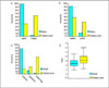
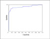
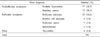
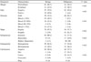
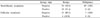
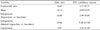
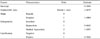
In a univariate analysis of the nonfollicular neoplasms, the depth/width ratio, shape, margin, structure, calcification and halo, and the homogeneity and echogenicity of the solid component were found to be significant parameters. The "sponge sign", a new parameter suggested by us, was found only in benign nodules. In a multiple logistic regression analysis, only the depth/width ratio, shape, presence of calcification and echogenicity of the solid component were significant parameters. According to the results of the multiple logistic regression analysis, the point and estimate of each characteristic of the significant parameters were found, and a formula for calculating a score for the prediction of malignancy computed. At a score of 0.44, the sensitivity and specificity of US were 85.9 and 88.7%, respectively. In a univariate analysis of follicular neop-lasms, the shape, calcification and echogenicity were found to be significant parameters.
Figures and Tables
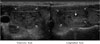
Fig. 1
Ultrasonographic finding of sponge sign (large arrow: loose density like a sponge due to diffusely distributed small sized cystic component and some hyperechoic dot in solid component)
References
1. Vander JB, Gaston EA, Dawber TR. The significance of nontoxic thyroid nodules. Final report of a 15-year study of the incidence of thyroid malignancy. Ann Intern Med. 1968. 69:537–540.
2. Ross DS. Editoroial: Nonpalpable thyroid nodules-managing an epidemic. J Clin Endocrinol Metab. 2002. 87:1938–1940.
4. Gharib H. Changing concepts in the diagnosis and management of thyroid nodules. Endocrinol Metab Clin North Am. 1997. 26:777–800.
5. Gharib H, Goellner JR. Fine-needle aspiration biopsy of the thyroid: an appraisal. Ann Intern Med. 1993. 118:282–289.
6. Garcia-Mayor RV, Perez Mendez LF, Paramo C, Luna Cano R, Rego Iraeta A, Regal M, Sierra JM, Fluiters E. Fine-needle aspiration biopsy of thyroid nodules: impact on clinical practice. J Endocrinol Invest. 1997. 20:482–487.
7. Burch HB. Evaluation and management of the solid thyroid nodule. Endocrinol Metab Clin North Am. 1995. 24:663–710.
8. Gharib H, Goellner JR, Zinsmeister AR, Grant CS, Van Heerden JA. Fine-needle aspiration biopsy of the thyroid. The problem of suspicious cytologic findings. Ann Intern Med. 1984. 101:25–28.
9. Mazzaferri EL. Management of a solitary thyroid nodule. N Engl J Med. 1993. 328:553–559.
10. Solbiati L, Volterrani L, Rizzatto G, Bazzocchi M, Busilacci P, Candiani F, Ferrari F, Giuseppetti G, Maresca G, Mirk P, et al. The thyroid gland with low uptake lesions: evaluation by ultrasound. Radiology. 1985. 155:187–191.
11. Rojeski MT, Gharib H. Nodular thyroid disease. Evaluation and management. N Engl J Med. 1985. 313:428–436.
12. Lin JD, Huang BY, Weng HF, Jeng LB, Hsueh C. Thyroid ultrasonography with fine-needle aspiration cytology for the diagnosis of thyroid cancer. J Clin Ultrasound. 1997. 25:111–118.
13. Hatada T, Okada K, Ishii H, Ichii S, Utsunomiya J. Evaluation of ultrasound-guided fine-needle aspiration biopsy for thyroid nodules. Am J Surg. 1998. 175:133–136.
14. Brkljacic B, Cuk V, Tomic-Brzac H, Bence-Zigman Z, Delic-Brkljacic D, Drinkovic I. Ultrasonic evaluation of benign and malignant nodules in echographically multinodular thyroids. J Clin Ultrasound. 1994. 22:71–76.
15. Koike E, Noguchi S, Yamashita H, Murakami T, Ohshima A, Kawamoto H. Ultrasonographic characteristics of thyroid nodules: prediction of malignancy. Arch Surg. 2001. 136:334–337.
16. Kim EK, Park CS, Chung WY, Oh KK, Kim DI, Lee JT, Yoo HS. New sonographic criteria for recommending fine-needle aspiration biopsy of nonpalpable solid nodules of the thyroid. AJR Am J Roentgenol. 2002. 178:687–691.
17. Koike E, Yamashita H, Noguchi S, Murakami T, Ohshima A, Maruta J, Kawamoto H. Effect of combining ultrasonography and ultrasound-guided fine-needle aspiration biopsy findings for the diagnosis of thyroid nodules. Eur J Surg. 2001. 167:656–661.
18. Peccin S, de Castsro JA, Furlanetto TW, Furtado AP, Brasil BA, Czepielewski MA. Ultrasonography: is it useful in the diagnosis of cancer in thyroid nodules? J Endocrinol Invest. 2002. 25:39–43.
19. Papini E, Guglielmi R, Bianchini A, Crescenzi A, Taccogna S, Nardi F, Panunzi C, Rinaldi R, Toscano V, Pacella CM. Risk of malignancy in nonpalpable thyroid nodules: predictive value of ultrasound and color-Doppler features. J Clin Endocrinol Metab. 2002. 87:1941–1946.
20. Takashima S, Fukuda H, Nomura N, Kishimoto H, Kim T, Kobayashi T. Thyroid nodules: re-evaluation with ultrasound. J Clin Ultrasound. 1995. 23:179–184.
21. Rago T, Vitti P, Chiovato L, Mazzeo S, De Liperi A, Miccoli P, Viacava P, Bogazzi F, Martino E, Pinchera A. Role of conventional ultrasonography and color flow-doppler sonography in predicting malignancy in cold thyroid nodules. Eur J Endocrinol. 1998. 138:41–46.
22. Camargo R, Tomimori E, Seidenberger K, Medeiros-Neto G. Diagnosis of benign thyroid nodules by ultrasound and its correlation with cytopathology and histopathology. Thyroid. 2004. 14(9):706.
23. Tollin SR, Mery GM, Jelveh N, Fallon EF, Mikhail M, Blumenfeld W, Perlmutter S. The use of fine-needle aspiration biopsy under ultrasound guidance to assess the risk of malignancy in patients with a multinodular goiter. Thyroid. 2000. 10:235–241.
24. Tessler FN, Tublin ME. Thyroid sonography: current applications and future directions. AJR Am J Roentgenol. 1999. 173:437–443.
25. Söderström N. Puncture of goiters for aspiration biopsy. A Preliminary report. Acta Med Scand. 1952. 144:237–244.
26. Baloch ZW, Sack MJ, Yu GH, Livolsi VA, Gupta PK. Fine-needle aspiration of thyroid: an institutional experience. Thyroid. 1998. 8:565–569.
27. Sabel MS, Staren ED, Gianakakis LM, Dwarakanathan S, Prinz RA. Use of finde-needle aspiration biopsy and frozen section in the management of solitary thyroid nodules. Surgery. 1997. 122:1021–1026.
28. Seiberling KA, Dutra JC, Grant T, Bajramovic S. Role of intrathyroidal calcifications detected on ultrasound as a marker of malignancy. Laryngoscope. 2004. 114:1753–1757.
29. Kakkos SK, Scopa CD, Chalmoukis AK, Karachalios DA, Spiliotis JD, Harkoftakis JG, Karavias DD, Androulakis JA, Vagenakis AG. Relative risk of cancer in sonographically detected thyroid nodules with calcifications. J Clin Ultrasound. 2000. 28(7):347–352.
30. Gandolfi PP, Frisina A, Raffa M, Renda F, Rocchetti O, Ruggeri C, Tombolini A. The incidence of thyroid carcinoma in multinodular goiter: retrospective analysis. Acta Biomed. 2004. 75:114–117.
31. McHenry CR, Slusarczyk SJ, Khiyami A. Recommendations for management of cystic thyroid disease. Surgery. 1999. 126:1167–1172.
32. Meko JB, Norton JA. Large cystic/solid thyroid nodules: a potential false-negative fine-needle aspiration. Surgery. 1995. 118:996–1004.
 XML Download