

 PDF
PDF Citation
 Print
Print


INTRODUCTION
Endometrial cancer (EC) patients with positive nodal status show remarkable differences in outcome [1] with an estimated 5-year disease-specific survival ranging from 10% to 75% [2]. Thus, considerable heterogeneity exists in node-positive patients based on various clinicopathologic characteristics [13].
The majority of patients who eventually die of disease have positive lymph nodes (LNs) and/or adverse prognostic factors such as older age, higher tumor grade, or non-endometrioid histology [4]. In current clinical practice, patients with positive LNs are offered adjuvant platinum based chemotherapy with or without external beam radiation therapy [5]. Combination of radiation therapy with chemotherapy (chemoradiotherapy [CRT]), (sequential/concurrent), in the adjuvant setting for stage IIIC EC seems to be a safe and effective modality [6]. However, CRT has been shown to improve cancer specific survival, but not overall survival (OS) [7]. Positive LN status seems to be one of the most important prognostic factors in EC. The Gynecologic Oncology Group analysis showed the 5-year progression-free survival (PFS) as 90%, 75%, and 38% in EC patients with negative LNs, patients with stage IIIC1 and patients with stage IIIC2, respectively [8].
There has been recent interest in using lymph node ratio (LNR) as a prognostic tool in node-positive EC [2910]. LNR is defined as the number of metastatic LNs divided by the total number of LNs removed. Previous studies have found LNR to be associated with a worse PFS [2910], and OS [910] in EC.
It has been suggested that LNR is most meaningful when comprehensive lymphadenectomy is utilized routinely in surgical practice [10]. Additionally, it has been reported that LNR must be tied to adjuvant therapy in this patient population [2]. However, previous studies which have investigated the prognostic significance of LNR in EC were hampered by the limited number of median LNs removed [2910], lack of adjusted adjuvant therapies [910], and inclusion of non-endometrioid [10] and mixed [2] histologies. The prognostic impact of LNR in pure endometrioid EC has not been clearly delineated.
Given the limited number of previous studies on the impact of LNR on survival in EC [2910], we conducted this multicenter retrospective analysis aiming at a better understanding of the prognostic significance of LNR in endometrioid EC. The purpose of this study was to investigate the prognostic value of LNR in patients with stage IIIC [11] pure endometrioid EC.
MATERIALS AND METHODS
1. Study design and eligibility
After Institutional Review Board approvals, patients with pure endometrioid EC who underwent primary surgical treatment between January 1998 and December 2016 at 6 gynecologic oncology centers from Turkey were retrospectively reviewed. All patients gave informed consent for the surgical procedure and research use of their medical information at admission.
The study population included women with endometrioid type EC having positive nodal status at the end of final pathology report. Women with non-endometrioid type EC, patients with mixed histologies, those with a total number of LNs removed <10 at the end of final pathology report, and women with stage IV disease were excluded from the study. We also excluded patients with incomplete medical records as well as those with synchronous malignancies.
2. Clinical information
Patient data were extracted from 6 institutions with maintained EC databases. With the eligible cases, demographic characteristics were abstracted from medical records. Tumor characteristics were abstracted from original pathology reports, and the following data were recorded: grade, depth of myometrial invasion (MMI) (<50% or ≥50%), presence of lymphovascular space invasion (LVSI) (yes or no), cervical stromal involvement (yes or no), adnexal metastasis (yes or no), the status of peritoneal cytology examination (negative or positive), and stage of disease. The date of diagnosis, adjuvant treatment modality (radiotherapy, CRT, or chemotherapy), recurrence (if applicable), length of follow-up and survival were noted. Data were collected from centers with an online standardized form.
Surgical staging consisted of total hysterectomy, bilateral salpingo-oophorectomy, pelvic and para-aortic lymphadenectomy, and peritoneal washings. All operations were performed by gynecologic oncologists. Data on the extent of surgery included number of total LNs harvested, number of pelvic LNs removed, number of para-aortic LNs removed, and number of metastatic LNs (total, pelvic, and para-aortic).
All surgical specimens were examined and interpreted by gynecologic pathologists. All tumors were staged according to the International Federation of Gynecology and Obstetrics (FIGO) staging system [11]. Architectural grading was defined by standard FIGO criteria. Notable nuclear atypia inappropriate for the architectural grade raised grade 1 or grade 2 tumors by one grade. LVSI was defined as the presence of adenocarcinoma of any extent, in endothelium lined channels of uterine specimens extracted at the time of surgery [12]. In patients treated before 2009, stage was determined retrospectively on the basis of surgical and pathologic assessment.
The treatment policies were decided by the attending physician or by the multidisciplinary tumor board at each participating institution. Adjuvant therapy was administered to all patients. Adjuvant treatment was given based on the following guidelines: 1) extended-field irradiation with concurrent/sequential chemotherapy or only chemotherapy for para-aortic LN involvement; 2) whole pelvic irradiation for disease limited to the pelvis with negative para-aortic LNs; 3) vaginal brachytherapy was offered for all patients undergoing external beam therapy. None of the patients in our cohort received adjuvant hormonal therapy. The median radiation dose prescribed was 45 Gy for whole pelvis and 54 Gy for extended field. The standard primary chemotherapy regimen included paclitaxel 175 mg/m2 plus carboplatin dosed at an area under curve of 5 or 6 every 21 days for 6 cycles. The adjuvant treatment modalities were not standard within or among the institutions participated in the study. CRT was delivered in one of the 2 ways: 3 cycles of chemotherapy upfront, followed by radiotherapy, followed by 3 additional cycles of chemotherapy (“sandwich” CRT); or radiotherapy with concurrent cisplatin 40 mg/m2 weekly followed by carboplatin area under the curve 5 and paclitaxel 175 mg/m2 × 4 cycles. Concurrent chemotherapy with irradiation consisted of cisplatin 40 mg/m2 once a week.
Postoperative cancer surveillance included follow-up visits quarterly for the first 2 years, and biannually thereafter. A chest radiograph and vaginal smears were obtained once a year. The visits included a gynecologic medical history and a gynecologic examination that was further supplemented with biopsies in case of suspicious findings and imaging studies in case of suspicion of distant metastases. If an isolated recurrence was diagnosed, treatment with curative intent was initiated unless precluded by the patient or disease factors. All women included in the study were followed until death or to the end of study period (31st December 2016). The survival status of the patients was determined as alive or dead at the time of the last follow-up. For all study subjects with a recorded death, this was confirmed by performing a social security death index search.
3. Definitions
Lymphadenectomy was defined as the performance of pelvic and para-aortic LN dissection at the same time. We defined pelvic lymphadenectomy as removal of the lymphatic tissue in the external, internal and common iliac and obturator regions. Para-aortic lymphadenectomy was defined as removal of the lymphatic tissue over the inferior vena cava and aorta beginning at the level of aortic bifurcation up to the left renal vessels.
A systematic lymph node dissection was defined as removal of more than 20 nodes [13]. An adequate pelvic lymphadenectomy was defined as the removal of at least 10 pelvic LNs, and an adequate para-aortic lymphadenectomy was defined as the removal of at least 5 para-aortic LNs [1415].
After initial diagnosis, recurrence was defined as documentation of metastasis with physical examination and/or imaging techniques after a PFS ≥3 months. Progressive disease was defined according to the RECIST 1.1 criteria [16]. PFS was defined as the time from surgery to the first identification of recurrence or progression, or death from any cause, whichever occurred first, or the date of last contact for patients remaining alive without recurrent disease. OS was calculated as the time period between initial surgery to the date of death or the last contact. Surviving patients were censored at their last known follow-up.
4. Statistical analysis
LNR, defined as the percentage of positive LNs to total nodes recovered, was stratified into 2 groups: LNR1 (≤0.15), and LNR2 (>0.15). The LNR was arbitrarily set to 0.01 units and the log-rank test was performed with OS to determine the appropriate cut-off value [17]. Based on the results of these analyses, we used a cut-off value of 0.15, which yielded the most significant result and we divided patients into 2 groups according to the LNR, LNR1 (≤0.15), and LNR2 (>0.15).
Statistical analyses were performed using the statistical software package SPSS version 23.0 (IBM Corp., Armonk, NY, USA). The data were expressed as median and range for continuous variables. Age ≤60 and >60 years was chosen as age-adjusted variables for both univariate and multivariate analyses based on prognostic implications previously reported in the literature [18]. Binary variables were reported as counts and percentages.
Survival curves were generated using the Kaplan-Meier method, and the differences between survival curves were calculated using the log-rank test. In order to evaluate the prognostic factors for PFS and OS, a univariate Cox regression model was used. Any p-value of less than 0.05 in the univariate analysis was subjected to multivariate analysis. A p-value <0.05 was considered to indicate statistical significance.
RESULTS
During the study period, a total of 236 endometrioid EC with stage III disease were treated at 6 participating centers. We excluded 10 patients who had less than 10 LNs removed, 1 with synchronous breast cancer and 18 women with incomplete medical records. Therefore, the present analysis addresses the remaining 207 women with node-positive endometrioid EC.
One hundred and one patients (48.8%) were classified as stage IIIC1 and 106 (51.2%) as stage IIIC2. The median age at diagnosis was 58 (range, 30–82) and the median duration of follow-up was 40 months (range, 1–228 months). Table 1 demonstrates the clinical and pathological characteristics of women with stage IIIC pure endometrioid type EC.
Table 1
Baseline characteristics of the patients
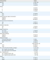
The median number of total LNs harvested was 45 (range, 10–134). The median number of pelvic and para-aortic LNs removed was 32 (range, 4–76), and 14 (range, 1–57), respectively. There were 196 women who received systematic LN dissection (94.7%) while 141 women (68.1%) had adequate pelvic and adequate para-aortic lymphadenectomy. There were 182 patients with pelvic LN involvement (87.9%), and 106 patients with para-aortic LN metastases (51.2%). Isolated pelvic LN metastasis was detected in 101 women (48.8%). There were 25 patients with isolated para-aortic LN metastasis (12.1%). Eighty-one patients (39.1%) had pelvic and para-aortic LN metastases at the same time. The median number of metastatic pelvic and para-aortic LNs was 2 (range, 0–37) and 1 (range, 0–46), respectively. Finally, the median number of total metastatic LNs was 3 (range, 1–64).
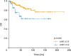
For the entire cohort, the median LNR was 0.054 (range, 0.006–1.0). There were 167 (80.7%) women with LNR1 (≤0.15) and 40 (19.3%) women with LNR2 (>0.15). As depicted in Fig. 1, the 5-year PFS rates for LNR ≤0.15and LNR >0.15 were 76.1% and 58.5%, respectively (p=0.04). Additionally, an increased LNR was associated with a decrease in 5-year OS from 87.0% for LNR ≤0.15 to 62.3% for LNR >0.15 (p=0.005) (Fig. 2).
Fig. 1
PFS analyses with regard to LNR (LNR1 ≤0.15 [n=167], LNR2 >0.15 [n=40]).
LNR, lymph node ratio; PFS, progression-free survival.
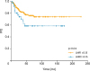
Fig. 2
OS analyses with regard to LNR (LNR1 ≤0.15 [n=167], LNR2 >0.15 [n=40]).
LNR, lymph node ratio; OS, overall survival.
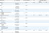
Univariate analysis revealed age ≥60 years (p=0.002), cervical stromal involvement (p=0.02) and LNR >0.15 (p=0.02) as significant prognostic factors for decreased 5-year PFS (Table 2). At the end of multivariate analysis, age ≥60 years (hazard ratio [HR]=2.96; 95% confidence interval [CI]=1.58–5.5; p=0.001), cervical stromal involvement (HR=2.45; 95% CI=1.33–4.52; p=0.004) and LNR >0.15 (HR=2.05; 95% CI=1.07–3.93; p=0.03) persisted as independent prognostic factors for decreased PFS in women with stage IIIC pure endometrioid EC (Table 2).
Table 2
Univariate and multivariate analyses of all patients for PFS
Univariate analysis revealed age ≥60 years (p=0.002), cervical stromal invasion (p=0.006) and LNR >0.15 (p=0.002) as significant factors for decreased OS (Table 3). At the end of multivariate analysis, age ≥60 years (HR=3.87; 95% CI=1.74–8.62; p=0.001), cervical stromal invasion (HR=3.57; 95% CI=1.7–7.46; p=0.001) and LNR >0.15 (HR=3.35; 95% CI=1.57–7.19; p=0.002) remained as independent prognostic factors for decreased OS (Table 3).
Table 3
Univariate and multivariate analyses of all patients for OS
The 5-year PFS rates for stage IIIC1 and IIIC2 disease were 74.5% and 68.0%, respectively (p=0.30). Similarly, there was no significant difference in terms of 5-year OS rates between stage IIIC1 and IIIC2 disease (86.7% vs. 75.6%, respectively; p=0.24). After stratification by substage of the disease, the 5-year PFS rate for women with stage IIIC1 disease and a LNR ≤0.15 was 74.8% compared to 66.7% for women with stage IIIC1 disease and a LNR >0.15 (p=0.67). These figures were found to be 74.2% and 55.7%, respectively for stage IIIC2 disease (p=0.18). The 5-year OS rates did not differ significantly between women with LNR ≤0.15 and LNR >0.15 in stage IIIC1 disease (88.1% vs. 66.7%, respectively; p=0.21). However, the 5-year OS rate for women with stage IIIC2 disease and a LNR ≤0.15 was significantly greater than that of women with stage IIIC2 disease and a LNR >0.15 (84.1% vs. 59.8%, respectively; p=0.03).
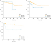
After stratification by adjuvant therapy, there was no significant difference in women receiving chemotherapy alone and CRT in terms of PFS and OS with regards to LNRs (Figs. 3 and 4). However, LNR >0.15 was significantly associated with a poorer 5-year OS when compared to LNR ≤0.15 in patients who received postoperative radiotherapy alone (93.3% vs. 61.4%, respectively; p=0.02; Fig. 4).
Fig. 3
PFS analyses stratified by adjuvant therapy with regard to LNR (LNR1 ≤0.15 [n=167], LNR2 >0.15 [n=40]). (A) Chemotherapy alone, (B) CRT, (C) radiotherapy alone.
CRT, chemoradiotherapy; LNR, lymph node ratio; PFS, progression-free survival.
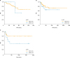
Fig. 4
OS analyses stratified by adjuvant therapy with regard to LNR (LNR1 ≤0.15 [n=167], LNR2 >0.15 [n=40]). (A) Chemotherapy alone, (B) CRT, (C) radiotherapy alone.
CRT, chemoradiotherapy; LNR, lymph node ratio; OS, overall survival.
There were no patients with progressive disease. However, we have detected 49 recurrences during the study period. At the time of reporting, of 207 node-positive women with pure endometrioid EC, 177 (85.5%) were alive and 30 (14.5%) were dead.
DISCUSSION
The key findings of the current study indicate that an increased LNR seems to be associated with a decrease in 5-year OS from 87% for LNR ≤0.15 to 62.3% for LNR >0.15 in women with stage IIIC pure endometrioid EC. Women with LNR >0.15 were 2.05 times more likely to have recurrent disease and 3.3 times more likely to die of their tumors when compared to women with a LNR ≤0.15. Age ≥60 years, and cervical stromal invasion seem to be the other independent prognostic factors for decreased PFS and OS in women with stage IIIC pure endometrioid EC.
The prognostic concept of LNR in EC has emerged during the last decade [2910]. LNR is a parameter based on 2 variables; the number of metastatic LNs, and the number of LNs removed. If the number of involved LNs is high when the number of removed LNs remains relatively limited, then the LNR would appear increased although the quality of LN dissection seems questionable. Because of that, we only included women who underwent surgical staging with ≥10 nodes removed in the current study. This cut-off (10 as the minimum number of LNs removed) has been already used in the subgroup analyses of previous studies evaluating the prognostic impact of LNR in EC [210]. The number of patients with ≥10 nodes removed was 81 (65.3%) in the Fleming study [2] whereas the corresponding figure was 123 (56.9%) in the study by Polterauer et al. [10]. However, it is important to note that the number of nodes recovered may reflect a more extensive dissection, comprehensiveness of pathologic evaluation, variations in number of nodes of each patient, and difficulties in performing LN dissection due to medical comorbidities [19].
Although sentinel LN navigation surgery is still under validation process [20], sentinel LN mapping is currently gaining more respect as an alternative to systematic LN dissection in EC. Many gynecologic oncology centers question the necessity of extensive LN dissection in patients with stage IIIC disease [2122]. The Gynecologic Oncology Group recommends that a positive pelvic or para-aortic node should indicate no further sampling from that area is necessary and that only palpably positive nodes must be sampled for histology [23].
On the contrary, some retrospective studies have shown that more extensive LN dissection led to improved survival outcomes pointing out the potential therapeutic benefits of comprehensive lymphadenectomy in EC [2425]. Additionally, recent studies reported a survival benefit of resection of LN metastasis and extensive LN dissection for patients with advanced disease and LN metastasis [262728]. Although, the nodal status for EC is categorized into stage IIIC1 and IIIC2 based on the metastatic LN location, neither the number of the harvested nor the number of the positive LNs have prognostic significance in the current FIGO staging system [11].
The prognostic significance of LNR in EC has been addressed in 3 previous studies which have revealed that LNR can improve prognostic information [2910]. Chan et al. [9] reported from a National Cancer Institute Registry study that an increasing LNR (≤10%, 10%–50%, and >50%) in 1,222 node-positive endometrioid ECs (stage IIIC and IV) was associated with a decrease in survival from 77.3% to 60.7% to 40.9%, respectively. For patients with stage IIIC disease, survival decreased from 78.6% to 66.5% to 65.3%, with regard to the 3 LNR groups. However, Chan et al. [9] did not provide details on adjuvant therapy modalities and recurrences. They only reported that 63% of their patients received adjuvant radiotherapy. No additional information was available on the types of adjuvant therapies received [9].
Polterauer et al. [10] reported a decrease in OS of 79.0%, 60.6%, and 35.8% with increasing LNRs of ≤10%, 10%–50%, and >50%, respectively. In multivariate analysis, only LNR was associated with both PFS and OS [10]. In the subgroup of patients with stage IIIC EC who had at least 10 LNs examined by pathology (n=123), LNR was shown to be a significant prognosticator for PFS and OS, whereas the number of removed LNs was not [10]. Polterauer et al. [10] included all histologies, of which 30% were non-endometrioid. This may have led to a variance in LNR outcomes reported in that study. Additionally, only 13% (n=28) of their patients had a LNR >50%; a finding leading to difficulties in order to make any comparisons.
Fleming et al. [2] reported that a LNR >50% showed a significantly worse PFS compared to those with LNR ≤10% or 10%–50% in patients with stage IIIC endometrioid EC who underwent surgical staging with ≥10 nodes removed. The authors have suggested that LNR may define a subgroup of stage IIIC ECs at increased risk of recurrence. It should be noted that the median number of pelvic nodes was 9 whereas the median number of para-aortic nodes was only 3 in that study. It should be emphasized that women with mixed histologies were included if non-endometrioid histology was a minor component (<50%) in the study by Fleming et al. [2]. Additionally, the number of patients with a LNR >50% was so small in order to draw any definitive conclusions.
While unresectable residual nodal disease in patients with stage IIIC EC is associated with rapid progression, the administration of external-beam radiotherapy alone or with chemotherapy is associated with an independent survival advantage [29]. Multimodal therapy with both radiation and chemotherapy is now frequently used for women with nodal metastases [30]. Accordingly, most of our patients (46.4%) received CRT in the current study. We were not able to show the prognostic significance of LNR in women treated with CRT or chemotherapy only. However, in the subgroup analysis of women who received only radiotherapy as adjuvant treatment, LNR >0.15 was associated with a poorer OS when compared to women with a LNR ≤0.15. Since LNR inherently must be tied to adjuvant therapy in this patient population [2], we think that stratification of patients with regard to different adjuvant treatment modalities is necessary while evaluating the prognostic impact of LNR in endometrioid EC. However, the data presented here do not allow us to draw definitive conclusions regarding the impact of adjuvant treatment modalities on prognosis in this patient population.
We should underline some limitations of the current study. First; the retrospective nature of the study cannot exclude any bias. Second, lack of central pathology review seems to be an important limitation. Third, adjuvant therapy was administered individually and was not standard during the study period. Those shortcomings of our study need to be kept in mind while interpreting our findings. Despite above limitations, our study provides additional information to the body of knowledge on this topic.
The strengths of the current study lie in its multicenter nature with a large number of patients with stage IIIC pure endometrioid EC, performance of uniform staging procedures with qualified gynecologic oncologists, and a very high-rate of systematic LN dissection. Our study is one of the largest retrospective cohorts associated with the prognostic significance of LNR in women with stage IIIC pure endometrioid EC.
In conclusion, the results of our study suggest that LNR is a meaningful prognostic factor in stage IIIC pure endometrioid EC. Larger studies including only women who have undergone adequate pelvic (at least 10 pelvic nodes removed) and adequate para-aortic (at least 5 para-aortic nodes removed) lymphadenectomy are needed in order to confirm or deny our findings.
 XML Download