

 PDF
PDF Citation
 Print
Print


INTRODUCTION
Cervical cancer is the second most commonly diagnosed cancer in women worldwide, with approximately 500,000 new cases of cervical cancer and 20,000 related deaths in 2010 [1]. However, since the introduction of the Papanicolaou (Pap) test several decades ago as a cervical cancer screening test, the survival rate of cervical cancer has increased markedly and the 5-year survival rate for early-stage cervical cancer now exceeds 90% [23].
Although the Pap test has been the core of cervical cancer screening programs and has contributed considerably to the early detection of cervical cancer, its variability and relatively low sensitivity (50% to 80%), depending on the performance of the health care infrastructure, has limited its efficacy as a single screening method [4]. To overcome the limitations of morphologic diagnosis, molecular diagnostic tests have been developed as a complementary form of testing and the human papillomavirus (HPV) test has been most widely evaluated [5]. Persistent infection with oncogenic HPV is the most important causal factor for cervical cancer development and the HPV test, when used in conjunction with a Pap test or when used alone, significantly improves the sensitivity to 95% to 100% [6]. However, as only a small fraction of high-risk HPV-positive women have clinically relevant cervical lesions, the HPV test also has a drawback of low specificity which results in unnecessary referrals to colposcopy.
DNA methylation of C-phosphate-G (CpG) islands in promoter regions of tumor suppressor genes has been frequently observed in human cancers [7]. Based on the observation that DNA methylation events occur in the early stage of carcinogenesis, DNA methylation has been extensively investigated as a potential biomarker for cancer detection [8]. Likewise, DNA methylation of various host cell genes has been detected in cervical cancer and in a subset of high-grade cervical intraepithelial neoplasia (CIN 2/3) [9]. In addition, aberrant DNA methylation was shown to have an increasing trend proportional to the degree of cervical lesions [1011]. The methylation status of several genes, including cell adhesion molecule 1 (CADM1) and myelin and lymphocyte (MAL), has also been tested as a potential triage tool among high-risk HPV-positive women [12]. However, most of the studies have been performed by a few distinct research groups in moderate-sized populations using different analysis methods.
Therefore, in this study, we aimed to evaluate the methylation profiles of four genes, including adenylate cyclase activating polypeptide 1 (ADCYAP1), paired box 1 (PAX1), CADM1, and MAL, which were described as promising biomarkers in earlier studies [11121314], from residual Pap test samples using pyrosequencing and to assess their diagnostic value in a Korean population.
MATERIALS AND METHODS
1. Patients
After obtaining approval of the institutional review board, residual liquid-based Pap (LBP; Surepath, BD Diagnostics, Oxford, UK) test samples were collected during 2011 in a single institution (Cheil General Hospital & Women's Healthcare Center). Inclusion criteria were as follows: (1) cases with a sufficient amount of residual sample for DNA methylation analysis, (2) HPV positive cases, and (3) cases showing a concordant biopsy result with Pap test result when biopsy was performed. Consequently, a total of 205 patients with negative (n=26), atypical squamous cells of undetermined significance (ASC-US, n=39), low grade squamous intraepithelial lesion (n=44), high grade squamous intraepithelial lesion (HSIL, n=48), and carcinoma (n=48) results in LBP tests were included in this study and all patients were HPV positive. Except for 35 women with normal (n=23) or ASC-US results (n=12), cervical biopsies were performed in 170 patients by colposcopic-directed biopsy or conization for histologic diagnosis. The histologic diagnoses matched with the Pap test results and were as follows: benign/chronic inflammation (n=39); condyloma/CIN 1 (n=45); CIN 2 (n=8); CIN 3 (n=40); and cancer (n=48) (Supplementary Table 1).
2. HPV DNA test
HPV detection and genotyping were performed using either PCR-based Seeplex HPV4 ACE screening (Seegene, Seoul, Korea; from 31 May 2011) or the GG HPV Genotyping Chip Kit (Goodgene, Seoul, Korea; to 30 May 2011). The classification of HPV types as high risk or low risk was based on a previous epidemiologic study [15]. High-risk types included HPV 16, 18, and other common types (31, 33, 35, 39, 45, 51, 52, 53, 56, 58, 59, 66, 68, and 82).
3. Bisulfite treatment and DNA purification
Specimens were stored at 4°C prior to DNA isolation using a DNeasy Blood and Tissue Kit (Qiagen, Valencia, CA, USA) according to the manufacturer’s instructions. Isolated DNA was stored at –20°C. Genomic DNA was chemically modified using sodium bisulfite, which converts all unmethylated cytosines to uracil, but leaves methylated cytosines unmodified. To this end, we used an EZ DNA methylation kit (Zymo Research, Irvine, CA, USA) according to the manufacturer’s instructions. In brief, genomic DNA was treated with sodium bisulfite for 2.5 hours at 65°C and desulfonation was performed for 20 minutes at room temperature. Bisulfite-converted DNA was purified using a Zymo-Spin IC column (Zymo Research) and eluted with 10 μL of distilled water. The eluted DNA was either used immediately for methylation analysis or was stored at –20°C until further use.
4. Quantitative bisulfite pyrosequencing analysis
To quantify the methylation levels of target genes in cervical cells, we performed quantitative bisulfite pyrosequencing [16]. Bisulfite PCR and pyrosequencing primers were designed using PyroMark Assay Design software ver. 2.0 (Qiagen, Hilden, Germany) to amplify two to five CpG dinucleotide sites in the target sequences. The primer sequences used are listed in Supplementary Table 2. Briefly, 20 ng of bisulfite-modified DNA was amplified in a 25-mL reaction volume with gene-specific primers and Taq polymerase (Solgent, Daejeon, Korea). Samples were heated to 95°C for 10 minutes and were then amplified for 45 cycles at 95°C for 45 seconds, at optimal annealing temperature for 45 seconds, and at 72°C for 60 seconds. All reactions were then incubated at 72°C for 10 minutes for final extension. Pyrosequencing was performed using a PyroGold kit and a PyroMark Q96 ID instrument (Qiagen, Hilden) according to the manufacturer’s instructions. The methylation indexes for each region of interest and for each sample were calculated as the mean percentage of methylated cytosine for all examined CpGs. Methylated non-CpG cytosines were used as internal controls, and to check the fidelity of bisulfite conversion.
5. Statistical analysis
The Kruskal-Wallis test performed using the MedCalc program (http://www.medcalc.be) was used to find the significance of DNA methylation according to the progression of cervical lesions.
A chi-square test for trend was used to analyze the status of DNA methylation in different groups. To evaluate the diagnostic performance of the four methylated genes for cancer detection, receiver operating characteristic (ROC) curves were drawn for each gene. Sensitivities and specificities were also tested using cutoffs determined from the ROC curves. All differences were considered statistically significant at p<0.05. Statistical analysis was performed using IBM SPSS ver. 19.0 (IBM Co., Armonk, NY, USA).
RESULTS
The distribution of HPV types, stratified according to Pap test results, is summarized in Table 1. When assessing the methylation status of the HPV-infected cervical cells from LBP samples (n=205), cervical cells from women with cervical cancer showed dramatically increased methylation levels for the four genes analyzed (ADCYAP1, PAX1, MAL, and CADM1) (Fig. 1). ADCYAP1 and PAX1 also trended toward elevated methylation levels in HSIL samples, although the levels were much lower than those in cancer cells (Table 2).
Table 1
HPV types according to cytology results

Values are presented as number (%).
ASC-US, atypical squamous cells of undetermined significance; HPV, human papillomavirus; HRC, high-risk common; HSIL, high grade squamous intraepithelial lesion; LR, low-risk; LSIL, low grade squamous intraepithelial lesion.
*HRC, high-risk common types including HPV-31, -33, -35, -39, -45, -51, -52, -53, -56, -58, -59, -66, -68, and -82. †LR, low-risk types including HPV-6, -11, -30, -42, -43, -44, -54, -70, -72, -81, and -90.
Fig. 1
Methylation status of (A) ADCYAP1, (B) PAX1, (C) MAL, and (D) CADM1 according to Pap test results. ASC-US, atypical squamous cells of undetermined significance; ADCYAP1, adenylate cyclase activating polypeptide 1; CADM1, cell adhesion molecule 1; HSIL, high grade squamous intraepithelial lesion; LSIL, low grade squamous intraepithelial lesion; MAL, myelin and lymphocyte; PAX1, paired box 1.
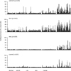
Table 2
Methylation levels of ADCYAP1, PAX1, MAL, and CADM1 according to cytologic categories

Values are presented as mean±SD.
ADCYAP1, adenylate cyclase activating polypeptide 1; ASC-US, atypical squamous cells of undetermined significance; CADM1, cell adhesion molecule 1; HSIL, high grade squamous intraepithelial lesion; LSIL, low grade squamous intraepithelial lesion; MAL, myelin and lymphocyte; PAX1, paired box 1.
*p-value was calculated by Kruskal-Wallis test.
To evaluate the diagnostic performance of the four methylated genes, ROC curves were drawn for each gene (Fig. 2). Methylated ADCYAP1 and PAX1 demonstrated relatively better discriminatory ability for cancer detection than did methylated MAL and CADM1 (area under the curves 0.911 and 0.916 vs. 0.854 and 0.756, respectively) (Table 3). The sensitivities of methylated ADCYAP1, PAX1, MAL, and CADM1 at the cut-offs of 13.26%, 17.92%, 4.20%, and 4.53% were 79.2%, 75.0%, 70.8%, and 52.1%, and the specificities were 92.0%, 94.0%, 94.7%, and 94.0%, respectively (Table 3).
Fig. 2
Receiver operating characteristic curves for cancer detection according to the methylated genes analyzed. ADCYAP1, adenylate cyclase activating polypeptide 1; CADM1, cell adhesion molecule 1; MAL, myelin and lymphocyte; PAX1, paired box 1.
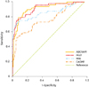
Table 3
Sensitivity and specificity of ADCYAP1, PAX1, MAL, and CADM1 for cancer detection

To validate our findings in biopsy-proven cases, the positivity rates of methylated genes were assessed according to pathologic diagnoses (n=170) (Table 4). Consistent with the methylation status according to Pap tests, methylated ADCYAP1, PAX1, MAL, and CADM1 were more frequently observed in cervical cells from women diagnosed with invasive cancer.
Table 4
Frequency of methylation of ADCYAP1, PAX1, MAL, and CADM1, according to pathologic diagnosis (n=170)

Values are presented as number (%).
ADCYAP1, adenylate cyclase activating polypeptide 1; CADM1, cell adhesion molecule 1; CIN, cervical intraepithelial neoplasia; CIS, carcinoma in situ; MAL, myelin and lymphocyte; PAX1, paired box 1.
*Frequencies were calculated for cases with valid methylation assay results, excluding cases with missing values.
DISCUSSION
In the current study, quantitative measurements of DNA methylation within the promoter regions of four genes, ADCYAP1, PAX1, MAL, and CADM1, in cervical cells retrieved from LBP samples demonstrated significantly increased methylation levels in cervical cancer cells. This provides additional support for earlier studies on the potential of DNA methylation as a biomarker for the early detection of cervical cancer. Of the four genes, ADCYAP1 and PAX1 also trended toward elevated methylation levels in HSIL samples and demonstrated relatively better discriminatory ability for cancer detection than did methylated MAL and CADM1.
PAX1, MAL, and CADM1 have been suggested to play roles as tumor suppressor genes in cervical cancer [1113]. Although there has been no convincing evidence of a tumor suppressive role, transcriptional silencing of ADCYAP1 through promoter hypermethylation has also been implicated in cervical cancer development [14]. The specificity of DNA methylation of these genes ranged from 90% to 95% in the present study, suggesting a potential role for DNA methylation testing in cervical cancer screening. However, due to the low sensitivity of <80%, the utility of DNA methylation as a single screening tool is limited. A concurrent or sequential screening strategy in combination with a highly sensitive test, such as the HPV test, may be a reasonable screening option, as also suggested by Hesselink et al. [12] who demonstrated that combined methylation analysis of CADM1/MAL could be an objective triage tool for high-risk HPV-positive women.
Of note, the discriminatory ability for cancer or CIN 3+ detection of methylated MAL and CADM1 was shown to be lower than for ADCYAP1 and PAX1, in contrast to previous studies which demonstrated that CADM1 and MAL methylation levels had excellent diagnostic performance [1117]. This discrepancy may have originated either from differences in study design or from differences in the study populations (ethnicity, HPV type distribution, etc.). However, in our subgroup analysis stratified by the pathologic diagnoses, no significant differences in methylation status were observed according to the infecting HPV type (data not shown).
The finding that DNA methylation levels increased in high-grade lesions may have two different implications. On the one hand, elevated levels are suggestive of progressive CIN disease. However, on the other hand, they may also reflect the size of the underlying CIN. Several studies have demonstrated that high-grade cytology results correlated with lesion size, thereby supporting the hypothesis that a greater number of abnormal cells might be exfoliated from larger high-grade lesions [111819]. The higher number of abnormal cells from larger lesions might, in turn, facilitate the detection of DNA methylation. Further studies are needed to determine a more appropriate cutoff to better discriminate a small CIN 3+ lesion from a benign/CIN 1 lesion.
Our study has several limitations, including that biopsy-matched LBP samples, rather than population-based screening samples, were used to investigate the overall methylation status of HPV-infected cervical cells according to their cytological and histological grade, thereby precluding the evaluation of its triage potential. Moreover, due to the lack of follow-up data, we could not evaluate the prognostic value of DNA methylation. Therefore, a larger population-based screening or triage trial is warranted to determine the actual diagnostic and prognostic performance of DNA methylation in conjunction with cytology and/or HPV testing. In addition, although the four genes were selected through extensive literature review and our own experience, a more comprehensive and unbiased assessment of DNA methylation patterns using novel technologies, such as genome-wide methylation analysis, may facilitate a better understanding of the clinical implications of DNA methylation with respect to the development of a more effective screening strategy or the discovery of potential therapeutic targets [8]. Genome-wide approach to DNA methylation profiling has enabled to identify unexpected non-classical carcinogenic pathways and also led to a more precise designation of a particular tumor type through a pathognomonic DNA methylation profile.
In summary, this biopsy-matched validation study of DNA methylation analysis in HPV-infected cervical cells from residual LBP samples provided the overall profiles of DNA methylation in a Korean population and showed that methylation levels were significantly elevated in cancer samples, irrespective of the infecting HPV type. These findings may provide useful baseline data for designing a cervical cancer screening strategy with improved efficacy in the Korean population. From the observation that the DNA methylation analysis showed a relatively improved level of specificity compared to the HPV test, an additive and complementary role of the DNA methylation test, particularly methylated ADCYAP1 and PAX1, to the conventional cervical cancer screening program may be proposed and needs to be validated in prospective population-based studies.
 XML Download