

 PDF
PDF ePub
ePub Citation
 Print
Print


INTRODUCTION
Congenital hypopituitarism is the deficiency of one or more anterior pituitary hormones,1 manifested either as an isolated growth hormone deficiency (IGHD) or combined pituitary hormone deficiencies (CPHD) when two or more pituitary hormones are affected.12 CPHD rarely occurs, at an estimated incidence of 1:8000 births, and is generally sporadic, although nearly 5–30% of all cases are familial.3 Congenital hypopituitarism can be caused by mutations in pituitary transcription factor genes involved in the anterior pituitary development and the hypothalamic-pituitary axis, leading to inherited forms of these disorders.4
Pituitary transcriptional factors are part of the homeobox gene family and are key regulators of the multistep process for morphogenesis and organogenesis of the pituitary gland. This involves highly complex developmental steps in a temporally- and spatially-specific manner. At the level of the anterior pituitary gland, mutations in the genes encoding key transcription factors, hypothalamic releasing and inhibiting hormones or receptors, and the pituitary hormones themselves can all result in the loss of action of one or more of the specialized hormone-secreting cell types.2 Several pituitary transcription factor genes have been linked to the proliferation and terminal differentiation of the anterior pituitary gland, including POU1F1, PROP1, LHX3, LHX4, OTX2, SOX2, SOX3, GLI2, and HESX1.56
The clinical phenotype of congenital hypopituitarism has been found to be associated with structural abnormalities of the hypothalamus and pituitary gland. The expression patterns of the pituitary transcription factors exhibit a phenotype when gene encoding the relevant transcription factor becomes mutated.37 The phenotype of subjects carrying mutations within transcription factors involving early pituitary organogenesis may lead to extrapituitary manifestations [e.g., septo-optic dysplasia (SOD) or holoprosencephaly in the case of HESX1 or GLI2 mutations, respectively]. Other extrapituitary manifestations (e.g., Chiari malformation, corpus callosum agenesis or hypoplasia, hearing loss, and skeletal abnormalities) are associated with LHX3 and LHX4 mutations. In contrast, POU1F1 and PROP1 are late-acting transcription factors, involving terminal cell differentiation. Mutations in these genes generate a pituitary-specific phenotype that is characterized by multiple hormone deficiencies without relevant extrapituitary phenotypes.
Mutation frequencies of genes involved in CPHD are low and vary substantially between ethnicities. Our group reported on a mutation analysis of the POU1F1, PROP1, LHX3, and HESX1 genes in a limited number of patients and showed that the mutation frequency of pituitary transcription factor genes is extremely small.8 This study was undertaken to compare the clinical, endocrinological, and radiological features of patients with IGHD or CPHD and to investigate the frequency of mutations in the most relevant transcription factor genes (i.e., POU1F1, PROP1, LHX3, LHX4, and HESX1) in a relatively large cohort of Korean patients with CPHD and IGHD.
MATERIALS AND METHODS
Subjects
This study included 27 unrelated patients (16 males, 11 females) with IGHD or CPHD. The following parameters were analyzed retrospectively: age at diagnosis, height, weight, perinatal history, family history, combined anterior pituitary function tests, and sellar magnetic resonance imaging (MRI) findings.
Diagnosis of IGHD or CPHD was based on history, auxological data, hormonal investigations, and neuroradiological imaging. GHD and a deficiency of one or more of the other anterior pituitary hormones constituted the criteria for the diagnosis of CPHD. Patients with acquired hypopituitarism caused by a brain tumor, cranial irradiation, inflammation, or trauma were excluded. This study was approved by the Institutional Review Board at the Asan Medical Center, Korea, and informed consent was obtained from all patients or parents.
Endocrinological evaluation
Pharmacological growth hormone (GH) stimulation was tested using the levodopa (125–500 mg) and insulin tolerance tests (regular insulin 0.1 U/kg). A combined anterior pituitary function test was also performed using combined infusion of regular insulin (0.1 U/kg), gonadotropin-releasing hormone (Relefact ®, Sanofi-Aventis, Frankfurt, Germany; 60 µg/m2), and thyrotropin-releasing hormone (TRH; Preline®, Ferring Pharmaceuticals Ltd., Kiel, Germany; 250 µg/m2). Basal and stimulated levels of GH, adrenocorticotropic hormone (ACTH), cortisol, thyroid stimulating hormone (TSH), prolactin (PRL), luteinizing hormone (LH), and follicle-stimulating hormone (FSH) were determined at sequential time points (15, 30, 60, 90, and 120 min post-stimulation). GH was measured using a monoclonal immunoradiometric assay (IRMA; Diagnostics Systems Laboratories, Inc., Webster, TX, USA). Measurements of insulin-like growth factor (IGF)-1 and IGF binding protein-3 levels were conducted using IRMA (Immunotech, Marseilles, France). All other hormones were also assayed by IRMA.
A peak GH level below 7 ng/mL in two separate tests was regarded as GHD, and TSH deficiency was diagnosed on the basis of a free T4 level less than 0.9 ng/dL in combination with an inappropriately low serum TSH level (<5 mU/L).9 An ACTH deficiency was diagnosed based on a basal cortisol level of less than 5 µg/dL and a peak cortisol level of less than 18 µg/dL, correlated with a failure to increase ACTH levels.10 Hypogonadotropic hypogonadism was defined as delayed, or absent, pubertal development with low serum testosterone or estradiol levels, as well as a blunted LH/FSH response to GnRH stimulation.11 A PRL deficiency was defined as abnormal PRL levels during random or TRH stimulation tests.12 Bone age was assessed using the Greulich-Pyle method.13
Mutation analysis of the pituitary transcription factor genes
Genomic DNA was extracted from peripheral blood leukocytes using a Puregene DNA isolation kit (Gentra, Minneapolis, MN, USA). Direct sequencing of the POU1F1, PROP1, LHX3, LHX4, and HESX1 genes was performed using genomic DNA from peripheral blood leukocytes. All coding exons and exonintron boundaries of the genes were individually amplified by polymerase chain reaction (PCR) using primers designed from the flanking regions of each gene. Amplified PCR products were directly sequenced using the BigDye Terminator v.3.1 Cycle Sequencing Kit (Applied Biosystems, Foster City, CA, USA) and ABI3130x1 Genetic Analyzer (Applied Biosystems, Foster City, CA, USA).
Statistical analysis
Data are expressed as the mean±standard deviation (SD), and the statistical analyses were performed using SPSS version 21.0 for Windows (SPSS Inc., Chicago, IL, USA). Height and weight are expressed as standard deviation scores (SDSs) using Korean references.14 The Mann-Whitney U test was used for a comparison between IGHD and CPHD patients. p values less than 0.05 were considered statistically significant.
RESULTS
Clinical, endocrinological, and radiological features
IGHD was observed in 4 patients (1 male, 3 females), and CPHD was observed in 23 patients (16 males, 7 females). Age at diagnosis was 8.28±7.25 years (range: 0.2–16.9 years) for IGHD patients and 13.48±10.46 years (range: 0.2–35 years) for CPHD patients (p=0.37) (Table 1). Height-SDS at diagnosis was -3.15±0.44 for IGHD and -3.54±1.10 for CPHD (p=0.39). Breech presentation at birth was documented in 10 patients with CPHD (43%), whereas it was not found in IGHD patients. There was no family history of congenital hypopituitarism in any patient.
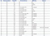
Endocrine characteristics, sellar MRI findings, and a molecular analysis of each patient are summarized in Table 2. Serum IGF-1 levels were 44.48±29.36 ng/mL (9.6–76.7 ng/mL) for IGHD and 10.92±9.73 ng/mL (1.0–35.7 ng/mL) for CPHD (p=0.047). The peak GH levels following GH stimulation tests were 2.96±2.69 ng/mL (0.83–6.9 ng/mL) for IGHD and 0.52±0.65 ng/mL (0.1–2.6 ng/mL) for CPHD (p=0.002) (Table 1). In CPHD patients, endocrine dysfunctions other than GHD included central hypothyroidism in 19 patients (82.6%), ACTH deficiency in 18 patients (78.3%), hypogonadotropic hypogonadism in 17 patients (73.9%), and central diabetes insipidus in 3 patients (13.0%).
The sellar MRI findings demonstrated structural abnormalities in 3 patients with IGHD (75%) and 21 patients with CPHD (91.3%) (p=0.62). In the IGHD group, pituitary hypoplasia was observed in one patient, and an ectopic posterior pituitary was found in two patients. In the CPHD group, pituitary hypoplasia was found in 11 patients, pituitary stalk interruption syndrome in 9 patients, ectopic neurohypophysis in 6 patients, SOD in 3 patients, and empty sella syndrome in 1 patient.
Molecular analysis of pituitary transcription factor genes
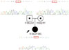
Mutation analysis identified homozygous c.326G>A (p.R109Q) mutations in the HESX1 gene in subject 21 with CPHD (Fig. 1, Table 2), reported to be pathogenic by an in vitro functional analysis.15 This patient was born after 37 weeks of gestation and had a birth weight of 2.56 kg (-1.77 SDS) after an uncomplicated pregnancy and delivery. She presented with a short stature at the age of 2.8 years. The height and weight at diagnosis were 77.5 cm (-4.16 SDS) and 8.6 kg (-4.56 SDS), respectively. Physical examination revealed a prominent forehead and small chin, while a combined anterior pituitary function test demonstrated GHD, ACTH deficiency, and central hypothyroidism. Brain MRI revealed an ectopic posterior pituitary and anterior pituitary hypoplasia, while optic nerve hypoplasia was not evident. The parents were non-consanguineous and phenotypically normal; the father was 179-cm tall (0.99 SDS) and the mother was 165-cm tall (0.83 SDS). Both parents were heterozygous carriers of a p.R109Q mutation in HESX1. No mutation was identified in the POU1F1, PROP1, LHX3, LHX4, and HESX1 genes in the other patients.
DISCUSSION
This study described the clinical and endocrinological features of patients with IGHD and CPHD and demonstrated that mutation frequencies of the POU1F1, PROP1, LHX3, LHX4, and HESX1 genes are rare in sporadic cases of congenital hypopituitarism in Korea. There were no significant differences in sex, age at diagnosis, height, weight, breech presentation, pituitary gland abnormalities, and optic nerve hypoplasia between patients with IGHD or CPHD. However, serum IGF-1 and peak GH levels after the GH stimulation tests were significantly lower in patients with CPHD than in those with IGHD (p<0.05) (Table 1).
Although the effects of these transcription factors in pituitary development are well-defined, neuroanatomy as delineated by neuroimaging in patients with and without genetic hypopituitarism can be highly variable. In a previous report, abnormal sellar MRI findings were more frequently found in CPHD than in IGHD cases.16 However, the present study showed that the frequency of structural abnormalities in the pituitary gland was not significantly different between IGHD and CPHD patients and that structural abnormalities were not related to the severity of GHD.
This study identified that only one CPHD patient without SOD phenotypes harbored homozygous p.R106Q mutations in the HESX1 gene. This mutation was reported to abrogate DNA-binding ability and was unable to repress PROP1-mediated activation.15 Arginine 109 is located in the homeobox domain, critical for binding to target DNA sequences, and is a highly evolutionarily conserved region. The parents of this patients, who carried the same mutation in a heterozygous state, were of normal adult height and clinically normal. However, the heterozygous p.R109Q mutation in HESX1 was identified in a patient with CPHD without SOD phenotypes.15 Variable penetrance of the p.R109Q mutation in HESX1 suggests that the impact of other genes in pituitary development or environmental factors can influence the clinical phenotype.
The HESX1 gene is essential for forebrain development and is one of the earliest transcription factors involved in pituitary organogenesis.17 Indeed, HESX1 has been shown to function as a repressor of PROP1-mediated gene stimulation,18 and 23 mutations in this gene have been identified to date (http://www.hgmd.org). The human HESX1 mutation was first reported in sibling cases of CPHD with SOD and agenesis of the corpus callosum in the homozygous state,19 while subsequently, heterozygous HESX1 mutations were shown to be associated with CPHD or IGHD, with or without SOD.202122 Interestingly, homozygous mutations in HESX1 are implicated in SOD in both humans and animal models,19 whereas heterozygous mutations were found to cause a wide spectrum of congenital hypopituitarism and midline defects with variable penetrance across families. These include unaffected mutation carriers and milder phenotypes, consistent with haploinsufficiency of the HESX1 protein.20
The endocrine phenotypes of the POU1F1, HESX1, PROP1, LHX3, and LHX4 mutations tend to overlap, and these mutations are associated with variable degrees of anterior pituitary hormone deficiency (ranging from IGHD to the complete failure of all five anterior pituitary cell lineages).23 This complex phenotype is further complicated by the evolution of the hormonal make-up over the lifespan of the patient. Therefore, a detailed endocrine investigation at the time of diagnosis is not sufficient to suggest specific genetic screening. Pituitary transcription factor genes should be investigated in CPHD patients with specific conditions and/or extrapituitary findings, e.g., SOD for HESX1, limited neck rotation and sensorineural hearing loss for LHX3, and poorly developed sella turcica for LHX4.23 An algorithm was suggested to look for the candidate genes for CPHD,11 although no genetic defects have been established in most CPHD patients, because of a wide range of phenotypes. The most significant indication for genetic screening of CPHD is a family history of CPHD.11
The advent of next-generation sequencing technologies has greatly reduced sequencing costs and significantly increased throughput, allowing for simultaneous analysis of several genes in singularly targeted platforms. In future studies, it would be worthwhile to consider the use of targeted platforms containing all pituitary transcription factor genes to improve the mutation detection rate and to detect multiple mutations in cases of oligogenic inheritance.
Environmental factors may also contribute to the variability of phenotypes. A causal relationship between gestational-perinatal complications and hypopituitarism has been suggested.24 However, it is difficult to differentiate whether perinatal insults (e.g., breech delivery) lead to hypopituitarism via a traumatic disruption of the pituitary stalk or whether hypopituitarism with structural hypothalamic-pituitary defects results in an increased prevalence of perinatal complications.
The frequency of mutations in sporadic patients with CPHD was much lower than that in familial patients.25 Mutation frequencies in genes encoding pituitary transcription factors vary substantially between populations. The frequency of defects in the genes encoding pituitary transcription factors is extremely low in western European countries, particularly in sporadic patients.23 The worldwide mutation frequency for the POU1F1, HESX1, PROP1, LHX3, and LHX4 genes is 12.4%, ranging from 11.2% in sporadic to 63% in familial cases.23 Mutations in PROP1 are most frequent, particularly in familial cases and certain geographical areas.23 Therefore, as previously described, genetic analysis for CPHD appears beneficial in familial subjects with CPHD.
In conclusion, patients with CPHD had more severe GHD than those with IGHD. The frequency of defects in the genes encoding pituitary transcription factors is extremely low in Korean patients with congenital hypopituitarism, as is the case in western European countries. Patients with GHD should be evaluated for other endocrine dysfunctions via ophthalmologic examination, sellar MRI, and combined anterior pituitary function tests. Further research is needed to investigate other causative genes involved in pituitary organogenesis or environmental factors.

 XML Download