

 PDF
PDF ePub
ePub Citation
 Print
Print


INTRODUCTION
It is crucial for the survival of patients with cardiac arrest that healthcare providers perform correct chest compressions (CC). However, the optimal CC depth and rate remain unclear.12 Unlike other parameters for high-quality CCs (e.g., compression rate, chest recoil, and hand position), CC depth is linearly related to the perfusion of vital organs. It has been shown that a 5 mm increment in CC depth is associated with a 2-fold increase in success of defibrillation during in-hospital and out-of-hospital cardiopulmonary resuscitation (CPR).34
American Heart Association (AHA) and European Resuscitation Council (ERC) guidelines (2010) recommended that CC depth should be at least 5 cm (with or not exceed 6 cm) for high-quality CCs during CPR.56 Reports have indicated that the quality of several CPR parameters often does not meet with the published guideline recommendations during both out-of-and in-hospital cardiac arrest situations.78 CC depth is influenced by the surface on which the patient is placed, especially during CPR performed in a hospital.91011 To compensate for differences in surfaces, it has been suggested that the patient should be placed on a rigid surface, that a backboard should be used to decrease mattress compression (MC), or that a feedback device reflecting MC depth using a dual accelerometer or magnetic sensor should be used.1213141516 However, the use of a backboard has yielded varying results regarding its effectiveness for high-quality CCs. New techniques that could compensate for MC depth have been introduced and used.141516171819 Considering an MC depth of approximately 1–1.5 cm,131415161718192021 performing CCs to a depth of approximately 6–7 cm depth could also compensate for MC during in-hospital CPR and effectively compress the actual chest depth at least 5 cm. To accurately apply this method, healthcare providers who are trained according to the 2010 guidelines need to compress the chest to a depth of at least 6.5 cm feedback if they could use a feedback device with an accelerometer or a pressure sensor during CPR in the hospital. Alternatively, healthcare providers working in the hospital could be trained according to the new training program that requires a CC depth of at least 6.5 cm.
No study on the effectiveness of training healthcare providers to use a CC depth of 6–7 cm has been conducted yet, because current manufactured manikins have a chest depth of ≤6.5 cm. In this study, we used a manikin with a chest depth of 8 cm for CPR training purposes. We hypothesized that training healthcare providers to use a CC depth of 6–7 cm (instead of 5–6 cm) on a manikin placed on a bed during CPR in the hospital setting might improve their CC depth and the proportion of CCs with accurate compression depth.
MATERIALS AND METHODS
Study design and setting
This prospective, randomised controlled trial was conducted at one tertiary medical center (Seoul, Korea) from 20th February to 20th April 2013. Written informed consent was obtained from all study participants under 'Ethic, consent and permission' and 'to publish' before enrolment. Study design was approved by the local ethic committee at our medical center (approval date: January 2013, ref. no. 2013-01-003) and the protocol was registered in Clinical trials before study initiation (ClinicalTrials. gov: NCT01936402).
Study participants
Sixty-six premedical students participated voluntarily in this study. Students who had been previously trained in CPR and those with heart, wrist, or lower back disease were excluded. We determined that a minimum sample size of 60 participants was needed to detect a difference in CC depth of 5 mm between two groups, through a pilot study with 10 participants who were not included in this study using the software package (G-power 3.1.2®, Heine Heinrich University, Düsseldorf, Germany) with an α error of 0.05, β power of 0.8, and considering a drop rate of 10%.
Materials
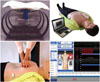
A standard hospital bed frame (Transport stretcher®, 760×2110 mm, 228 kg; Stryker Co., Kalamazoo, MI, USA), foam mattress (660×1920×80 mm, soft foam with polyurethane coverage; Stryker Co.), and BT-CPEA® manikin (17 kg; BT Inc., Wonju, Korea) that could measure a CC depth from 0.0 to 80.0 mm were used in this study (Fig. 1). We added weight to the manikin to achieve a total weight of approximately 40 kg, thus simulating the upper body weight of an adult human when the manikin was placed in a bed.17 We decided not to place a backboard between the manikin and the mattress as we considered the plate of the manikin's back sufficiently stiff.
Data collection
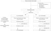
The participants were divided into 2 groups by randomly drawing lots in concealed envelop. Students drawing odd and even numbers were allocated to the control and experimental groups, respectively. The control group was trained to use a CC depth of 5–6 cm (G 5–6), whereas the experimental group was trained to use a CC depth of 6–7 cm (G 6–7). Other CPR parameters (e.g., CC posture, compression rate, complete chest recoil, hand position, etc.) were equally taught to both groups according to the 2010 AHA guidelines. An AHA basic life support faculty member trained each participant who did not know his/her allocated group during a standard 30 min lecture and hands-on practice for only continuous CCs with a kneeling posture and an audio-visual feedback beside a manikin on the floor.21 All participants passed a confirmation test with the manikin placed on the floor immediately after training. After the students passed this test, they performed CCs on a manikin placed on the bed 1 hour and again 4 weeks after the training to assess for the long-term effect of the training (Fig. 2). When the study participants performed CCs on the bed, the height of the manikin's back was adjusted to the height of the provider's upper border of the patella using the bed and step stools for height adjustment.122223 CCs were continuously performed for 2 min without an audio-visual feedback system of a manikin.
Primary outcomes
We measured CC depth, CC rate (the number of compressions per minute), the proportion of CCs with accurate compression depth (%ACD, % of number of CCs with an accurate compressions depth of ≥5 cm/total number of CCs).23 Graphs and data were produced and analysed using the BT-CPEA system (BT Inc., Wonju, Korea). For subgroup analysis, the participants of each group were stratified by gender and body mass index [BMI; low body weight (LBW), BMI <18.5 vs. no low body weight (N-LBW), BMI ≥18.5], and CC parameters were compared between the subgroups.
Statistical analysis
All data were compiled using a standard spread sheet application (Excel, Microsoft, Redmond, WA, USA) and analysed using the Statistical Package for the Social Sciences (SPSS) 18.0 KO for Windows (SPSS Inc., Chicago, IL, USA). We generated descriptive statistics, and present them as frequencies and percentages for categorical data, either medians and interquartile ranges (IQR) (non-normal distribution), or means and standard deviation (SD) (normal distribution) for continuous data. Participant characteristics and CC parameters were compared using either the Mann-Whitney U test (non-normal distribution) or an independent t-test (normal distribution) for continuous measures, and the χ2 test for categorical measures. p<0.05 was considered statistically significant.
RESULTS
Participant characteristics
Sixty-six students were enrolled in our study. One participant from each group did not show up at the 4-week follow-up test, and data of 2 additional participants of the G 6–7 group were excluded because of recording errors. Thus, data of 62 students were analysed (Table 1).
Comparison of chest compression depths
During the first test (1 hour after the training), median CC depths (IQR) were 47.0 mm (43.0–50.0 mm) among students of the G 5–6 group and 57.0 mm (50.0–61.0 mm) for those of the G 6–7 group (p<0.001). After 4 weeks, median CC depths were 48.0 mm (43.0–53.0 mm) and 59.0 mm (52.0–64.0 mm) among students of the G 5–6 and G 6–7 groups, respectively (p<0.001) (Fig. 3).
Comparison of chest compression rates
We found mean CC (SD) rates of 118.0 times/min (8.1) and 120.0 times/min (10.8) in the G 5–6 and G 6–7 groups, respectively, when the manikin was placed on the bed 1 hour after training (p=0.40). During the second test (4 weeks after the training), mean CC rates were 104.3 (18.1) times/min and 102.0 (13.2) times/min, respectively (p=0.57) (Fig. 3).
Comparison of the proportion of chest compression with accurate compression depth
%ACD was 29.0% in participants of the G 5–6 group and 78.8% in those of the G 6–7 group 1 hour after the training. Four weeks after the training, the proportion was 43.2% in the G 5–6 and 83.4% in the G 6–7 group (p<0.001) (Fig. 3).
Subgroup analysis with participants stratified by sex and body mass index
Both men and women of the G 6–7 group performed deeper CCs and a higher performed CPR with a higher %ACD than those of the G 5–6 group during both tests (p<0.001) (Fig. 4). Both LBW and N-LBW of the G 6–7 group compressed deeper than students of the G 5–6 during both tests (p<0.001) (Fig. 5). %ACD was higher in the G 6–7 than the G 5–6 group during both tests (p<0.001) (Fig. 5). We found no difference in the CC rates between the two groups, irrespective of gender and BMI (p>0.05) (Table 2).
DISCUSSION
Healthcare providers who work in critical places within a hospital (e.g., emergency departments, intensive care units, or geriatric wards where cardiac arrests frequently occur) might not correctly perform CCs because of MC.131415161718 In the current study, the participants of the G 6–7 group compressed the chest deeper than the participants of the G 5–6 group when the manikin was placed on a bed. Moreover, the proportion of CCs with accurate compression depth was higher in the G 6–7 than the G 5–6 group. This effect was still seen 4 weeks after the training. Training of healthcare providers working in a hospital setting to perform a CC of 6–7 depth cm could therefore be an alternative method to compensate for MC and an appropriate solution. If healthcare providers use an audio-visual feedback device with a pressure sensor or an accelerometer during CPR in the hospital, a CC with a real-time, 6–7 cm depth feedback might have the same effect as the training to use a CC depth of a 6–7 cm.
CCs can result in complications such as rib fractures, sternal fractures, hemothorax, and pneumothorax.24 CCs with a depth of >6 cm carry an increased risk of complications; however, it is crucial not to a cause fatal complication.25 Complications of CPR are also influenced by other factors such as experience of the performer, hand position, and patient characteristics.2425 We believe that performing high-quality CCs outweighs the risk of complications during CPR.
The ERC guidelines and a study by Stiell, et al.2 recommend that the CC rate should be ≤120/minute, because CC depth might decrease as the CC rate increases.6 We found that the CC rate among participants in the G 6–7 group was not different from that of participants in the G 5–6 group.
Female CPR providers with LBW should be especially careful to ensure that CCs are performed according to the 2010 ERC guidelines.26 In our study, participants with LBW were able to perform deeper CCs when they were in the G 6–7 as opposed to the G 5–6 group. Both women and men of the G 6–7 group did CC depth and %ACD better than those in the G 5–6 group. Training healthcare providers to use a CC depth of 6–7 cm education is not to decrease the MC, but to compress the chest deeply. If MC does not reduce, healthcare providers with LBW may be unable to perform CCs sufficiently, even when being trained to use a CC depth of 6–7 cm. Moreover, CPR providers with LBW might get easily tired when using a CC depth of 6–7 cm and might not therefore correctly perform high-quality CPR. However, if a sufficient number of CPR providers are on site, this problem may be solved by a frequent and short rotation of providers, resulting in minimised time of hands-off.
Our study has several limitations. Despite our adjustment of the manikin's weight, the manikin used in our study differed from actual human patients in weight, height, and back rigidity. We did not know the effect on perfusion, cardiac output, and clinical outcomes of deeper compressions for patient on mattresses. Moreover, current commercial manikins that are used for CPR training have a chest depth of <6.5 cm. To successfully apply our novel training method, the manikin's chest depth must be adjusted to 7–8 cm. We investigated CC depth using a manikin that was placed on one type of bed frame and foam mattress without a backboard. Therefore, we could not analyse if CC depth would have differed between the 2 groups if a different surface or a backboard were used. We believe that the use of a backboard would not have changed our results, as the manikin's back was considered sufficiently stiff. Our study participants were young premedical students. A previous study showed that lay elderly first responders could perform CCs to a depth of 4–5 cm.27 Thus, CPR providers of other (especially older) ages might not be able to compress the chest deeper than 6 cm. Further studies are required to assess the ability of older healthcare providers to perform CCs of 6–7 cm. In our study, we did not assess the difference between performing CCs with a depth of 6–7 cm education method and performing CC using the push hard method. However, we believe that this unqualified technique might cause CPR providers to lose the rhythm of high-quality CPR and experience fatigue faster than when using CCs with a depth of 6–7 cm. Finally, we assessed only the parameters of CCs 1 hour and 4 weeks after the training. It is possible that the effect of the training decreased within a few months, and that CC parameters would not have been different between the 2 groups at this time. The long-term effect of the training method described herein requires further studies.
In conclusion, young healthy healthcare providers who were trained to perform CPR with a CC depth of 6–7 cm could compress the manikin's chest deeply and accurately when it was placed on a mattress as it would be in a hospital CPR setting. Our data, therefore, suggest that training healthcare providers working in a hospital setting to perform a CC of 6–7 cm depth might compensate for MC and improve CC depth when performing CPR in hospitals.


 XML Download