

 PDF
PDF ePub
ePub Citation
 Print
Print


INTRODUCTION
Since the first laparoscopic cholecystectomy was performed in 1897, it has come to be accepted as the gold standard treatment of benign pathologic conditions of the gallbladder.1 Many previous studies have clearly shown that laparoscopic surgery resulted in a shorter hospital stay, less pain, a better cosmetic effect, a more rapid return to normal daily activity, and improved immunologic responses compared with the outcome of conventional surgical techniques.2-4 However, many surgeons also admit that there are some critical disadvantages to using laparoscopic techniques, for example, limitations in the degree of motion of instruments, loss of three-dimensional visualization, and loss of touch sensation.5
Currently, an increasing number of surgical procedures are being performed with an emphasis on minimizing trauma to the patient. To accomplish this, more complex and precise movement of the laparoscopic instruments are required. The introduction of robotic and computer-assisted surgical systems has allowed for tremendous progress in the field of minimally invasive cardiac surgery6 and has the potential to allow surgeons to overcome several difficulties encountered during laparoscopic surgery.
We herein report the first Korean experience of laparoscopic cholecystectomy assisted by a da Vinci system (The Intuitive Surgical Endoscopic Instrument Control System), focusing on the introduction of the system and perspectives on robotic surgery.
CASE REPORT
Patient
A 56-year-old patient with upper right quadrant discomfort for 2 months was admitted to our department. She had already been diagnosed with gallstone disease at a local hospital. She had no unusual medical history of past illness. She weighted 75kg and was 160cm tall (Body Mass Index = 29.3). Other laboratory evaluations including complete blood count, SGPT, SGOT, total/direct bilirubin, alkaline phosphatase, and GGT were all within normal limits. An abdominal ultrasound scan performed at the local hospital showed multiple gallbladder stones ranging in size from 0.5cm to 1.5cm. She underwent laparoscopic cholecystectomy using a da Vinci robotic surgical system on the second day after admission.
Introduction of the da Vinci system
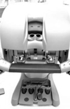
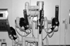
The da Vinci robotic surgical system has three components. The first is the vision cart that holds the dual light source and image processors for the dual three-chip cameras. The dual cameras are mounted on an endoscope which is placed in the camera arm, and provide three-dimensional images. The second component is the surgical console (Fig. 1), where the operating surgeon sits and manipulates the robotic arms. This console contains image processing computers that combine the images into a true 3-D image with depth of field, and the view port screening through which the surgeon can view the 3-D operative field. There are two control grips and several foot pedals to control the movement of robotic arms at the patient's side, other robotic laparoscopic instruments, camera focus and instrument/camera arm clutches. The last component is the surgical robotic cart (Fig. 2). The three instrument arms and one camera arm are mounted on this robotic cart. The laparoscopic instruments with uniquely designed instrument tips can be equipped to the three instrument arms, thereby creating flexible movement with seven degrees of freedom like that of a human hand. Each arm has several articulates by which it is controlled to adjust its location appropriate to respective trocar sites.
Operation and outcomes
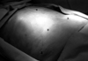
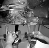
In this time, only three robotic arms including camera arm were used. The da Vinci system was prepared behind sterile drapes before the patient arrived at the operating room. The patient was placed in the supine position on the operating table and underwent general endotracheal anesthesia. The abdomen was prepared and draped in the usual sterile fashion. The 12-mm subumblical camera port① was placed through mini-laparotomy and pneumoperitoneum was achieved by CO2 insufflation. Under direct vision, the two 8-mm robotic instrument ports were placed in the standard position for laparoscopic cholecystectomy; one② at the right midclavicular line in the subcostal region and the other③ in the epigastric region. Another 5-mm accessory port④ was placed lateral and inferior to the robot's right instrument arm port (Fig. 3). The patient was placed in the steep reverse Trendelenburg position with her left side tilted. The da Vinci system was rolled into position, 40˚-45˚ off of the right head of the table. The robotic camera arm and instrument arms were then connected to their respective ports. The right accessory port was used to place the grasper that would retract the gallbladder upward and outward, operated by the patient-side assistant surgeon. At this point, the surgeon sat down at the surgical console located about 3m from the operating table. The patient-side assistant surgeon was positioned on the patient's right to retract the gallbladder using a 5-mm grasper and then change the robotic instrument. The operating surgeon controlled the da Vinci robotic surgical system in the dissection of the gallbladder, including Calot's triangle, and its complete removal (Fig. 4 and 5)
Total operation time (beginning with minilaparotomy for robotic port insertion and ending with completion of wound dressing) was 110 minutes. Total robot time (beginning when the surgeon sat at the console and ending when the surgeon completed robotic manipulation) took 45 minutes. The dissection time of Calot's triangle (measured as starting when the grasping forcep and hook cautery were introduced and concluding when cystic artery and cystic duct were completely ligated) was 14 minutes. Gallbladder dissection time (beginning when the hook cautery and grasping forcep were in place to dissect the gallbladder out of the liver bed and ending when the gallbladder was put into an Endopouch bag) was 14 minutes. After inspection of the liver bed and dissected area to confirm no injuries, the abdomen was desufflated. The portside incision was closed by several stitches with a skin stapler. The patient was able to have an oral diet on the first postoperative day, and was discharged without problems on the second postoperative day. The delivered gallbladder had the appearance of chronic cholecystitis with wall thickening and several cholesterol stones reaching up to 2.5cm in size.
DISCUSSION
Since 1921 when the Czech playwright Karel Capek introduced the term "robot" in his play, 'Rossum's Universal Robot', it has been a popular term.7 In his play, the term simply referred to a machine performing simple repetitive tasks in the place of human beings. Now, there have been so many advancements in science and technology that extremely delicate and complex motions and missions can now be performed with robotics.
The history of robotics in surgery begins with the Puma 560, a robot used in 1985 to perform neurosurgical biopsies with improved precision.8 A few years later, transurethral resection of the prostate was performed using the same robot system.9 This system eventually led to the development of PRPBOT, which was designed specifically for transurethral resection of the prostate. ROBODOC, a robot system designed to machine the femur with greater precision in hip replacement surgeries,10 was the first surgical robot approved by the FDA.
Much intensive research on robot systems to develop telepresence surgery, and efforts to overcome the limitations produced by conventional laparoscopic surgery, eventually lead to the introduction of robotics into the surgical community. The Automated Endoscopic System for Optimal Positioning (AESOP), a robotic arm just for holding a laparoscopic camera controlled by a surgeon's voice command, was introduced and commercialized in the mid-1990s. Computer Motion, Inc. of Santa Barbara, CA developed and marketed a surgical robotic system (ZEUS) equipped with AEOSOP.11 Notably, Integrated Surgical System (now Intuitive Surgical) of Mountain View, CA, licensed the SRI Green Telepresence Surgery System, which was extensively redesigned and reintroduced as the current da Vinci surgical system, with a full seven degrees of freedom of motion at the instrument tip and 3-D visualization. This system is unique in the current field of robotic surgery due to its great advantages over the ZEUS system. In 1997, Himpens et al.12 performed the first telemanipulative laparoscopic cholecystectomy with its use. In the US, the da Vinci system received FDA approval for general use in abdominal laparoscopic procedures in late July 2000, and Korean FDA approval was confirmed on July 13, 2005. Two days later, we performed the first case of telemanipulative laparoscopic cholecystectomy using the da Vinci in Korea.
The several serious limitations of conventional laparoscopic surgery are loss of dexterity, haptic feedback, natural hand-eye coordination (fulcrum effect), and movement based on a 2-D video monitor, which are all somewhat counterintuitive. Physiologic tremors in the surgeon are transmitted through the length of the rigid instrument. Most laparoscopic gastrointestinal operations are difficult to learn, master, and perform routinely and surgeons have to face a long period of learning curve. Finally, poor ergonomic position for the surgeon is another problem for laparoscopic surgery. These limitations make delicate dissection and anastomosis more difficult, if not impossible. However, the present da Vinci system was developed to overcome these problems. One of the most outstanding points of the da Vinci system is that the tips of the laparoscopic equipment have seven degrees of freedom of motion, which means that the same exact movements as those of a human hand are possible. Another sure advantage is the 3-D visualization of the operative field that the da Vinci system provides, compared to conventional minimally invasive procedures (Table 1).
However, there are limitations of robot-assisted surgery that need to be resolved, namely the long preparation time, high cost and complete absence of touch sensation. In this case, it took about 40 minutes to prepare the system prior to surgery, but it is certain that preparation time will shorten with experience. The cost for robotic-assisted surgery ranges from about form 7 million to 15 million won($6,900 to $18,750; exchange rate $1.00 = 1035won, December 1, 2005), which is another obstacle to overcome in the popular application of this system for laparoscopic surgeries.
Since 1987, laparoscopic cholecystectomy has been widely attempted in almost all medical centers. With the accumulation of laparoscopic experience in cholecystectomy, great advances of laparoscopic ability in the local anatomy around Calot's triangle made complete laparoscopic cholecystectomy possible (without help of a robotic surgical system). In terms of cost-effectiveness, the da Vinci robot system is not suitable for cholecystectomy and does not provide any additional value to surgeons or patients. However, taking this system's several critical advantages over conventional laparoscopic surgery into consideration; its value may be realized in cases of more complex gallstone disease, such as Mirrz syndrome. It has the potential to reduce the incidence of accidental and unexpected injury to surrounding organs during laparoscopic cholecystectomy, including to the bile duct, bowel and major vascular structures because of the robot's providing better visual image and precise movements. Furthermore, laparoscopic cholecystectomy using the da Vinci system can be regarded as an ideal teaching tool for both surgeons just starting to learn robotic-assisted surgery and residents who will most likely have this technology as a standard procedure in their future careers.13
In order to properly establish this robotic-assisted surgery in one institution, we would like to emphasize the need for not only surgeons but also scrub nurses to be trained and educated as part of a "Robot Team." Surgeons need to become well aware of the differences in robot-assisted surgery from conventional laparoscopic surgery. All team members should learn how to connect the robotic arms to each trocar how to connect instruments to the robotic arms, how to disconnect the arms and how to adjust the arms to respective trocar sites when needed. Through virtual reality training prior to actual intervention, all members can discuss issues that need to be corrected and become more familiar with the robot system. In our case, three experienced laparoscopic surgeons and two nurses specially chosen for robot surgery participated in the educational programs provided by Intuitive Surgical Inc. in the US. They educated other team members in Korea by performing several simulated operations using the da Vinci system. This direct and indirect experiences with robot- assisted surgery is likely to be of great aid to understanding the system and achieving successful first laparoscopic cholecystectomies.
From the end of July 2005 to the end of this year, more than twenty surgeries using the da Vinci system were performed at our institution, including cholecystectomy, correction of choledochal cyst, gastrectomy, prostatectomy, partial bladder excision, and excision of a mediastinal tumor. According to our early experiences, the 3-D visualization and seven degrees of motion of the laparoscopic equipment have definitively provided more precision in delicate laparoscopic procedures, especially when dissecting soft tissues around major vessels, performing hepaticojejunostomy, and maneuvering in narrow spaces like the pelvic cavity. We are preparing a report of the preliminary our early experiences with laparoscopic surgery using the da Vinci system.
In summary, due to advancements in robotic technology, the first Korean robot-assisted laparoscopic cholecystectomy was performed in the Yonsei University Medical Center. The feasibility of the da Vinci system has been shown in accordance with our early experiences.14-17 This telemanipulative robotic system has the potential to expand surgical treatment modalities beyond the limits of human ability. Further trials of robot-assisted surgery are needed in our country and research on evaluating both the efficacy and safety of the procedure as well.

 XML Download