

 PDF
PDF ePub
ePub Citation
 Print
Print


INTRODUCTION
FcεRI is a high-affinity immunoglobulin E (IgE) receptor I that contains four distinct polypeptide chains: an extracellular α-chain, a β-chain, and a dimer of γ-chains.1 The extracellular binding domain of the α-chain binds with high affinity to the Fc region of IgE, whereas the other chains are responsible for the transduction of initial cross-linking signals into the cell through phosphorylation of their intracytoplasmatic immune-receptor tyrosine-based activation motif.234 Elevated serum IgE levels upregulate FCεRI, increase the sensitivity of these cells to allergen-triggered activation, and enhance cell survival.56789 When an antigen cross-links with an IgE bound to its FcεRI on these cells, an allergic reaction is initiated, resulting in the synthesis and release of a variety of allergic mediators. A number of these mediators (e.g., histamine, neutral proteases, proteoglycans, proinflammatory lipid mediators, and cytokines) are released from the mast cells and basophils.71011 These cytokines and mediators continuously induce IgE production in immunoglobulin class-switching B cells9 and induction of eosinophil infiltration as well as airway hyperresponsiveness.12
Since allergen cross-linking with FcεRI-bound IgE is considered the initial factor in the allergic cascade, therapeutic approaches intervening the binding of IgE to its receptor have been repeatedly proposed. Antibodies have been developed to prevent IgE from binding to FcεRI, by blocking the ability of IgE to induce upregulation. The first approved therapeutic antibody was a humanized monoclonal anti-IgE antibody (omalizumab), which prevents IgE from binding to receptors in order to suppress the allergen-mediated degranulation of mast cells and basophils.13141516 Another possible target of a therapeutic antibody could be FcεRI. However, a full antibody structure could lead to an agonist-like effect; hence, we designed a monoclonal antibody of the Fab fragment to block FcεRIα instead.
In this study, we developed NPB311, a humanized monoclonal antibody Fab fragment containing FcεRIα-binding sites but lacking the Fc portion, and investigated the affinity of NPB311 for recombinant FcεRIα by surface plasmon resonance (SPR). We also examined whether NPB311 could inhibit β-hexosaminidase, histamine and Ca2+ release in vitro by using rat basophilic leukemia (RBL-SX38) cells17 that express human FcεRIα. From these and additional studies using human FcεRIα-transgenic mice, we concluded that NPB311 has the ability to regulate the IgE-FcεRIα complex in response to IgE in IgE-dependent passive cutaneous anaphylaxis (PCA) mice.
MATERIALS AND METHODS
Phage display against human FcεR1α
Human recombinant FcεR1α-Fc fusion protein (Origene Technologies, Rockville, MD, USA) was coated on a plate and the human scFv phage library was added. After incubation, bound phages were detached by 0.1 M triethylamine treatment. Selected phages were added to the FcεR1α-Fc fusion protein-coated plate, followed by incubation with horseradish peroxidase (HRP)-conjugated anti-M13. Luminescence was measured with the LumiGLO chemiluminescence kit (KPL, Gaithersburg, MD, USA) according to the manufacturer's instructions.
The IgE-binding competitive activity of the selected antibody was measured by competition enzyme-linked immunosorbent assay (ELISA). A plate was coated with FcεR1-Fc fusion protein (0.1 µg/mL), and selected phages were added by panning. After washing, 0.1 µg/mL of human IgE (hIgE) (Abcam, Cambridge, MA, USA) was added, followed by HRP-conjugated mouse anti-hIgE, and luminescence was detected.
IgG conversion and digestion
From the selected scFv expressed in Escherichia coli, the heavy chain and light chain were synthesized by polymerase chain reaction and ligated into the human IgG2 constant domain-expressing vector. The reconstructed DNA was transformed to DH5 competent cells. Antibodies were obtained by transient transfection in 293E cells and quantified by direct ELISA. Plates were coated with either anti-human lambda or kappa antibodies, respectively. Diluted (1:200) conditioned medium from a culture of transient transfection cells was added to each well. After 1 h incubation, HRP-conjugated anti-hIgG was added and luminescence was detected.
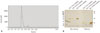
The Fab fragment was prepared by papain digestion of the NPB311-IgG complex, using a Fab preparation kit (Pierce, Thermo Scientific, Rockford, IL, USA). Papain digestion was carried out according to the manufacturer's instructions. Fractions collected during the papain digestion and Fab purification were analyzed under reducing conditions by sodium dodecyl sulfate polyacrylamide gel electrophoresis (SDS-PAGE). The Fc part and undigested IgG molecules were removed by affinity purification using a Protein A bead column (GE Healthcare Bio-Sciences, Pittsburgh, PA, USA). Size-exclusion chromatography using a Hiprep 26/60 Sephacryl S-200 HR column (GE Healthcare Bio-Sciences) in 50 mM Tris and 150 mM NaCl (pH 7.0) buffer was used as the final purification step of the Fab fragment.
Competition and binding assays
Candidate antibodies were produced by transient transfection in 293E cells and purified by protein A column chromatography (KPL). The purified proteins were added to a plate coated with 0.2 µg/mL FcεR1α protein and incubated for 1 h at 37℃. After washing, 0.2 µg/mL hIgE was added and the plate was further incubated for 1 h at 37℃. Competition was measured by reaction with HRP-conjugated mouse anti-hIgE and detected with an EnVision Multilabel Reader using the LumiGLO chemiluminescence substrate. Direct binding against FcεR1α on the cell surface was measured using RBL-SX38 coated on 96-well plates.
Affinity measurement by surface plasmon resonance
NPB311 was immobilized on a sensor chip (CM5; BiaCore, Giles, UK) in 10 mM sodium acetate (pH 5.0) to an immobilization level of 1000 response units. Coupling was performed using 1-ethyl-3-(3-dimethylaminopropyl)-carbodiimide/N-hydroxysuccinimide and 1 M ethanolamine (Biacore) according to the manufacturer's instructions. A negative reference cell was prepared by the same process but without NPB311. A serial dilution of NPB311 was made with HBS-EP sample buffer (HEPES-buffered saline with EDTA and P20 surfactant; Biacore) (0, 6.25, 12.5, 25, 50, and 100 nM, respectively). Association of NPB311 with hIgE were determined by injection of NPB311 solution for 4 min at a rate of 30 µL/min, followed by injection of buffer solution for 4 min at the same rate. Regeneration between each measurement was performed by injection of 10 mM glycine (pH 2.0) at 30 µL/min for 40 s, followed by 2 min of stabilization before the next injection.
β-hexosaminidase activity assay and histamine release measurement
For determination of NPB311 functional activity, IgE-induced β-hexosaminidase and histamine release from cultured RBL-SX38 cells were measured. Cells were seeded on plates and, after overnight incubation and washing, either diluted NPB311 or hIgG was added to the wells, followed by 4-hydroxy-3-nitrophenylacetyl (NP)-hIgE (0.5 µg/mL), and the plate was incubated at 37℃ for 2 h. Then, 0.1 µg/mL NP-BSA was added to each well and the plate was further incubated. Thereafter, the supernatants were collected, and residual cells were lysed with 0.1% Triton X-100 solution. The production of β-hexosaminidase in the medium and in the cell lysates was determined by a fluorometric assay, using 4-methylumbelliferyl-Nacetyl-β-D-glucosaminide as the substrate (0.1 mM in 100 mM citrate, pH 4.5). After 90 min, the reaction was stopped with 0.2 M glycine buffer and the plate was read on 380 nm excitation and 405 nm emission filters.
For the histamine release assay, RBL-SX38 cells were plated at 2×10⁵ cells/well and incubated with 0.5 mL of medium containing 0.5 µg/mL hIgE, with or without NPB311. After a 12 h incubation, the cells were washed with Tyrode's buffer (125 mM NaCl, 5 mM KCl, 0.4 mM MgCl2, 1 mM CaCl2, 5.6 mM glucose, 10 mM Hepes/Na+, and 0.1% BSA, pH 7.2) and then incubated in 0.5 mL of Tyrode's buffer containing 0.1 µg/mL NP-BSA for 30 min. The supernatants were collected and analyzed for histamine content, using a HTRF histamine assay kit according to the manufacturer's instruction.
Measurement of cytosolic Ca2+ concentrations
Cells were seeded at 5×10⁴ cells/well per 100 µL loading medium (RPMI 1640, 10% FBS) into 96-well black wall plates. After 24 h, medium was replaced with NPB311 followed by addition of NP-IgE. After 2 h incubation, antibody/IgE mixture was aspirated, leaving the cell monolayer intact, and replaced with 100 µL/well of FLUO-4AM loading buffer [Dulbecco's modified Eagle's medium with 0.1% FBS, 20 mM HEPES, 2.5 mM probenecid and 2 µg/mL FLUO-4AM (Invitrogen, Life Technologies, Carlsbad, CA, USA)] for 1 h at 37℃. Cells were washed with PBS and placed in 100 µL/well of Fluorometric Imaging Plate Reader (FLIPR) buffer (125 mM NaCl, 5 nM KCl, 1 mM MgCl2, 1.5 mM CaCl2, 30 mM HEPES, 2.5 mM probenecid, 5 mM glucose, 0.01% v/v fetal calf serum) with FLIPR 4 dye. Fluorescence of the FLUO-4AM dye was recorded every 2 sec.
Animals
B6.Cg-Fcer1atm1Knt Tg(FCER1A)1Bhk/J mice were purchased from Jackson Laboratory (Bar Harbor, ME, USA). C57BL/6 mice were purchased from Orient Bio (Seongnam, Korea). Animals were kept in a specific pathogen-free facility under alternate dark-light cycles of 12 h at room temperature. The care and treatment of all animals were done in agreement with the Institutional Animal Care and Use Committee of Yonsei University, Seoul, Korea.
Measurement of preventive effects on anaphylactic reactions in mice
Specific pathogen-free female wild-type and B6.Cg-Fcer1atm1Knt Tg(FCER1A)1Bhk/J mice were injected subcutaneously with 0.1 µg of NP-IgE, with or without NPB311. At 30 min or 24 h after sensitization, the mice were tail-intravenously challenged with 0.1 mL of 1% Evans blue (EB) dye solution containing 1 mg/mL of NP-BSA. Cutaneous anaphylaxis was assessed visually by the blue dye leakage from blood vessels into the skin.
Statistical analyses
Statistical analysis was carried out with Sigma plot 10.0 software (Jandel Scientific, Sausalito, CA, USA). All data are expressed as means±SDs and represent one of four independent experiments. Significant differences between two groups were estimated using the unpaired Student's t-test. Statistical significance was set at p≤0.05.
RESULTS
Development of an anti-FcεRI Fab fragment antibody
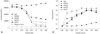
More than 3500 individual clones were isolated and subsequently characterized for their specificity, affinity, and ability to block IgE binding to FcεRIα. Biopanning against FcεRIα was performed using the human scFv phage display library, on which phage with a binding capacity to FcεRIα was enriched. The specificity of the polyclonal phage after panning was verified by ELISA using recombinant FcεRIα in microplates. After this panning, the affinities of candidates were increased. Among them, the anti-FcεRIα Fab fragment mAb NPB311, which showed high reactivity against human FcεRIα in direct ELISA and blocking of the IgE-FcεRIα complex in competition ELSIA, was selected (Fig. 1).
Effect of NPB311 on [Ca2+] in RBL-SX38 cells
The allergy profile is based on the crosslinking of IgE-antigen complexes to high-affinity IgE receptors (FcεRI) on immune effector cells, which leads to the release of variety of mediators such as a protein tyrosine kinase- and calcium (Ca2+)-dependent signal cascade.18 Therefore, we identified the inhibition effect of NPB311 on [Ca2+] release. As shown in Fig. 3A, incubation of RBL-SX38 cells with NP-IgE, followed by the treatment with NPB311, inhibited strong drop of florescence in cells dose-dependently. No increase of [Ca2+] florescence was observed by adding 100 nM NPB311 with NP-IgE. Next, we examined the NPB311 agonistic activity against hFcεRIα. Cultured RBL-SX38 cells were stimulated with NPB311, and then Ca2+ fluorescence was examined. At concentrations of 0 and 500 nM, NPB311 did not show the agonist-induced increase in the binding of NPB311 to hFcεRIα (Fig. 3B), showing that NPB311 have an inhibition effect on IgE binding against hFcεRIα, but no agonist activity against hFcεRIα.
NPB311 binding affinity to FcεRIα and inhibition of RBL-SX38 cell degranulation
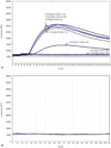
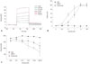
For evaluation of antigen binding affinity, SPR analysis was performed. Thus, SPR biosensors were utilized to derive the KD values for antibody against the immobilized FcεRIα ligand. Affinity measurement of NPB311 for hFcεRIα showed a KD value of 4 nM (Fig. 4A). To ascertain the inhibition effects of NPB311 on cell degranulation, we treated NP-hIgE-sensitized RBL-SX38 cells with NPB311, ranging from 0.001 to 20 µg/mL. NPB311 significantly suppressed β-hexosaminidase secretion (Fig. 4B) and histamine release (Fig. 4C) in IgE-sensitized RBL-SX38 cells.
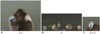
Effect of NPB311 treatment on passive cutaneous anaphylaxis
We assessed in vivo activity of NPB311 in transgenic double-mutant mice that expressed the human high-affinity Iα, instead of mouse FcεRIα, by performing PCA to test the NPB311 ability to block IgE-driven FcεRI-mediated mast cell release. Thus, we intradermally primed the transgenic mice with NP-hIgE with or without NPB311, and the intensity of the PCA at each site was assessed by the size of the skin turning a blue color after 30 min. Fig. 5 shows that the size and color intensity of the reaction at the sites of NPB311 injection were lower than sites injected with NP-hIgE/hIgG.
DISCUSSION
In this study, we have described the Fab fragment antibody NPB311, which is targeted against human FcεRIα. We determined that NPB311 could disrupt IgE binding to FcεRI through FcεRIα engagement, then reducing the release of inflammatory mediators in cells. These results came from the following findings: 1) NPB311 did not show the agonist-induced increase in the binding of NPB311 against hFcεRIα; 2) NPB311 inhibited IgE-induced histamine, β-hexosaminidase and Ca2+ release; 3) NPB311 reduced IgE-induced dye leakage in the PCA murine model.
Asthma is the most common chronic disease.19 In Korea, it is estimated that 2273290 patients have asthma in 2008, expending $831 million, with an average per capita cost of $366. Many clinical trials have shown that omalizumab reduces exacerbation risk and improves health-related quality of life related to asthma.20 However, omalizumab, is an expensive medication (US $1874 per 4 weeks on average).21 Clearly, new treatment options are required to improve the management of this common and troublesome condition. Antibodies and recombinant proteins have been developed to prevent IgE binding to FcεRIα and block the production of inflammatory mediators. Omalizumab binds to IgE, thereby hindering its binding to FcεRI and suppressing allergen-mediated degranulation of cells. However, omalizumab showed to increase circulating IgE level because of the formation of stable omalizumab/IgE immune complexes, and high IgE levels are sustained for several months after the cessation of drug treatment.22 Unlike omalizumab, however, antibody NPB311 reduces the level of IgE in the blood, can disrupt IgE-FcεRI binding through FcεRIα engagement and ultimately reduces the releases of inflammatory mediators in cells. Thus, NPB311 could be a new therapeutic strategy of allergic diseases of airway inflammation such as asthma.
Other possible target could be IgE-mediated atopic dermatitis (AD) and urticarial. AD is a chronic inflammatory cutaneous disease in childhood, in which the elevated and persistent production of IgE plays an important role.2324 Such a disorder is a possible target of anti-IgE treatment, but thus far there are scant data on the effects of omalizumab in AD. Recent studies described that anti-IgE therapy in patients with AD found symptomatic improvement with omalizumab.2526 Ozdemir, et al.27 showed that all patients receiving omalizumab had decreased levels of inflammatory mediators and interleukin-9 compared to placebo in their randomized, placebo-controlled clinical trial. In addition, patients on anti-IgE therapy had an improvement in clinical outcomes. In this regards, NPB311 could potentially be used as an alternative in the future, thereby sparing the use of high potency topical corticosteroids, oral steroid dosing, and systemic therapies.
Generally, the Fab fragment antibody has issues of a short half-life in circulation and lack of biological effector functions. Nevertheless, full antibodies do not always show optimal therapeutic effects. For example, inappropriate activation of FcεRI-expressing cells can lead to inflammatory mediator and cytokine release, a potentially fatal immune reaction caused by highly elevated levels of different cytokines.282930 Thus, we manufactured a Fab fragment structure (NPB311) as a potential solution to overcome the unwanted properties of full-structured antibodies. In our findings, NPB311 showed high affinity to FcεRIα (KD=4 nM), and hIgE-induced histamine, β-hexosaminidase (Fig. 4B and C) and Ca2+ release (Fig. 3A) were decreased in vitro. To evaluate the efficacy of NPB311 in vivo, we developed an IgE-mediated PCA model in transgenic mice expressing human FcεRIα. PCA is one of the most important local allergic reaction in vivo models. A local dye extravasation was induced by a local injection of dye-antigen mixture.31 The dye leakage caused by IgE-mediated PCA in mouse ear was suppressed by intradermally administered NPB311 (Fig. 5). Moreover, in the process of manufacturing NPB311, we observed that it recognized an epitope within the second domain of FcεRIα (data not shown). The binding site of NPB311 showed major overlap with FcεRI-binding residues on IgE, and thus it disrupted the IgE-FcεRI complex. These results indicated that NPB311 may suppress the allergen specific inflammatory response by inhibiting IgE-FcεRI interaction, regardless of Fab fragment and short half-life (Supplementary Method 1, Supplementary Figs. 1 and 2, only online).
Recently, various Fab fragment antibodies have been developed and studied in murine airway inflammation models.3233 A previous study demonstrated the effects of IL-13 Fab fragment treatment on the allergen-induced asthma mouse:34 inhaled anti-IL-13 Fab significantly reduces airway inflammation, hyperresponsiveness, and remodeling. Interestingly, anti-IL-13 Fab administrated by the in Expose system induced a significant decrease of the peribronchial smooth muscle cell layer but not in intravenous administration system. In this regard, the use of these alternative administrations could be a way to overcome the limited half-life of the Fab fragment in the chronic asthma model and help decrease the administered dose. However, because of their reduced size, antibody fragments usually penetrate tissues more rapidly and efficiently than full IgG, thus this benefit is counterbalanced by a very short serum half-life.35 Therefore, further investigation of alternative approaches is needed to increase the serum half-life of Fab fragment, the most promising one being the chemical addition, including conjugation of polyethylene glycol (PEG) residues,36 fusion of PEG-mimetic polypeptides and albumin-binding moieties.
In conclusion, anti-FcεRIα Fab fragment may be a useful therapeutic agent for the treatment of IgE-dependent inflammation. To overcome the limitation of its short half-life, the implementation of half-life extension might be needed. Also, further studies are needed to investigate the NPB311-mediated changes of decreased inflammation in the allergic diseases murine model.
 XML Download