

 PDF
PDF ePub
ePub Citation
 Print
Print


INTRODUCTION
Hepatocellular carcinoma (HCC), the most common primary malignant tumor of the liver, is one of the most lethal malignancies (1). It is the sixth most commonly diagnosed cancer worldwide and the third leading cause of cancer death (2, 3). Liver transplantation is currently the best treatment for HCC. However, the number of available donors is limited. Thus, hepatic resection remains the treatment of choice for potentially curable disease in most areas of the world (3, 4, 5). Curative therapies including liver transplantation and resection are applicable in only 30-40% of HCC patients. Therefore, most patients are suitable only for locoregional or palliative therapies (6, 7).
Transarterial chemoembolization (TACE) is one of the preferred treatments for patients with HCC who are not suitable for curative therapy (8, 9). TACE is also considered the standard of care for non-surgical patients with tumors limited to the liver because it can preserve liver function (4, 10, 11). Conventional TACE involves intra-arterial infusion of a viscous emulsion of an ethiodized oil (e.g., lipiodol) and a chemotherapeutic agent such as doxorubicin, followed by an injection of gelatin sponge particles or other agents to embolize the blood vessel (12). This embolization ensures that lipiodol is retained selectively in HCC, enhancing drug delivery to the tumor. The embolizing agent also reduces drug washout from the tumor and induces ischemic necrosis. Ideally, TACE should result in a maximum sustained concentration of chemotherapeutic agent within the tumor with minimal systemic exposure. Additionally, TACE should obstruct the tumor vessels without obstructing blood supply to the surrounding tissue (13, 14). Randomized and controlled studies have shown that TACE has survival benefits superior to those of supportive care in appropriately selected patients (4, 7, 10, 11, 15). Despite the clinical efficacy of TACE with lipiodol, there are recognized limitations. In some cases, HCC does not exhibit lipiodol retention which may result in decreased effectiveness of the treatment and increased risk of liver damage (16, 17).
To release cytotoxic drugs (e.g., epirubicin or doxorubicin) in a controlled and sustained manner, drug-eluting beads (DEBs) have been introduced to TACE for transarterial treatment of HCCs (18, 19). These microspheres allow local delivery of high concentrations of chemotherapeutic agents to the tumor, with systemic concentrations comparable to conventional chemotherapeutic regimens. The use of DEB can reduce the occurrence of common adverse events (AEs) such as abdominal pain, fever, nausea, and vomiting that typically occur with TACE when gelatin sponges or microspheres (Embosphere; Merit Medical Systems, South Jordan, UT, USA) are used (20, 21, 22). Studies highlighting the use of DEB with TACE for the treatment of HCCs have shown similar or better results compared to conventional TACE with lipiodol (14, 15, 18, 20). The goal of this study, therefore, was to compare transarterial chemoembolization using gelatin sponges or microspheres plus lipiodol-doxorubicin vs. doxorubicin-loaded beads for the treatment of hepatocellular carcinoma.
MATERIALS AND METHODS
Study Design
This is a retrospective study of consecutive HCC patients who received TACE at a single medical center from November 2010 to November 2011. The study was approved by the Institutional Review Board of the hospital. Because of its retrospective nature, the requirement of informed patient consent was waived. All patients underwent pretreatment assessment including a medical history, physical examination, laboratory assessment, and imaging studies (contrast-enhanced computed tomography [CT] or magnetic resonance imaging [MRI]). Inclusion criteria for the study were: 1) adult ≥ 18 years old with HCC diagnosed based on noninvasive criteria (4); 2) at least one tumor that was treatment-naïve and > 1 in diameter; 3) Barcelona Clinic Liver Cancer (BCLC) criteria A or B; 4) Eastern Cooperative Oncology Group (ECOG) performance score of 0 or 1; 5) serum creatinine < 1.2 mg/dL (normal range, 0.6-1.2 mg/dL); 6) aspartate aminotransferase (AST) and alanine aminotransferase (ALT) levels < 200 IU/L (normal range, 0-40 IU/L and 0-45 IU/L, respectively); and 7) total bilirubin < 3 mg/dL (normal range, 0.1-1 mg/dL). Exclusion criteria were: 1) if the tumor invaded the portal vein, hepatic vein, or biliary duct; 2) if the tumor had an extrahepatic arterial supply (drug-eluting beads are not recommended to be used on extrahepatic feeders); 3) if they were diagnosed with atypical HCC (e.g., infiltrative).
Treatment
Treatment with chemoembolization was planned by a multidisciplinary team. Patients were treated with either conventional TACE with a gelatin sponge (group A), TACE with Embosphere microspheres (Biosphere, Roissy, France) (group B), or chemoembolization with doxorubicin-loaded DEB (DC Beads; Biocompatibles, Farnham, United Kingdom) (group C). All patients included in this study received only one cycle of TACE during the data collection period. The attending physician explained the tumor response rate and complication rate of each method to the patient based on current published literature. The patient was asked to decide on the method based on the information they were provided.
On treatment day, a thorough diagnostic angiographic evaluation of the celiac trunk, superior mesenteric artery, and hepatic artery was performed to determine the vascular anatomy and to assess portal flow (23). Super-selective angiography was subsequently performed using a microcatheter to catheterize the segmental or subsegmental arteries feeding the tumor. In patients whose right hepatic artery arose from the superior mesenteric artery, the right hepatic artery was selectively when necessary. Embolization of the cystic artery and falciform artery was carefully avoided. The phrenic artery was studied when it was suspected to supply the target tumor.
Variations in the preparation of lipiodol-drug emulsion can affect the release of the drug into the systemic circulation, thus affecting the outcome of TACE (24). In group A and B, 50 mg of doxorubicin was mixed with 10 mL of lipiodol so that a consistent concentration was achieved. In group A, the lipiodol/doxorubicin was injected into a segmental or subsegmental artery, followed by an injection of 500-700 µm gelatin sponges (Spongostan standard, Johnson & Johnson, Gargrave, Skipton, United Kingdom). In group B, the lipiodol/doxorubicin was injected into a segmental or subsegmental artery, followed by injection of 100-300 µm Embosphere microspheres. In both groups, the amount of lipiodol/doxorubicin injected was based on tumor diameter as described previously (25). In both groups, the endpoint of embolization was stasis in the second- or third-order branch of the right or left hepatic artery (25).
In group C, 2 mL of DEB at 300-500 µm in diameter were loaded with 70 mg of doxorubicin and injected (15). If "near stasis" was not achieved after the injection of the first dose of DEB, an additional volume of DEB was injected until "near stasis" occurred in the artery, i.e., when the contrast column was found clear within 2 to 5 heartbeats (14). The amount of beads injected was based on the manufacturer's recommendation using the inscribed measurements on the injection syringe.
We chose 300 to 500 µm-sized DEB because the 100 to 300 µm-sized beads have not yet been approved for use in Taiwan. After binding the drug, the 300 to 500 µm-sized beads shrunk to 80% of the original diameter (i.e., 240 to 400 µm), which was close to the size of Embospheres.
Response Evaluation and Follow-Up
According to modified Response Evaluation Criteria in Solid Tumors (mRECIST) (26), patients received triple-phase contrast-enhanced CT at 3 months following the procedure. Tumor response was assessed every 3-4 months. If residual tumor was considered a partial response (PR) or stable disease by mRECIST criteria, the follow-up was continued every 3-4 months. If the residual tumor was enlarged (progressive disease by mRECIST criteria), another treatment was given to patients according to BCLC guidelines and disease status (27). Complete response (CR) was defined as the disappearance of any intratumoral enhancement at CT. PR was defined as at least a 30% decrease in the sum of the diameters of the visible target lesions compared to the baseline measurements. We defined progression based on mRECIST criteria. Target and non-target lesions were treated at the same time. Therefore, it was unnecessary to separate target tumor response from overall tumor response.
Progressive disease was defined as at least a 20% increase in the sum of the diameters of the visible target lesions compared to the smallest measurements recorded since the start of the treatment. Stable disease was defined as any case that did not meet the definition of either PR or progressive disease (26). Two experienced radiologists evaluated the images. Discrepancies were resolved by consensus.
Safety
Adverse events were categorized according to the clinical practice guidelines of the Society of Interventional Radiology (28). Major AEs included those needed increased level of care, major therapy, prolonged hospitalization, and those with in permanent sequelae or death. Minor AEs were defined as those required only nominal therapy or observation. The primary safety endpoint was liver toxicity defined as an increase in the levels of AST, ALT, or bilirubin at 48 hours after the procedure.
Statistical Analysis
Tumor response and complications within the three treatment groups were compared using Fisher's exact test. Normally distributed data were compared by one-way analysis of variance, with Bonferroni post-hoc tests for pair-wise groups. For data that had abnormal distribution, non-parametric Kruskal-Wallis test was used to compare the three groups. Mann-Whitney test was used for pair-wise group comparison with Bonferroni correction. Kaplan-Meier survival curves were plotted to describe the progression-free survival rates of the three groups. Log-rank test was performed to compare the Kaplan-Meier survival curves of the three groups. A two-tailed p value < 0.05 was considered statistically significant. All analyses were performed using SPSS version 20.0 software (IBM Corp., Armonk, NY, USA).
RESULTS
Patient Characteristics
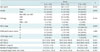
A total of 158 patients with HCC who met the inclusion criteria received TACE from November 2010 to November 2011, including 64 (40.5%) who received TACE with gelatin sponges, 41 (25.9%) with Embospheres, and 53 (33.5%) with DEB. The demographic and clinical characteristics of the patients are summarized in Table 1. The mean follow-up periods for group A, B, and C were 7.3 months (range, 2-17 months), 8.0 months (range, 4-16 months), and 10.8 months (range, 4-16 months), respectively. No significant difference was observed among the three treatment groups with respect to demographic characteristics, etiology of underlying liver disease, liver function, renal function, ECOG performance status, Child-Pugh score, or tumor burden. Significantly (p = 0.039) more group C patients had tumors classified as BCLC stage B compared to group A or B (100% vs. 90.6% or 97.6%) (Table 1).
The doxorubicin dosage, number of complications, laboratory data, and tumor response after TACE are summarized in Table 2. The doxorubicin dose used in group C (median, 50 mg) was significantly (p < 0.001) higher than that used in group A or B (31 mg and 25 mg, respectively). Significantly (p < 0.001) higher levels of AST, ALT, and total bilirubin were observed in group A compared to either group B or C at 48 hours post the procedure. However, post-treatment creatinine ratios were not significantly different among the three groups (Table 2).
Adverse Events
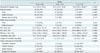
No major AE was observed in group C patients. Minor AEs were significantly (p < 0.001) more common in groups A and B patients compared to group C patients, with rates of 54.7%, 34.1%, and 5.7%, respectively (Table 2). Significantly (p < 0.001) more instances of fever and abdominal pain were observed in group A patients compared to that in group C patients (35.9% vs. 1.9%; 31.3% vs. 3.8%, respectively) (Table 2).
Progression-Free Survival and Tumor Response Over 16 Months
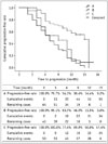
Significantly (p < 0.001) more patients in group C had a CR compared to group A or B (32.1% vs. 6.3% or 2.4%) (Table 2). The Kaplan-Meier survival curves were plotted to describe the progression-free survival rates of the three groups (Fig. 1). The median time to progression for groups A, B, and C were 7, 9, and 15 months, respectively. The 3- and 6-month progression-free rates for groups A, B, and C were 79.7% and 54.3% for group A, 95.1% and 53.7%, and 100% and 77.4%, respectively. Log-rank test showed that group C patients had significantly (p < 0.001) higher progression-free survival rates compared to group A or B.
DISCUSSION
This study demonstrated that TACE with DEB exhibited a better safety profile (fewer AEs) than conventional chemoembolization with either gelatin sponges or Embospheres. In addition, significantly more patients treated with DEB had CR compared to the other two groups of patients. The log-rank test showed that group C patients had significantly higher progression-free survival rates compared to group A or group B patients. Chemoembolization has been used for the treatment of HCC for decades. Its outcomes have been improved by advances in interventional techniques and refinement in patient selection (1). Commonly used embolic agents include gelatin sponges, polyvinyl alcohol particles, and microspheres. Gelatin sponges, the most widely used embolic agents, can be prepared in various forms such as particles, pellets, or fragments. The use of a gelatin sponge alone for embolization results in temporary occlusion of an artery with recanalization taking place within 2 weeks.
Lipiodol is an iodinated ethyl ester of poppy seed oil that selectively remains in tumor nodules from several weeks to over a year (9). A lipiodol-drug emulsion is injected into a vessel supplying the tumor. The anticancer slowly released from lipiodol remains at high concentrations within the tumor for a prolonged period (29). DEB is a drug delivery and embolization system composed of biocompatible, nonresorbable hydrogel beads that can be loaded with chemotherapeutic drugs. Studies have shown that TACE with DEB results in higher tumor drug concentrations and lower toxicity compared to intra-arterial doxorubicin and conventional TACE (18, 20, 30). Song et al. (31) recently reported that TACE with DEB was effective for HCC refractory to conventional TACE, with tolerable adverse effects. Kalva et al. (32) performed TACE with DEB loaded with doxorubicin in 80 patients with advanced stage HCC. They reported that the procedure was safe. ECOG performance status ≤ 1 and > 2 DEB-TACE procedures were associated with better overall survival. Our study represented a unique 3-way comparison among TACE with gelfoam, microspheres, and DEB in the treatment of HCC. Only two randomized prospective studies have previously compared the conventional TACE with DEB-TACE (21, 33). Only a few retrospective studies have attempted to evaluate the effectiveness of DEB-TACE vs. conventional TACE (15, 18, 20, 30, 31, 34). Lammer et al. (21) found that their DEB group showed higher rates of CR, objective response, and disease control compared to conventional TACE. Sacco et al. (33), on the other hand, reported no difference between DEB-TACE and conventional TACE groups in time to recurrence or local recurrence, radiologic progression, or survival. Our results were in consistent with the findings of Song et al. (15) who found that TACE with DEB loaded with doxorubicin offered a distinct advantage in objective tumor response rate compared to conventional lipiodol-based TACE in Asian patients with HCC. Dhanasekaran et al. (34) found a distinct survival advantage of DEB-TACE over conventional TACE in patients with unresectable HCC.
In this study, compared to patients who underwent TACE with gelatin sponges or Embospheres, patients treated with TACE using DEB received a higher dosage of doxorubicin. Although a higher dosage of doxorubicin was used in the DEB-TACE group, the degree of liver toxicity and the number of drug-related AEs were both reduced. A sustained concentration of doxorubicin within the tumor as a result of DEB delivery system might explain the better treatment effect, because minimal systemic efflux of doxorubicin is believed to reduce liver toxicity and drug-related AEs (19). Our results are consistent with many reports in the literature (18, 20, 21, 35, 36). These benefits may result in shorter length of hospital stay due to the improved efficacy and safety profiles of high-dose doxorubicin used in patients undergoing TACE with DEB, regardless of patients' baseline characteristics (15).
The most common complication of TACE is post-embolization syndrome. This syndrome consisting of transient abdominal pain and fever occurs in 60-80% of patients after TACE. Post-embolization syndrome is typically accompanied by an elevation of hepatic transaminase (37). It is unclear whether post-embolization syndrome is a result of damage to the normal liver tissue or tumor necrosis. Post-embolization syndrome is self-limiting within 3-4 days. However, hospitalization may be required for pain control and observation (37).
Based on the results of this study, TACE with DEB appeared to be better tolerated compared to conventional TACE using gelatin sponges with respect to liver enzyme elevation and drug-related AEs. These findings are in consistent with those of previous reports by Recchia et al. (38) and other studies (18, 20, 21, 39). Drug-related AEs were rare in our group C patients, although the dosage of doxorubicin in group C was greater than group A or group B. Embosphere and gelatin sponge had few drug delivery effects. Therefore, more AEs might have occurred if higher dosages were given to patients in groups A and B. A prior study by López-Benítez et al. (40) could support such theory to some degree. Despite the improved tolerability profile of TACE with DEB, interventional radiologists should be aware of potential risks of procedure-associated AEs. They should have sufficient knowledge to manage these complications appropriately (30).
There are several limitations in this study, including its retrospective nature. Previously, only two reports of randomized prospective studies compared lipiodol TACE vs. DEB-TACE (21, 33). All other studies were retrospective series. In addition, our sample size was limited and our follow-up period was too short to determine the potential long-term benefit of chemoembolization with DEB. Future prospective studies involving larger cohorts and longer follow-up periods are needed to confirm our findings.
In conclusion, in patients with HCC, TACE with DEB offered better safety and efficacy profiles compared to TACE using gelatin sponges or TACE with microspheres. Further investigations featuring long-term use of TACE with DEB are merited.
 XML Download