

 PDF
PDF ePub
ePub Citation
 Print
Print


INTRODUCTION
Therapeutic irradiation for head and neck cancer has increased recently and has wide applicability. Heterotopic calcification as a late complication of radiation therapy has been reported in cases of calcification related to radiation therapy, including breast cancer, seminoma, genitourinary malignancies, sarcoma, anal carcinoma, and lymphoma (1-5). Among these, only one case of subcutaneous calcification after radiotherapy in head and neck cancer has been described (6). According to these reports, heterotopic calcification generally develops as a late complication no sooner than 1 year after therapy (1).
Only one case report is available describing the long-term time course of heterotopic calcification following radiotherapy (4). Prevertebral soft tissue calcification as a late manifestation of a head and neck tumor has not been reported.
We present a case of growing prevertebral soft tissue calcification that developed in a patient with previous tonsil cancer who underwent chemotherapy and radiotherapy.
CASE REPORT
A 74-year-old female with rheumatoid arthritis was admitted to the department of orthopedic surgery with pyogenic arthritis in her right elbow. She had a history of chemotherapy and radiotherapy due to tonsil cancer 21 years ago. She was referred for an otolaryngologic consultation due to chronic neck pain and swallowing difficulty on hospital day 7. The patient's complete blood cell count showed leukocytosis of 14.4 × 10³/mm3 (normal range, 4.0-10.0 × 10³/mm3) on admission, which decreased gradually and normalized at the time of the otolaryngologic consultation. Her C-reactive protein level was 16.9 mg/dL (normal range, 0-0.3 mg/dL) on initial examination and had fallen to 5.0 and 2.0 mg/dL on a follow up study performed on hospital days 5 and 8, respectively. No clinical or laboratory evidence of chronic renal failure or hypercalcemia was detected. She had no history of cervical spine trauma or infection. No cervical motion limitation was observed on a physical examination. Pharyngo-laryngoscopic findings were also unremarkable.
A neck computed tomography (CT) scan revealed symmetric fatty atrophy of both the parotid and submandibular glands that seemed to be associated with the previous radiation therapy. Soft tissue swelling in the pharyngeal mucosal space, the retropharyngeal space, and the prevertebral space was observed with obliteration of the parapharyngeal space and fat planes around the carotid sheath. These findings suggested chronic radiation changes with fibrosis. The radiation changes were most prominent at the C2 and C3 levels, so these areas were thought to be included in the radiation field. The chronic neck pain and swallowing difficulty seemed to be due to radiation-induced changes in the pharyngeal mucosa and radiation-induced sialoadenitis. No evidence of tonsil cancer recurrence was detected.
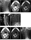
However, prevertebral soft tissue calcification was observed with a curvilinear configuration at the level of C2 and C3 (Fig. 1A-D). The calcification was about 1.4 × 0.6 × 2.1 cm in size. It was irregularly shaped and partly fragmented. Prevertebral soft tissue thickening was noted at the C2 and C3 level (at C2: 9.1 mm; at C3: 8.2 mm). Additional prevertebral calcification was found at the C3-4 and C6-7 intervertebral disc levels, and they were thought to be associated with degenerative changes in the intervertebral discs (Fig. 1D). The C2 and C3 bodies and posterior elements showed osteopenia and coarsening of trabeculae. suggesting radiation osteitis or osteoradionecrosis. Mild degenerative changes were observed in the lower cervical spine. No significant findings suggesting atlantoaxial involvement of rheumatoid arthritis were found.
A neck CT scan obtained 7 years ago demonstrated calcification at the same site (Fig. 1E-G). The calcification was about 0.9 × 0.5 × 1.5 cm in size and was ovoid-shaped and smooth-margined. The retropharyngeal and prevertebral soft tissue swelling was slightly more prominent compared with that of the later study. Findings of C2 and C3 osteoradionecrosis were not significant 7 years ago.
Follow-up C-spine lateral radiography after 1 year showed no change in size or configuration of the calcification (Fig. 1H). Although not histopathologically confirmed, the lesions were thought to be benign because they grew slowly and remained unchanged for at least 1 year. Based on the clinical course and association with previous radiation therapy, the diagnosis was heterotopic calcification in the prevertebral space as a late complication of radiation therapy.
DISCUSSION
Heterotopic mineralization may be due to calcification or ossification. Heterotopic calcification differs from heterotopic ossification. Heterotopic calcification is a condition in which calcium salts are deposited in previously normal or damaged tissues. Heterotopic calcification is called "metastatic calcification", and heterotopic ossification is called "dystrophic calcification". However, the term "ossification" refers to bone formation (calcification in a collagen matrix) (7). The mechanism of heterotopic calcification is not well understood. It can be caused by hypercalcemia, ischemia, trauma, inflammatory metabolic disorders, infection, or hereditary factors. The natural history of heterotopic calcification depends on causative conditions. For example, post-traumatic calcification usually stabilizes and may regress (8), suggesting that heterotopic calcification is not part of the aging process but a result of intracellular and extracellular metabolic processes. Some authors suggest that radiation-induced vascular damage may play a role in intracellular calcium deposits mediated by mitochondria (1, 2).
Radiation damage can be acute, early-delayed, or late delayed. Acute complications include hematologic toxicity, epithelial damage, and edema that appear during radiotherapy. An early delayed complication refers to complications that occur within 6 months to 10 years after radiation exposure. Hyperpigmentation, telangiectasia, atrophic dermatitis, fibrosis, necrosis, and ulcers are well known late complications of radiotherapy (2, 3). They are related to factors including total dose, fraction size, patient age, extent of disease, and pre-existing abnormalities (2). Radiation-induced changes in the head and neck include edema of superficial and deep soft tissues and thickening of the laryngeal and pharyngeal soft tissues in the acute phase. Edema diminishes gradually and progresses to fibrosis in the chronic phase. However, it can persist for several months or years after radiotherapy (9).
Heterotopic tissue calcification as a complication of radiation therapy occurs infrequently. This type of calcification can occur in previously malignant tissue such as a pathologic lymph node in patients with Hodgkin's disease (5). This generally develops no sooner than 1 year after radiation and often more than 5 years later. It seems to be similar to previously normal tissues incidentally radiated in the treatment. According to previous case reports, calcification tends to occur as a late complication of radiotherapy (1, 2, 4, 6). In a study by Carl and Hartmann (1), which is the largest study of heterotopic calcifications as a late radiation effect, the time interval between radiotherapy and the occurrence of heterotopic calcification was > 10 years, except for three cases (median, 19 years; range, 2-31 years). In that study, calcification was generally associated with other severe radiation damage, ulceration, and fibrosis following high radiation doses.
A major differential diagnosis of prevertebral soft tissue calcification is calcific tendinitis of the longus colli muscle, also known as retropharyngeal calcific tendinitis. The longus colli muscle is located on the anterior surface of the vertebral body and extends from the level of the anterior tubercle of the atlas to the level of the T3 vertebral body. In most cases of retropharyngeal calcific tendintis, amorphous calcification involves superior fibers of longus colli muscle tendons at the C1-C2 level. Only a few cases of retropharyngeal calcific tendinitis involving vertical fibers at the C4-C5 level have been reported. Calcific deposits and soft tissue swelling in retropharyngeal calcific tendintis tend to resolve on follow-up imaging (10). However, in our case, calcification had grown over 7 years and remained on a 1 year follow-up. Prevertebral soft tissue swelling had decreased slowly for 7 years, suggesting that the condition is not an acute process.
Ossification of the anterior longitudinal ligament (OALL) should be considered as a possible differential diagnosis. However, in reported cases of OALL (11), the ossifications are closely attached along the anterior aspect of vertebral bodies, whereas our case showed a gap between the calcification and the vertebral bodies.
Only one case report of soft tissue calcification in the neck is available following radiation therapy for laryngeal cancer (6). In that case, calcification was located in the subcutaneous layer of the pectoralis major myocutaneous flap and had been growing slowly for 2-3 months. Similarly, in most of the reported cases, calcification was located superficially, usually in subcutaneous tissues (2, 3). However, our case shows that calcification can also occur in the deep soft tissue included in the radiation field.
In conclusion, we describe the first case of heterotopic calcification in the prevertebral space of the cervical spine as a late complication of irradiation, which increased in size during a 7-year follow-up imaging study. We suggest that this case demonstrates the natural course of heterotopic calcification after radiation therapy.
 XML Download