

 PDF
PDF ePub
ePub Citation
 Print
Print


INTRODUCTION
Eccrine spiradenomas were first reported as rare, benign, adnexal tumors of the skin in 1956 (1). Eccrine spiradenomas typically present as painful, slow-growing, solitary masses on the head or upper trunk, and they usually occur during the fourth or fifth decade of life, and without a predilection for either gender (2). Eccrine spiradenomas in the breast are very rare (2) and little is known about the corresponding radiologic findings. We report here on the case of an eccrine spiradenoma in the breast of a 47-year-old woman and we present the mammography, ultrasound (US) and breast MRI findings.
CASE REPORT
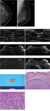
A 47-year-old woman sought evaluation at our hospital for a palpable and occasionally painful lump that had been gradually increasing in size and it had persisted for three years in the left breast. The physical examination revealed a tender, movable, palpable mass in the left upper breast. Mammography demonstrated a well-defined, isodense mass in the left upper center quadrant (Fig. 1A, B). A breast US exam showed three well-defined hypoechoic masses in the subcutaneous layer. The lesions had first been noticed three years previously, and at that time the lesions were thought to be benign masses such as epidermal inclusion cysts on breast US due to the imaging findings and their superficial location in the skin and subcutaneous layer (Fig. 1C). It was recommended that the patient undergo routine follow-up. The current US examination demonstrated an apparent increase in the size of the masses as compared to that seen on the breast US examination performed three years earlier (Fig. 1D), and the lesions were shown to be solid rather than cystic. The patient underwent an US-guided 14-gauge core needle biopsy for pathologic evaluation. The pathologic findings on core needle biopsy were consistent with an eccrine spiradenoma of the breast. A breast MRI exam using a dedicated bilateral breast coil showed low signal intensity on the T1-weighted images, low- to intermediate-signal intensity on the T2-wighted turbo spin echo images, high signal intensity on the turbo inversion recovery magnitude (TIRM) sequence and homogeneous enhancement on the gadolinium-enhanced T1-weighted images (Fig. 1E-H). The patient then underwent surgical excision for pain relief and due to the potential for malignancy. The surgical specimen showed superficially located masses in the breast that each measured approximately 1 cm in size (Fig. 1I). Microscopy demonstrated the presence of masses in the dermis without obvious continuity with the epidermis (Fig. 1J). A histologic examination showed a characteristic biphasic population of outer small cells with darkly staining nuclei surrounding larger cells with pale cytoplasm and tightly packed cells arranged in a jigsaw puzzle pattern without epidermal connections (Fig. 1K). A histopathologic examination confirmed the diagnosis of a benign, adnexal skin tumor in the breast, and this was all consistent with an eccrine spiradenoma.
DISCUSSION
Eccrine spiradenomas are benign tumors of the sweat glands and they are characteristically located in the superficial and deep dermis. Eccrine spiradenomas have no gender predilection, they usually occur on the trunk or extremities, they are 1-2 cm in size and they are occasionally associated with pain and tenderness (3). Eccrine glands are present throughout the skin, but they are most frequent on the palms, soles and axillae. The majority of eccrine spiradenomas involve the trunk and extremities; lesions involving the breast are very rare (4). Of note, malignant changes and systemic metastasis of eccrine spiradenoma of the breast have been reported (2, 3). Malignant changes of an eccrine spiradenoma are extremely rare (2) and they generally arise from long-standing benign eccrine spiradenomas. To diagnose an eccrine spiradenoma, a biopsy should be obtained for pathologic evaluation (5). An eccrine spiradenoma is a highly cellular, cutaneous and subcutaneous tissue tumor with clear margins and it is composed of two types of cells (small, dark-staining basaloid cells and larger pale-staining cells). At low power magnification, a spiradenoma appears as a solid neoplasm composed of a single mass or a few masses of basaliod cells. At higher magnification, two distinct populations of neoplastic epithelial cells can be seen as dark and pale cells (6).
A few classic cases of eccrine spiradenomas have been reported (3, 6). However, the imaging findings of eccrine spiradenomas of the breast have not been clearly demonstrated because of the rare incidence of eccrine spiradenomas and the lack of imaging studies. In 2008, Jin et al. (7) reported the US findings of an eccrine spiradenoma in the upper arm that revealed a well-defined lobulating mass with heterogeneous hypoechogenicity in the deep portion of the dermis and superficial subcutaneous fat layer without connections to the epidermis, and there was no extension into the muscular structures. The imaging findings of our case corresponded well with the imaging findings reported by Jin et al. (7). The US findings of the current case were characterized as well-circumscribed, oval-shaped, hypoechoic masses in the cutaneous and subcutaneous layer, and all the masses were surrounded by echogenic lines that represent the dermal layer, which are findings suggestive of a cutaneous origin. The lesion in our patient was first diagnosed as an epidermal inclusion cyst based on the images and the masses' superficial location. Epidermal inclusion cysts are well-defined, round- or oval-shaped, iso- or hyper-dense masses on mammography; the US findings also include round- or oval-shaped, well-demarcated masses with variable echogenicity (hypo- or hyper-echogenicity) due to variable amounts of keratin debris, and epidermal inclusion cysts are superficially located in the cutaneous or subcutaneous layers. Posterior acoustic enhancements are common, but epidermal inclusion cysts frequently mimic solid masses due to their variable echogenicity. It is difficult to differentiate eccrine spiradenomas from epidermal inclusion cysts based solely on mammographic and US imaging studies due to their similar appearance on imaging and their superficial location. Usually, epidermal inclusion cysts are not recommended for fine needle aspiration or a core needle biopsy because the contents, which can leak out after needle biopsy, often irritate the surrounding tissues and they evokes an inflammatory response, and an abscess may also occur; however, a biopsy is needed to pathologically diagnose an eccrine spiradenoma.
Only one study (6) has reported the MRI appearance of multiple eccrine spiradenomas that were distributed in the trunk, face and extremities. Cystic or solid masses with low signal intensity on the T1-weighted images and high signal intensity on the short T1 inversion recovery images were seen. Thus far, there have been no MRI reports of eccrine spiradenomas in the breast. In the case presented herein, the MR images showed low signal intensity on the T1-weighted images, high signal intensity on the TIRM images and intermediate- to low-signal intensity on the T2-weighted images with homogeneous enhancement on the T1-weighted enhanced images. In the differential diagnosis of epidermal inclusion cysts, Hong et al. (8) reported that subcutaneous epidermal inclusion cysts in the trunk and extremities showed slightly high signal intensity on the T1-weighted images with some bright foci, a high signal intensity background with variable low signal components on the T2-weighted images and peripheral thin rim enhancement on the fat-suppressed T1-weighted enhanced images; the study by Hong et al. (8) is the largest series of MR images of pathologically-confirmed epidermal inclusion cysts. The signal intensities of epidermal inclusion cysts might be the result of the cystic content filled with keratin materials and bounded by a wall of stratified squamous epithelium. An epidermal inclusion cyst is more likely than another subcutaneous lesion when the T2-weighted image shows variable low signal components within the lesion. According to Hong et al. (8) and the current case, the MRI findings of epidermal inclusion cysts and eccrine spiradenomas are different and so these findings might be used in making the differential diagnosis.
In summary, we have described a rare case of a sweat gland tumor in the breast, an eccrine spiradenoma, with the use of imaging modalities, including mammography, US and MRI. Although an eccrine spiradenoma may resemble an epidermal inclusion cyst on mammography and US, there is a difference between the MRI findings of epidermal inclusion cysts and eccrine spiradenomas.
 XML Download