

 PDF
PDF ePub
ePub Citation
 Print
Print


Primitive embryonic anastomoses including the trigeminal, otic, hypoglossal, and proatlantal intersegmental arteries, are temporary communicating arteries between the developing internal carotid and basilar arterial systems during the embryonic period. After the formation of the posterior communicating artery, these arteries are generally obliterated. Rarely, these primitive carotidbasilar anastomoses persist into adult life, with the most common of these the trigeminal artery, followed by the hypoglossal artery. The primitive otic artery (POA) is the rarest of the four anomalous vessels. In this article, a case of persistent primitive trigeminal artery (PTA) and POA associated with an unruptured giant aneurysm located in the right internal carotid artery (ICA) is presented. To the best of our knowledge, no such report exists in the English literature. We describe this rare entity revealed by a digital subtraction angiography and discuss its important clinical significance.
CASE REPORT
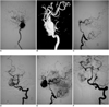
A 54-year-old woman with a right visual field defect was referred to the neurosurgery department of our hospital. Her MRI findings raised the suspicion of the presence of a giant cavernous aneurysm of the right ICA. Consequently, a cerebral angiography and balloon occlusion test were planned. The right ICA angiography disclosed a giant cavernous aneurysm (23×24×27 mm) with a communicating artery, which arose from the sac of the giant aneurysm and flowed to the posterior circulation (Fig. 1A, B). Despite this, the vertebrobasilar angiography was not remarkable (Fig. 1C). Next, a balloon occlusion test was performed in the right ICA. After expanding the balloon, the control angiography disclosed the anterior communicating artery in well patent, in addition to the posterior communicating arteries, which were also were patent but small in size. The basilar artery system was connected with the right internal carotid system by another two persistent anastomoses, which supplied a continuous blood supply to the giant aneurysm and then to the distal end of the right ICA (Fig. 1D-F). Consequently, the right cerebral hemisphere was likely in well perfusion based on these communication channels. Moreover, the patient passed the balloon occlusion test. As for the anastomoses, one arose from the aneurysm sac, near the normal origin of the meningohypophyseal trunk (Fig. 1B), and joined the basilar artery near the junction of the middle and upper thirds (Fig. 1D). As described in multiple studies, this represents the PTA (1-3). Another anastomoses was smaller in caliber than the PTA, and arose from the basal part of the giant aneurysm, but joined the basilar artery at the caudal point (Fig. 1F). Moreover, it was clearly visualized after occlusion of the PTA with detachable coils under general anesthesia on another day (Fig. 1F). The radiographic anatomy was consistent with the criteria of POA as described by Lie (4). In his article, Lie quotes that the POA arises from the carotid artery within the carotid canal, emerges from the internal acoustic meatus, and joins the basilar artery at a caudal point.
Obviously, these persistent anastomoses greatly complicated the endovascular therapy management. A second balloon occlusion test was performed after embolization of the PTA and the POA with detachable coils under general anesthesia. The tests still revealed a negative result. Then, the giant aneurysm was isolated by detachable balloons. The patient was discharged uneventfully on the seventh day following the procedure. Her right visual field defect improved without any evidence of clinically significant thromboembolic events at the six and 12 month follow-up.
DISCUSSION
Carotid-vertebrobasilar anastomoses, referred to as presegmental arteries in the embryonic period, supply blood from the ICA to the primitive vertebrobasilar system. At approximately the fifth week of gestational age, four pairs of presegmental arteries, which are named according neighboring structures, originate from the primitive ICA: the trigeminal, otic, hypoglossal, and proatlantal intersegmental arteries (1). At the sixth gestational week, the posterior communicating arteries develop from the caudal division of the ICA and begin to serve as the communication between the ICA and the arteries of the primitive hindbrain. Then, the normally transient intracranial carotid-vertebrobasilar anastomoses regress with development of the posterior communicating arteries and the basal sphenoid cartilage; beginning by the otic artery, followed by the hypoglossal artery, and lastly the trigeminal artery. After the involution of the other transient anastomoses, the proatlantal intersegmental artery and posterior communicating arteries supply the vertebrobasilar territory until the vertebral system is fully developed (5).
In certain cases, the embryologic carotid-vertebrobasilar anastomoses may persist into adult life. There is no sex predilection for persistent primitive anastomoses and they may be discovered in patients of any age, may be encountered on either side, and may be multiple. However, a persistent trigeminal artery is the most common case and accounts for approximately 80-85% of persistent anastomoses (1, 2). The otic artery only represents a minor contribution to the supply to the developing vertebrobasilar system and is the first presegmental artery to regress. Therefore, the otic artery is rarely present into adult life (1, 2).
However, not all the anastomoses can be disclosed in a routine cerebral angiography. They may exist larvaceously and can only be visualized in the case of a sudden and obvious bilateral pressure change, just as the POA case in this article. Awareness of such neuroanatomical variations is of paramount importance in the diagnosis and treatment in cerebral vascular lesions. These persistent anastomoses were important collateral pathways between the posterior and the anterior circulation. This is especially true in the setting of the carotid or vertebral artery diseases, where these persistent primitive neuroanatomical variations can serve as the compensation conduits.
These primitive carotid-vertebrobasilar anastomoses may cause complications during endovascular therapy or surgical interventions. As for unruptured giant cavernous aneurysms, sac embolization may deteriorate the visual field defect because of the concomitant mass effect. Hence, traditional endovascular approaches, such as aneurysm trapping or ipsilateral ICA occlusion with detachable balloons, may be performed if the patient is tolerant to the balloon occlusion test or an ICA occlusion combined with an extracranial-intracranial bypass operation if the patient cannot pass the balloon occlusion test. This criterion was inadequate in this particular case because of continued blood supply to the aneurysm sac via the PTA and the POA from the vertebrobasilar system. Furthermore, the result of the balloon occlusion test is indefinitely due to the interaction of these persistent anastomoses. Accordingly, a control occlusion test was required after embolization of the PTA and the POA with coils.
Cerebral vascular anomalies, including aneurysms, arteriovenous malformations, and neoplasms, have been described in patients with PTA or other persistent carotid-vertebrobasilar anastomoses in the literature (3, 6-8). Agnoli (9) reported that in 25% of patients with persistent PTA, and in 27% of patients with primitive hypoglossal artery, vascular malformations or a history of subarachnoid hemorrhage were found. However, Cloft et al. (10), reported that aneurysms involving the PTA itself occurred in approximately 2-3% of cases, arising most often from the ICA side of the PTA, which is consistent to the prevalence of aneurysms in the general population. However, most of these dates were based on case reports or groups of small sample size. Therefore, such an association may be dubious or perhaps coincidental. If a database of these variations and cases could be assembled, a metaanalysis could be performed on a satisfactory sample size to refine the present ambiguous classifications.
In conclusion, we report a case of a giant unruptured aneurysm of the cavernous sinus associated with the PTA and the POA in the English literature for the first time. It is important for clinical physicians to recognize these persistent neuroanatomic variations, and also be aware of their clinical implications for surgical and interventional neuroradiologic procedures.
 XML Download