

 PDF
PDF ePub
ePub Citation
 Print
Print


Breast metastases in cases of leukemia are very rare and occur primarily in patients with acute myeloid leukemia. Secondary acute lymphoblastic leukemia (ALL) involving the breast is uncommon (1, 2). We experienced a case of acute T cell lymphoblastic leukemia which was diagnosed by bone a marrow biopsy with breast metastases.
We report the mammographic and ultrasonographic findings of bilateral metastatic breast masses of ALL as well as a literature review.
CASE REPORT
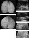
A 30-year-old woman was admitted to the Department of Radiology at Ondokuz Mayis University Hospital due to palpable masses in both breasts. We performed bilateral mammograms using the Mammo Diagnost UC (Philips, Hamburg, Germany) conventional mammography unit. The ultrasonographic evaluations were performed using a 12 MHz linear array probe on Logiq 5 Pro (GE Medical Systems, Milwaukee, WI) ultrasound equipment. The mammograms showed an extremely dense pattern with ill-defined, denser mass-like lesions in upper outer quadrants of both breasts (Fig. 1A, B). An ultrasonographic evaluation revealed lobular-shaped and partly ill-defined hypoechoic masses with a 3-4 cm maximum diameter and a multi-septated nodular (mottled) appearance without acoustic changes (3) (Fig. 1C, D). Both axillae indicated pathologically enlarged lymph nodes with skin thickening, increased echo texture in the breast parenchyma, and fine fluid between the fat lobules, which were consistent with edema of the breasts.
A bone marrow biopsy revealed acute T cell lymphoblastic leukemia. Following chemotherapy, the breast lesions disappeared (Fig. 1E-H). Consistently, the axillary lymph nodes also decreased in number and size.
DISCUSSION
Metastatic breast leukemia is very rare and occurs primarily in patients with acute myeloid leukemia (4-7). An ultrasonography is useful in young patients who usually have dense breasts (1). However, in patients with extreme breast edema, a mammography might not be helpful in representing breast masses. Hence, in such cases, the ultrasonography would be performed as an alternative modality. Previous studies suggest that the reduced diagnostic capability of the ultrasonography may be improved by Doppler flow mapping, by depicting the hypervascular stroma as highly resistive index values (1).
Past studies examining the sonographic features of breast metastases range widely, from hypoechoic to hyperechoic findings (1, 4, 8, 9). The involvement of the breasts in cases of acute myeloid leukemia has been described as areas of mixed echogenicity, with or without acoustic shadowing (2, 10). Another study described the microscopic results of breast involvement in an acute leukemia case study of an autopsy series (1). In most described cases of acute myeloid leukemia involving breasts, the disease appeared bilaterally as multiple nodules (3). In addition, unilateral breast involvement in a case of acute myeloid leukemia could be seen as a single mass (6).
The mammographic findings in cases of leukemia in the breasts can be normal (5, 6), enlarged with diffusely coarse breast parenchyma (2), ill-defined margin with an irregular shaped mass (5, 6), or well-defined bordered lesions with a benign appearance (6).
The extremely rare involvement of breasts in cases of leukemia can be the only complaint at initial presentation or seen as relapse. Moreover, radiotherapy performed on breast malignancies can also cause acute leukemia in breast tissue (2). Following a detailed examination, this condition should be added to the series of findings including lung consolidations, pleural effusion, a mass in the mediastinum, or a hepatosplenomegaly which resembles systemic disease. Our study subject was diagnosed with breast metastases of ALL, which markedly improved as demonstrated by mammographic and sonographic findings following chemotherapy.
 XML Download