

 PDF
PDF ePub
ePub Citation
 Print
Print


Mesenchymal benign tumors originating from the pancreas, such as a fibroma and neurilemmoma, are exceedingly rare. Their incidence is known to be less than 1% of all pancreatic tumors (1). Most benign tumors of mesenchymal origin in the pancreas present on CT and ultrasonography as a sharply-dermarcated mass with or without a cystic portion. A solitary fibrous tumor (SFT) is a type of mesenchymal tumor and is very rare in the pancreas. We report here on imaging findings of an SFT arising from the pancreas.
CASE REPORT
A 54-year-old man was referred to our hospital for further evaluation of an asymptomatic pancreatic mass that had been found incidentally on ultrasonography during a routine health examination at a private clinic. On admission, the patient had neither abdominal pain nor any other symptoms. A physical examination and laboratory data were unremarkable. Levels of tumor markers, including carcinoembryonic antigen (CEA) and carbohydrate antigen (CA) 19-9, were within normal ranges.
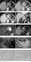
After admission, pancreatic dynamic CT was performed. The dynamic CT demonstrated the presence of a large, well-defined mass with a lobulate contour between the pancreatic body and the lower body and antrum of the stomach. The mass showed heterogeneous isoattenuation with the pancreas parenchyma on unenhanced CT. On contrast-enhanced CT scans, the mass showed progressive enhancement from arterial phase to portal venous phase with a large and multiple small non-enhancing portions and a well-enhancing thin capsule (Figs. 1A-D). The mass was based on the pancreatic body and partial invasion of the stomach was considered. The mass was hypointense to the pancreas parenchyma on T1-weighted MR images and hyperintense on T2-weighted MR images (Fig. 1E). The non-enhancing portions within the tumor on CT revealed a bright signal intensity that was almost isointense to that of the cerebrospinal fluid on T2-weighted MR images (Fig. 1E). On gadolinium-enhanced MR images during the arterial phase, the mass was heterogeneously hypointense to the pancreas parenchyma, and during the portal venous and 2-minute delayed phase, the mass was more enhanced than the pancreas parenchyma and had non-enhancing cystic portions and a well-enhancing capsule (Fig. 1F). MR cholangiopancreatography (MRCP) showed no dilatation of the main pancreatic duct. Regional lymphadenopathy was not observed on the CT and MR images. Endoscopic ultrasonography (EUS) revealed a large, well-defined, ovoid, echogenic mass in the pancreas body (Fig. 1G). The mass had a cystic portion with an irregular internal surface and protruding solid portions. The cystic portion had heterogeneous internal echogenecities. Based on these imaging findings, the mass was suggested to be an exophytic-growing non-functioning islet cell tumor or a solid pseudopapillary tumor (SPT), or less likely, a gastrointestinal stromal tumor of the stomach or neurogenic tumor of the lesser omentum. The patient underwent median segmentectomy of the pancreas and anastomosis of the pancreas and small bowel. At surgery, the mass was noted to arise from the pancreas body and had neither adhered to nor invaded the neighboring organs. The resected mass measured 7.6×6 cm and was an ovoid shaped, lobulate, firm mass with a well-capsulated, smooth margin. The cut surface of the mass was yellowish-gray and smooth with hemorrhage and cystic changes (maximum diameter, 4.5 cm) (Fig. 1H). Microscopically, the mass was a mesenchymal tumor composed of spindle-shaped cells and collagen (Fig. 1I). Based on immunohistochemical analysis, the tumor cells were strongly positive for cluster of differentiation (CD) 99 (Fig. 1J) and vimentin, and were less strongly positive for CD 34, and were negative for cytokeratin, S-100, CD 117, and Ki-67. The tumor was diagnosed as an SFT of the pancreas.
DISCUSSION
An SFT is a submesothelial neoplasm that usually affects the visceral pleura. In 65% of SFTs, they arise from the pleura, but they can also be found at other sites such as the lung, mediastinum, pericardium, mesothelium, peritoneum, extraperitoneal space, nose, and paranasal sinus. The incidence of an SFT is very low (approximately 2.8/100,000). From 1931 to 2002, approximately 800 cases of SFTs in the pleura have been reported in the clinical literature. Over 50% of the affected patients were middle-aged women. These tumors were not associated with exposure to asbestos fibers. SFTs that originate in the peritoneum or mesentery are referred to as extrapleural SFTs. Extrapleural SFTs have been reported to occur slightly more frequently in men than in women, and at a mean age of 54 years (2). The symptoms appear according to the location and size of the mass. The natural history of an SFT is unknown. Grossly, an SFT is a well-defined, firm mass with a pale, cut surface; it may have the appearance of whorled fibrous tissue (2, 3). An intact layer of mesothelium overlying the tumor histologically characterizes an SFT. The tumor is composed of spindle-shaped cells resembling fibroblasts with varying amounts of hyalinized collagen. On radiological images, an SFT appears as a well-defined, solid tumor containing cystic portions (4). An SFT is noted to have low signal intensity on T1-weighted MR images and mixed signal intensity on T2-weighted MR images (5).
Only two cases of an SFT originating in the pancreas have been reported in the clinical literature (1, 6). The tumors were 5.5 cm and 2.0 cm in the greatest diameter, respectively. Ultrasonography revealed a neoplastic enlargement of the corpus of the pancreas and a hypoechoic mass in the pancreatic body, respectively. CT showed a neoplasm with contrast enhancement during both the arterial and venous phases, suggesting an endocrine neoplasm, and a subtly hyperattenuating mass, respectively. There was no pancreatic duct obstruction in both cases. However, radiological images of those cases were not available (1) or were limited (6). In the present case, the tumor was an echogenic mass with cystic portions in the pancreas body as seen on EUS and a well-defined enhancing mass with cystic portions as seen on CT and MR images. These imaging findings are similar to those of pleural or extrapleural SFTs.
If there is a well-defined, solid pancreatic tumor with cystic portions, a non-functioning islet cell tumor or SPT must be differentiated from an SFT (7, 8). A non-functioning islet cell tumor usually manifests as a well-defined tumor which does not invade the peripancreatic vessels. On CT, small non-functioning islet cell tumors show moderate or strong homogeneous enhancement, while large non-functioning islet cell tumors show heterogeneous enhancement with central necrotic cystic changes (7, 8). An SPT is a well-circumscribed mass with a fibrous capsule. An SPT has various extents of hemorrhage or cystic changes within the tumor. Approximately 30% of SPTs have peripheral calcifications, and approximately 20% of SPTs have a fluid-fluid level caused by hemorrhage and cystic degeneration (9, 10).
In conclusion, we report a very rare case of an SFT of the pancreas that appeared as a well-defined mass with cystic portions on radiological images. It is difficult to differentiate an SFT from a nonfunctioning islet cell tumor or SPT of the pancreas.
 XML Download