

 PDF
PDF ePub
ePub Citation
 Print
Print


A side from the small bowel series examination, color Doppler ultrasound (CDUS) findings have been reported as reliable evidence for diagnosing intestinal malrotation based on the "whirlpool" appearance of the omphalomesenteric vessels (i.e., superior mesenteric artery and vein) in adult patients with atypical presentations (1-3). The diagnosis of intestinal malrotation with volvulus by way of the catheter angiography method is no longer the modality of choice, probably due to its invasive nature (4). With the advance of multi-row detector technology, the computed tomography (CT) angiography can reliably be used to demonstrate the vascular findings described by ultrasound. This is the first report, to our knowledge, that depicts both the digital subtraction angiography (DSA) and multidetector CT (MDCT) angiography findings in an adult patient with mildly symptomatic intestinal malrotation proven by a gastrointestinal barium series.
CASE REPORT
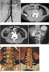
A 37-year-old man was seen for complaints of intermittent abdominal pain, which usually occurred after meals. The initial US and CDUS screening revealed counterclockwise superior mesenteric vein twisting around the superior mesenteric artery (SMA). However, the etiology underlying this appearance was not recognized and the patient was referred for an angiography due to the suspicion of a congenital vascular anomaly or variation as a reason to explain his post-prandial abdominal pain. In the catheter angiography study, the SMA was found to be twisted (Fig. 1A). Subsequent to this study, the patient was taken into suites for upper GI series and CT examinations, with the suspicion of intestinal malrotation. Although the angiography was fairly diagnostic, a MDCT was performed to document the state of the superior mesenteric vein and the vascularity of the subtended bowel. A CT angiography was performed with a 16-detector row computed tomography (Philips Medical System, MX8000IDT, Haifa, Israel) with the following parameters; kV: 100, mAs: 158, collimation: 16 × 0.75 mm, slice thickness: 1 mm, slice increments: 0.5 mm, and pitch: 0.9. One hundred twenty milliliters of Iohexol 300 mgI/ml (Omnipaque, Amersham Health, Cork, Ireland) was administered via the antecubital vein at a flow rate of 4 mL/sec. In addition, the automated bolus tracking method was used for imaging in the arterial phase. Moreover, portal venous scans were obtained with a 60-second delay after the initial injection. The arterial phase axial CT images showed that the superior mesenteric vein and artery had an inverse relationship. The three-dimensional reformatted images from the CT angiography demonstrated the mesenteric vessels to be rotating with an eventual tortuous and dilated superior mesenteric vein (Figs. 1B-E). Otherwise, the CT imaging results were normal, except for the absence of the uncinate process of the pancreas. This was likely related with the well-known generalized problem of counterclockwise rotation in malrotation patients, which is the uncinate process formed from the ventral pancreatic bud after its rotational migration around the duodenum and fusion with the dorsal pancreas. The small bowel series revealed that the duodenojejunal junction was on the right side of the midaxis and inferior to the duodenal bulb. The proximal jejunum was also observed to be rotated around its axis (corkscrew sign) (Fig. 1F). As a result of this diagnosis, the patient was offered surgical correction of the volvulus and release of predisposing fibrous bands or adhesions that might have developed in the long run secondary to the congenital malrotation (Ladd's procedure). However, the patient did not consent to surgery and was lost to follow-up.
DISCUSSION
The condition of intestinal malrotation is characterised with a narrow mesentery and poor fixation of the midgut, as well as representing a major diagnostic challenge in children and moreso in adults. Midgut volvulus followed by subsequent bowel necrosis secondary to the torsion of the mesenteric vessels is a complication of intestinal malrotation mostly observed in infancy. However, not all midgut malrotation cases have full-blown obstructive volvulus. In such patients, vague symptoms extending till adulthood have been reported (5), as is the case with our case study. The incidental discovery of intestinal malrotation is not uncommon in adults (6). This is probably related with the nature of this developmental abnormality since it has a wide spectrum of patterns, which also vary from one author to another. A simple and reasonable way of classifying intestinal anomalies of rotation and fixation would be as non-rotation, malrotation (incomplete rotation), and reversed rotation (7, 8). The pathogenetic mechanisms underlying this spectrum basically encompass the timing of return of the midgut into the peritoneal cavity after certain degrees of rotation (less than the normally expected 270° counterclockwise) during intrauterine developmental stages. The interested reader should refer to Berrocal et al. or Long et al. for the details of this embryopathogenetic process, which are beyond the scope of this report (7, 8). More relevant with this case report, is the positioning of the omphalomesenteric vessels (future superior mesenteric artery and vein) in relation to the malrotating intestine.
Since the normal 270° rotation of the midgut during embryological development is "counterclockwise" (as observed on axial images from superior to inferior), twisting of the superior mesenteric vein on superior mesenteric artery in this direction may have a normal radiographic appearance or variant (9). However, the "clockwise" whirlpool or barber's pole sign is a color Doppler US and angiography finding of symptomatic intestinal malrotation and describes the twisting of the superior mesenteric artery and vein in the opposite direction as an end-result of midgut volvulus (which happens in the clockwise direction secondary to shortened mesenteric attachment and poor fixation of the intestines) in such patients (1, 2, 4). In addition, the twisting of the dilated mesenteric vein on the mesenteric root has been reported as a sign of malrotation with volvulus (4). In our case study, the imaging findings are consistent with the aforementioned configuration abnormalities of the mesenteric vessels.
Although there is no report on the utility of the MDCT angiography to demonstrate the malrotation of the mesenteric vessels, there are axial CT studies that have been used to diagnose either the intestinal complications of malrotation like volvulus and herniations, or the vascular complications like twisting or malpositioning of the mesenteric vessels in relation to each other and subsequent bowel ischemia (10, 11).
The "whirl sign", as seen on abdominopelvic CT examinations, refers to a whirling or spiral shape of the mesenteric vessels, which may or may not accompany the intestinal loops and their feeding vessels. The sensitivity in the detection of intestinal malrotation is high and its specificity with regard to symptomatic volvulus increases when accompanied with intestinal loops and the whirling vessels feeding them (12). As an ultrasound counterpart of it in the imaging of intestinal malrotation, one can see whirling mesenteric arteries and veins (as in our case), or inverted positions of the vessels in relation to each other (e.g., superior mesenteric vein on the left and the mesenteric artery on the right or vein positioned anterior to artery, at least) (13). The specificity of this sonographic sign with respect to the midgut volvulus is high if the symptoms are accompanied with a fully whirling mesenteric vessel pattern. Moreover, the specificity of the diagnosis decreases only if the anterior position of the vein is noted (14).
Sometimes, the prognostic significance of the findings from the upper gastrointestinal tract examinations are unclear and the ultrasound has been suggested as perhaps a more reliable means of diagnosing intestinal malrotation with volvulus (15). However, the subjective US and objective documentation with a catheter angiography or MDCT angiography (plus 3D reconstruction) would be more acceptable for a clinician who seeks an objective demonstration of the process before deciding on whether surgery is the most viable option. Although the angiography was fairly diagnostic, we performed a MDCT to document the state of the superior mesenteric vein and the vascularity of the subtended bowel. The three-dimensional reconstruction assisted MDCT angiography correlated, accompanied with a catheter angiography as a means to demonstrate midgut malrotation of the mesenteric vessels in the had not been reported so far.
In conclusion, although a DSA is not feasible in every suspected case of intestinal malrotation due to its invasive nature and high cost, a MDCT angiography is a good way of preoperative evaluation and an alternative to depict both the clockwise "whirlpool" and clockwise "barber's pole" intestinal configuration in patients with volvulus secondary to malrotation.
 XML Download