

 PDF
PDF ePub
ePub Citation
 Print
Print


Castleman disease, or giant lymph node hyperplasia, is a rare disorder most often located in the visceral mediastinum (1, 2). We present a patient in whom a double-contrast esophagography and contrast-enhanced computerized tomography (CT) revealed a polypoid mass in the esophageal wall that, based on pathology, was diagnosed as a Castleman disease.
CASE REPORT
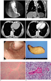
A 62-year-old man was referred to our hospital for further examination and treatment of an asymptomatic esophageal mass (1, 2). At the time of admission, he had no dysphagia or any other digestive tract symptoms, but an endoscopic examination for a routine medical health exam showed a protruding mass in the distal esophagus. He was a heavy alcohol drinker. A physical examination and laboratory data showed mild hepatic dysfunction, and an abdominal sonography revealed a liver with heterogeneous echotexture and blunt margins, suggestive of chronic liver disease. A double-contrast esophagography showed a 2.5×6 cm polypoid mass arising in the distal esophageal wall, located just above the gastroesophageal junction (Fig. 1A). Contrast-enhanced chest CT demonstrated a 2.5×6 cm, well-defined lesion in the esophagus. A coronal image showed that the tumor was located in the esophagus and showed a well-circumscribed soft-tissue density mass with a homogeneous enhancement after the injection of contrast material. The mean density of the mass was 50 HU at precontrast scan and 100 HU at postcontrast scan. CT depicted no lymph node enlargement in the mediastinum or upper abdomen (Figs. 1B-D). Endoscopic observation revealed one column of elevated sausage-like lesion at approximately 28 cm from the incisor teeth of the esophagus, a possible varix, leiomyoma, or submucosal hemorrhage. The surface of the proximal portion of the tumor was slightly reddish, but smooth and covered with normal mucosa (Fig. 1E). Preoperative preliminary diagnosis was an esophageal submucosal lesion, and a leiomyoma was strongly suspected based on the incidence of such lesions. After incision of the esophageal muscle layer, the mass was exposed and enucleated from the esophageal wall. The lesion was located in the submucosal layer of the esophagus. The specimen was reviewed by two pathologists (with 10 and 25 years of experience in pulmonary and lymphoreticular pathology, respectively) and they agreed with the diagnosis of Castleman disease. The excised mass showed a multilobulated mass with whitish color and a smooth margin (3×3×7 cm) at the distal esophagus (27-34 cm from incisor). On sectioning, the mass had a yellowish gray multinodular appearance (Fig. 1F). Microscopically, the esophageal mass revealed nodular lymphoid areas, a marked expansion of the mantle zone, and small, relatively inconspicuous germinal centers (Fig. 1G). The follicles show marked vascular proliferation with hyalinization (Fig. 1H). The final diagnosis of this tumor was Castleman disease of the hyaline vascular type, lymphoid subtype.
DISCUSSION
Castleman disease is a disease of unknown etiology, and also referred to as angiofollicular lymph node hyperplasia, angiomatous lymphoid hamartoma, and giant mediastinal lymph node hyperplasia. Based on histological differences, the disease has been divided into two variants, and the hyaline vascular variant comprise 91% of cases and is usually asymptomatic. Typically this variant appears as a hypervascular mass with strong enhancement on CT. The plasma cell variant of Castleman disease is often concomitant with systemic manifestations, such as fever, anemia, hypergammaglobulinemia, and increases in acute phase proteins (3).
Pathologically, the hyaline vascular type Castleman disease is characterized by small hyaline vascular germinal centers and interfollicular capillary proliferation. However, the proportion of the two components - abnormal lymphoid follicles and increased interfollicular vascularity - may vary, from cases featuring predominantly large mantle zones with inconspicuous germinal centers ("lymphoid variant") to cases with a predominantly vascular component and fibrosis (4).
The most common locations of Castleman disease are the thorax (63%), abdomen (11%), and axilla (4%), although extrathoracic sites have been reported in the neck, axilla, shoulder, orbit, pelvis, pancreas, leptomeninges, and retroperitoneum (3, 5-8).
There are some reports of gastrointestinal involvement in castleman disease, such as gastric or colonic erosion, gastric outlet obstruction, intestinal obstruction or, rectal cancer (1, 2). Most cases are caused by pressure from abdominal, retroperitoneal lymph node enlargement. To our knowledge, Castleman disease of the esophagus has not been previously reported in the literature. There were two reports of mediastinal Castleman disease with esophageal varices, which formed in response to copious blood flow (because of increased blood drainage) from the angiomatous tumor to the esophageal veins (9, 10). In our case, esophagography and CT manifestations were similar to those considered characteristic of leiomyoma. It seems to be difficult to diagnose submucosal Castleman disease without any histological findings.
In conclusion, the imaging findings of Castleman disease localized in the esophagus were homogeneous well enhancing mass at CT. From a clinical standpoint, establishing an accurate preoperative diagnosis is difficult, and Castleman disease should be included in the differential diagnosis of asymptomatic, well-enhancing esophageal mass.
 XML Download