

 PDF
PDF ePub
ePub Citation
 Print
Print


INTRODUCTION
Breast cancers are increasing rapidly worldwide, with a 20% increased incidence in 2012 compared to that in 2008. Moreover, in Korea, breast cancer incidence in 2000 was 5906 and has increased steadily, and reached 18381 in 2014 (12). Therefore, breast cancer is the second most common cancer among women in Korea, following thyroid cancer. According to the Ministry of Health and Welfare's National Cancer Registration Project, breast cancer accounted for 19.3% of all cancers in female patients in 2014 (3). This increase in breast cancer incidence in Korea is probably caused by the effects of Westernized diets including high amounts of fat and calories, and a rise in total estrogen exposure due to decreased frequency of pregnancy and breastfeeding, as well as increased age at childbirth, early menarche, late menopause, and obesity. With public interest in women's health rising, breast cancer screening has become a part of the government-sponsored cancer-screening program. As a result, the frequency of early detection of breast cancer has increased. The proportion of patients with early breast cancer (stage 0 or stage 1) from 32.6% in 2000 to 60.6% in 2015 (4). As the number of screening programs has increased, the number of breast imaging examinations has also increased. Although the number of breast examinations has risen, there have been no reports of actual practices in breast imaging in Korea, for example, the daily workload, standardization of imaging interpretation, performance of quality assurance (QA), medical auditing, and so on. The purpose of our study was to report the current practices in breast imaging among Korean radiologists.
MATERIALS AND METHODS
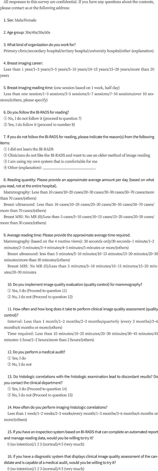
From October 2017 to December 2017, we invited 312 members of the Korean Society of Breast Imaging (KSBI) working as breast radiologists to participate in a survey by e-mail on the current practices in breast imaging. An e-mail message from the investigators with a link to a web survey was sent to members of the KSBI with known e-mail addresses via a list server. Follow-up emails were sent to non-respondents. Follow-up telephone calls were not made. The survey instrument was a 21-question online questionnaire implemented through the SurveyMonkey online survey tool (SurveyMonkey, Portland, OR, USA). The questions were designed by two investigators and revised several times before the administration of the instrument to ensure question clarity and utility. This survey consisted of 21 questions divided into four categories (Appendix 1): questions about the baseline characteristics of the respondents, daily practices in breast imaging, performing medical audits and QA, and interest in a dedicated breast imaging reading system incorporating QA and medical auditing.
RESULTS
BASELINE CHARACTERISTICS OF THE RESPONDENTS
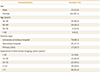
One hundred and sixteen individual responses to the survey were received. Most respondents were below 50 years of age (82.7%) and women (87%). The types of hospitals that the breast radiologists who participated in the survey worked at were as follows: 79 (68.1%), university or tertiary hospitals; 20 (17.2%), secondary hospitals; and 17 (14.7%), primary clinics. Four respondents whom were classified as working in primary clinics mentioned that they are worked at screening centers. The respondents' experience during their breast imaging career was over 20 years in 11, 10–20 years in 34, 5–10 years in 34, and 5 years or less in 36. One respondent skipped this question. Table 1 shows the detailed baseline characteristics of the respondents.
DAILY WORKLOAD
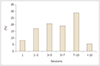
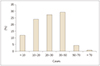
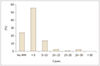
Regarding dedicated working time in breast imaging, most radiologists commonly worked 7–10 sessions per week (32, 28.8%) followed by 3–5 sessions (23, 20.1%) (Fig. 1). One-hundredfourteen of 116 (96.6%) respondents followed the Breast Imaging Reporting and Data System (BI-RADS). The physicians in their 50s and 30s working at the primary institution did not use BI-RADS because they used another system more familiar to them. The daily numbers of mammograms read were 30–50 for 34 radiologists (29.3%), 20–30 for 32 (27.6%), 10–20 for 28 (24.1%), fewer than 10 for 16 (12.1%), and over 70 for one (0.9%) (Fig. 2). The daily numbers of breast ultrasound examinations performed were 10–20 for 50 radiologists (43.1%), 20–30 for 32 (29.3%), fewer than 10 for 25 (21.6%), and 30–50 for seven (6%) (Fig. 3). The daily numbers of breast MRI scans read were less than five for 65 radiologists (56%), 5–10 for 16 (13.8%), 10–15 for three (2.6%), 20–30 for three (2.6%), 15–20 for one (0.9%), and zero for 28 (24.1%) (Fig. 4).
The average reading durations for mammography were 1–2 min for 50 radiologists (44.6%), 30 sec to 1 min for 35 (31.3%), 2–3 min for 14 (12.5%), less than 30 sec for 7 (6.3%), 3–4 min for three (2.7%), 4–5 min for three (2.7%). Four radiologists skipped the question. The average duration required to perform and interpret ultrasound scans were 5–10 min for 50 (45.5%), 10–15 min for 28 (25.5%), less than 5 min for five (23.6%), and 15–20 min for six (5.5%); six radiologists skipped the question. The average reading durations for MRI were 10–15 min for 33 radiologists (39.3%), 5–10 min for 29 (34.5%), 15–20 min for 12 (14.3%), less than 5 min for four (4.8%), while 32 did not perform MRI interpretation.
MEDICAL AUDITING AND QA
Most respondents (91, 82%) performed QA for mammography, and 20 (18%) did not; five respondents skipped the question. The intervals for performing QA were every 3 months for 57 radiologists (63.3%), 3–6 months for 14 (15.6%), less than 3 months for 13 (14.4%), 1 year for four (4.4%), 6–12 months for two (2.2%), and one respondent skipped the question. The average durations for performing QA were less than 10 min for 29 radiologists (34.9%), 10–20 min for 27 (32.5%), 20–30 min for 19 (22.9%), 45–60 min for five (6%), 30–45 min for three (3.6%), and eight skipped the question.
In contrast, most respondents (66, 61.1%) did not perform a medical audit. Forty-two (38.9%) performed medical auditing and nine skipped the question.
Most respondents (83, 76.9%) performed imaging-histologic correlation and discussed the findings with the clinician in cases of discordance; 25 (23.2%) reported not performing imaging- histologic correlation and eight skipped the question. The intervals for performing imaging- histologic correlation were 1–2 weeks for 37 radiologists (41.1%), less than 1 week for 30 (33.3%), 2–3 weeks for eight (8.9%), 1 month for six (6.7%), 1–3 months for two (2.2%), 3–6 months for one, over 6 months for 1, no regular interval for five, and 26 skipped the question.
INTEREST REGARDING A DEDICATED BREAST IMAGING READING SYSTEM INCORPORATING QA AND MEDICAL AUDITING
Most respondents (108, 97.3%) were willing to try out a BI-RADS-based interpretation system that can complete automated reports and manage data that have been read, and three (2.7%) were unwilling; five skipped the question.
Most respondents (109, 97.3%) were willing to use a reading system that displays candidates for clinical image QA, and is capable of medical auditing and three (2.7%) were unwilling; four skipped the question.
DISCUSSION
According to the statistical data provided by the Health Insurance Review and Assessment Service in Korea, breast MRI frequency increased twofold from 27072 cases in 2013 to 40286 cases in 2016, and breast ultrasound frequency increased more than tenfold from 12004 cases in 2013 to 126132 cases in 2016 (5). Despite a dramatic increase in the frequency of breast cancer imaging owing to the increased breast cancer prevalence and public and media attention toward breast cancer, there has been little consideration of medical auditing, or other issues related to breast imaging interpretation.
From this survey, most of them (28.8%) worked 7–10 sessions per week. The median daily workload was 30 routine four-view mammography procedures, 20 whole-breast ultrasound procedures, and five breast MRI procedures. An exact comparison of this workload to those in other countries is not possible because our performance and medical insurance systems are different. However, when the data were converted to work relative value units (RVU) from the USA, then 45.55 RUV would be the 50th percentile according to the Association of Administrators in Academic Radiology (AAARAD) survey conducted in the fiscal year 2016 (67). When converting individual responses to RUV, the mean was 50.8 RUV (range, 7.6–123.4; 114 responses available). The mean RUV at a university hospital or tertiary hospital (58.1, n = 79) and mean RUV at a primary or secondary hospital (33.1, n = 35) were statistically significantly different (p < 0.001, independent t-test). In reference to the AAARAD survey from the fiscal year 2016, 39 RUV/day was the 25th percentile, 47 was the 50th percentile, 59 was the 75th percentile, and 75 was the 90th percentile. The survey responses showed that the lower 36% corresponded to the 25th percentile, 14% to the 50th percentile, 33.3% to the 75th percentile, and 15.8% to the 90th percentile (67). These comparisons did not include biopsies with higher RUV than the routine image interpretation; thus, the true RUV for Korean breast radiologists are greater than that shown in this survey's results. Moreover, biopsy is more commonly performed at university and tertiary hospitals, and the difference in RUV values between hospital types is more significant.
Among the respondents, 18% did not perform QA. This is because there was another colleague responsible for QA. The most common interval was 3 months because the clinical imaging evaluation and phantom imaging evaluation performed by physicians were done in this time interval. Over 80% of respondents were conduction QA because of the legal requirement for the mammography QA; however, medical auditing was performed by fewer than 40%. Fortunately, 76.9% of respondents performing imaging-histologic correlation discussed findings with clinicians in discordant biopsy cases. The false-negative rate of breast imaging, which is the main problem in breast cancer screening, is known to be 10–30%, and it is known that the density of the breast parenchyma (breast density), the quality of mammography, and the quality of the reader affect mammography accuracy (8). In May 2011, the Ministry of Health and Welfare and National Cancer Center conducted a “Cancer Conquest Forum,” which revealed that only 0.6% of patients with cancer were found to have breast cancer (9). As a measure to supplement the low breast cancer detection rate in national breast cancer screening, continuous education is being provided for mammography screening, but no medical auditing has been conducted for the readings. In the United States in the early 1980s, the Quality Assurance Certification Program for mammography institutes was initiated under the leadership of the Society of Radiation Oncology and with the Food and Drug Administration's approval. In 1992, the mammography quality standard law was enacted and the mammography facility, radiation dose, worker regulations, regular image quality evaluations, and certification systems were implemented (10). The content of the breast readings should be qualified by examinations only for those who have been educated for a certain period of time or more; this stipulates rigorous requirements for equipment, standardizes reading methods, daily, weekly, 3 months, 6 months, designates one person who is responsible for image quality management for each institution, designates one radiologist who is responsible for image quality management activities, and conducts medical audits of the reading results (10). Medical auditing provides an ideal target for mammography screening results, includes the accuracies of surveillance by the radiologist and the medical audit, and notifies the patient of the results. As a result, in the United States, improvements in quality control compared to that in the 1960s increased the incidence of breast cancer detected by mammography compared with findings by promotion, and especially the discovery of small breast cancers less than 1 cm was increased (1112). In Korea, regulations were revised and newly established on the safety management of diagnostic radiological apparatus by the Ordinance of Health and Welfare on January 13, 2001, and include matters concerning X-ray field examinations, the compression band size test, the average wire dose test, the phantom image evaluation test, and so on.
Image quality control for mammography has been implemented in Korea since 2004, but the inspection standard is not strict compared to that in the USA. Furthermore, there is no regulation of medical auditing. During the last 20 years, there has been greatly increased breast imaging according to the numbers; however, presently, improvement of quality is warranted.
This study has several limitations. First, only 37.2% of candidates responded. Further, this study may demonstrate selection bias, because only those who responded voluntarily were involved. In addition, many respondents worked in university or tertiary hospitals, and thus the results are not reflective of the practices in primary clinics. In addition, since most respondents were working in referral hospitals, most of the examinations were probably performed for diagnostic purposes; however, the proportion of diagnostic and screening examinations have not been investigated. Second, the survey was distributed only to KSBI members. The practices of other doctors including breast surgeons in large, private breast clinics were not reflected, and hence it is difficult to represent all the current breast imaging practices. Third, in the comparison of breast radiologist workloads, we could not reflect differences in the number of patients or breast radiologists in each hospital and did not include tasks such as biopsy, outpatient consultations, or conferences.
Despite these limitations, this study could be important in providing information on the current practices of breast imaging in Korea including how imaging is performed and interpreted, and the performance of QA, medical auditing, and imaging-histologic discordance. The employing institutions and working patterns of breast radiologists were diverse. Most respondents followed BI-RADS when interpreting breast imaging and performed QA and imaging- histologic correlation, although many were not conducting medical audits.

 XML Download