

 PDF
PDF ePub
ePub Citation
 Print
Print


INTRODUCTION
Breast lymphomas constitute about 0.04 to 1% of malignant mammary neoplasms; approximately 1.7% to 2.2% of all lymphomas occur in the breast. Breast lymphoma may occur as a primary extranodal involvement of the breast or as secondary infiltration by systemic disease at the time of either initial diagnosis or disease recurrence. Primary breast lymphoma in a male patient is a rare clinical entity and altogether, only 1% of all malignant breast tumors occur in male patients. Patients having either primary or secondary immunodeficiency states are predisposed to developing non-Hodgkin's lymphoma (1). Here, we report a case of primary breast lymphoma in a secondary immunocompromised male patient.
CASE REPORT
A 63-year-old male noticed a palpable mass in the left breast for one month. He had undergone a living donor kidney transplant from his spouse 11 years earlier. His immunosuppressive regimen included cyclosporine and prednisolone. On clinical examination, there was an approximately 2.5 cm sized, painless, palpable, mobile mass in the left breast. The patient did not have ipsilateral axillary lymphadenopathy. The hilar lymph nodes were not visible on chest radiography. Mammography was not performed for this patient.
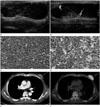
Ultrasound (US) was performed with a 12-5 MHz probe (IU22, Philips Medical Systems, Bothell, WA, USA) equipped with broadband linear array transducers. A 5 cm heterogeneous, hypoechoic mass was located in the left subareolar area. The mass had an oval, circumscribed margin without any posterior acoustic enhancement. Increased vascularity was seen within the lesion on color Doppler examination (Fig. 1). There was no abnormal US finding in the right breast or axillary fossa.
To distinguish breast lymphoma from invasive breast cancer in male patient, we next performed US-guided core-needle biopsy. Four passes were performed with a 14-gauge automated gun biopsy. The pathological diagnosis was diffuse large B cell lymphoma with CD20 and CD3 positive, and EBV mRNA negative on immunohistochemical analysis (Fig. 1D).
After a confirmed diagnosis of lymphoma, multi-detector computed tomography (MDCT) and positron emission tomography (PET)/computed tomography (CT) scans were applied for grading the disease. On contrast enhanced MDCT scan (Brilliance CT 64-channel, Cleveland, OH, USA), a left subareolar mass measuring 4.5 × 2.0 × 4.1 cm was noted (Fig. 1E). The lesion was a solitary lobular mass with well-defined margins, showing moderate homogenous contrast enhancement.
The 18F fluorodeoxyglucose PET/CT (Biograph 6, Siemens Medical Solutions, Malvern, PA, USA) scan showed an intense homogeneous hypermetabolic mass involving the left subareolar region of the breast with a maximum standardized uptake value of 14.40 (Fig. 1F). There was no evidence of abdominal, mediastinal, or cervical lymphadenopathy.
The patient underwent chemotherapy with a rituximab-cyclophosphamide, doxorubicin hydrochloride, vincristine sulfate, and prednisone regimen. After 10 months of follow-up CT and PET/CT scans, he achieved complete remission. There was no evidence of recurrent disease on follow-up examinations, until he experienced lower motor weakness after a month of complete remission. Brain CT and MRI were performed for evaluation, and they revealed a brain metastasis.
DISCUSSION
Lymphoma is a malignant disease arising from the lymphatics. Breast lymphoma is classified into three major groups, based on their diagnosis: primary breast lymphoma (44%), breast involvement from disseminated disease at the time of initial diagnosis (29%), and recurrence of preexisting lymphoma to the breast (27%) (2). Primary breast lymphoma is an uncommon disease accounting for 0.85 to 2.2% of extranodal lymphomas. Secondary breast lymphoma is more common than primary breast lymphoma. Clinical criteria for the classification of primary breast lymphoma are as follows (3): 1) adequate pathologic evaluation, 2) mammary tissue in close association with lymphomatous infiltrate, 3) no evidence of disseminated lymphoma, other than simultaneous ipsilateral lymph node involvement, 4) no prior diagnosis of lymphoma. In the present case, only a left subareolar breast mass was present, without involvement of any other lymph nodes, thus indicating a primary breast lymphoma.
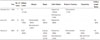
To the best of our knowledge, there have been only three original articles published about the US features of breast lymphoma (Table 1) (456). Yang et al. (6) reported the largest series of primary breast lymphoma cases with US features, including 22 tumors. Of all evaluated tumors, the most common US features were solitary (79%), irregular shape (45%), indistinct margins (59%), hypoechoic (59%), lack of posterior acoustic phenomena (64%), and tumor vascularity (64%). Liberman et al. (4) reported the common US imaging characteristics in eight cases of non-Hodgkin lymphoma of the breast. Lymphomas were commonly solitary (71%), hypoechoic (100%), homogeneous (75%), and exhibited posterior acoustic enhancement (71%). Lyou et al. (5) reported that patients presented with a solitary (75%), hypoechoic (87.5%), oval-shaped (50%), circumscribed mass with microlobulated margins (50%), and posterior enhancement (75%). These findings coincided with some common reported US findings of breast lymphoma. In agreement with these previous studies, US features of the current case included an oval, circumscribed mass with hypoechoic, heterogeneous echogenicity, and without posterior acoustic phenomena. There is no general agreement, however, regarding the US features of primary breast lymphoma in male patients. These features should be clarified in future studies.
Of the differential diagnoses considered based on US features, invasive ductal carcinoma represents the majority of male breast malignancies. Most invasive ductal carcinomas in male patients present as unilateral, eccentric, subareolar, lobulated, hypoechoic masses. However, unlike invasive ductal carcinoma, primary breast lymphoma usually does not exhibit calcification, surrounding parenchymal distortion, or spiculated margins. Concordant with the previous studies, our case did not demonstrate intratumoral calcification or surrounding architectural distortion.
Post-transplant lymphoproliferative disorder represents a heterogeneous group of abnormal lymphoid proliferations that occur after solid organ transplant or hematopoietic transplantation (7). Although the frequency of all cancers does not always increase post-transplantation, the incidence of breast cancer in women is 1.1 fold higher in the first 3 years after kidney transplantation than in the general population (89). Breast cancer in male transplant recipients is 4.0 fold higher than in the general population (9); the risk of lymphoma after transplantation increases 20-30 times (18). The current patient was in an immunosuppressive state for 11 years after renal transplantation, and therefore lymphoma was included as a possible diagnosis.
CT is a reliable, popular tool for grading of lymphoma. However, recognition of a positive lymph node, or detection of bone marrow or extranodal tissue involvement, may be limited on CT. Because PET/CT can indicate the overall level of metabolic activity of lymphoma, it has a greater sensitivity for extranodal involvement than CT alone (10). In this case, transverse MDCT images showed a homogeneous enhancing mass in the subareolar portion of the left breast that corresponded to lymphoma. Transverse PET/CT scans revealed hypermetabolism in the left breast. Absence of uptake in the abdominal, mediastinal, or cervical regions indicated the absence of systemic disease or distant metastases. MDCT and PET/CT are important in the diagnosis of primary breast lymphoma and the exclusion of sec-ondary breast lymphoma.
Microscopic examination of the tumor with US-assisted gun biopsy revealed CD20-positive large atypical lymphocytes. The breast tumor in our case was diagnosed as diffuse large B cell lymphoma. Major surgical (excisional or open) biopsy can be avoided through use of US-guided CNB.
In conclusion, although primary breast lymphoma in a male patient is extremely rare, it should be included in the differential diagnosis of breast masses in immunocompromised male patients.
 XML Download