

 PDF
PDF ePub
ePub Citation
 Print
Print


INTRODUCTION
An epithelioid hemangioendothelioma is an extremely rare tumor classified as an intermediate grade vascular tumor. This tumor can arise in soft tissue and deep organs such as the lung and liver, and in rare cases, within bone (1). In this article, we describe a case of epithelioid hemangioendothelioma of the femur that was mistaken for a benign lesion, such as a simple bone cyst and fibrous dysplasia, because the tumor showed an unusual cystic appearance on MR imaging which to the best of our knowledge has not yet been reported in the current literature.
CASE REPORT
A 77-year-old female presented with pain and swelling in her right thigh after falling down and complained of resting pain of the right thigh for 2 years before the time she was injured. A plain radiography showed a comminuted fracture with displacement in the middle of the femur shaft and an underlying osteolytic lesion of the femur shaft. She did not have any underlying disease or history of cancer. She had total knee replacement arthroplasty of the right knee 11 years ago and the radiograph obtained at that time was not available.
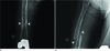
Radiography of the right femur demonstrated an osteolytic lesion involving almost the entire right femur, except for the head and trochanters. The lesion was confined to be in the medullary cavity. Cortical bone was markedly thinned, but there was neither cortical disruption nor a periosteal reaction. The lesion showed a lobulated contour and was septated into multiple lobules by thin bony septae. The border was well demarcated and slightly sclerotic (Fig. 1).
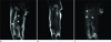
MR images of the right thigh showed a mass, measuring about 2.8 × 2.4 × 29 cm, replacing most of the normal medulla of the femur. The mass appeared to arise from the medullary cavity and extend to the adjacent cortex. There was no apparent soft tissue involvement, but focal cortical disruption was noted on a few axial images. The margin of the lesion was distinct and lobulated. The mass was septated by incomplete thin septa. Femoral shaft fracture and associated findings such as intraosseous hemorrhage and soft tissue edema were found around the fracture site. Regarding signal intensity, the mass was isointense to muscle on the T1-weighted image and hyperintense on the T2-weighted image. The rim and septum were hyperintense on the T1-weighted image and hypointense on the T2-weighted image. After injecting gadolinium, the rim and septum were enhanced, whereas the mass itself did not show any enhancement (Fig. 2). The radiologic differ ential diagnoses included a simple bone cyst, fibrous dysplasia with cystic degeneration, or cystic angiomatosis.
The surgical plan was made to perform curettage, because the tumor was thought to be a benign lesion. On pathologic evaluation, there were aberrant vessel formations and extravasated red blood cells within the tumor. No involvement of skeletal muscle and adipose tissue was noted and increased cellularity and moderate nuclear pleomorphism were seen with mitosis of up to 12/10 high power field (Fig. 3). Surprisingly, necrosis was present in less than 10% of the tumor. The initial differential diagnosis included angiosarcoma. Immunohistochemistry results revealed a malignant tumor with expression of endothelial markers CD31 and CD34. The final histopathologic diagnosis was malignant epithelioid hemangioendothelioma of bone. The patient underwent adjuvant radiotherapy after pathologic confirmation, and showed no evidence of disease at 2 years of follow-up.
DISCUSSION
Epithelioid hemangiothelioma (EH) is an extremely rare vascular tumor. EH is classified as an intermediate-grade vascular tumor, whose clinical course and prognosis are somewhere between hemangioma and angiosarcoma. In 1982, Weiss and Enzinger first used the term epithelioid hemangioendothelioma to describe a vascular tumor originating from the endothelial cell of epithelioid appearance (1). Liver and lung are the most frequently involved sites, but EH can be found in soft tissues and bones. The peak incidence occurs in the second and the third decades, but EH can occur at almost any age (7-76) (2). Unlike EH in other locations, osseous EH has a predilection for occurrence in males (male to female ratio is 2 : 1).
Osseous EH involves skull, axial skeleton, and the lower extremities in most cases. The most frequently involved long bones are the tibia (23% of cases), femur (18%), and humerus (13%) (3). In over 50% of cases, lesions are multifocal, especially with involvement of the lower extremity. Multifocal lesions have a tendency to involve bones of the same region. Synchronous involvement of contiguous bones is common, such as the tibia and fibula, but in some cases, separated tumor foci are present in distant bones.
Clinically, local pain and swelling that last weeks to years are the most common chief complaints. A pathologic fracture can be associated in 10% of patients (3). Constitutional symptoms, such as hemolytic anemia and consumption coagulopathy are reported but rare (4).
Radiologically, EH presents as osteolysis without mineralization. Calcifications and periosteal reaction are rare. Lesions may be well defined or poorly demarcated. Expansile remodeling, cortical disruption and soft tissue extension can be seen. Most of them develop in the metaphysis and diaphysis, but the epiphysis can be involved. Multifocal neoplastic foci in cortical or medullary bones can appear as a bubble-like pattern (5). In CT scans, a soft tissue mass shows isoattenuation to muscle with homogeneous enhancement on a contrast enhanced CT scan. The signal intensity on MR imaging is not specific. EH has been reported to show low to intermediate signal intensity on T1-weighted image and high signal intensity on T2-weighted images. After injecting gadolinium contrast agent, the mass has been reported to show homogeneous enhancement (5). The presence of flow voids, suggestive of vascular channels, may represent a neoplasm of vascular origin, but this finding does not indicate hemangioendothelioma, but rather should suggest other diagnosis such as hemangiopericytoma (5).
The gross features of EH is a bright red hemorrhagic tumor with irregular scalloped borders. Microscopically, the EH usually consists of an anastomosing cord of epithelioid cells that occasionally form poorly defined vascular channels. Cells are plump with abundant, granular, eosinophilic cytoplasm. The vacuolization of cytoplasm is characteristic, presenting attempts to form a primitive vascular lumen. The nuclei are round with prominent nucleoli. The individual epitheliod cell cytoplasmic lumen may contain red blood cells. Mitotic activity is usually low with 1 to 2 mitoses per 10 high power fields. Epithelioid cells express endothelial markers such as factor VIII-related antigen, Ulex europaeus lectin, as well as CD31 and CD34 (6).
The clinical course and prognosis of EH is somewhere between that of hemangioma and angiosarcoma. Most cases show locally destructive indolent behavior, but the prognosis of EH of bone is hardly predictable because prognosis cannot be made on the basis of histologic grade alone (4). Visceral involvement seems to be the most important indicating poor prognosis (7). There is no established standard treatment of EH of bone. The number, size, location, and presence of metastasis determine the treatment. For localized disease, wide resection followed by radiation therapy is recommended (8). Radiation therapy or chemotherapy has been used for multicentric disease.
In our case, EH showed a cystic appearance with rim and septal enhancement, which is unusual in a vascular tumor. Radiologists should be aware of this cystic benign-appearing malignant bone tumor, as this benign cystic appearance can lead to misdiagnosis and improper treatment of the patient.

 XML Download