

 PDF
PDF ePub
ePub Citation
 Print
Print


Abstract
Purpose
To evaluate the characteristics of tracheal and main bronchial diverticula in relation with emphysema.
Materials and Methods
A total of 967 CT images were reconstructed with 1.25 mm axial images over 2 months. The incidence, size, number, and location of the tracheal and main bronchial diverticula were analyzed using 3D medical software (Seoul, Korea). The incidence of emphysema and the relationship between emphysema and the size of the diverticula were analyzed.
Results
In total, 50 patients (5.1%) showed tracheal diverticula in the right posterolateral wall. In addition, 51 patients (5.2%) showed 89 (9.4%) main bronchial diverticula in the inferior wall, while 68 (72%) showed diverticula in the left posterolateral wall. Tracheal diverticula (6.4 ± 5.0 mm, 1.0 ± 0.2) were larger and fewer than the main bronchial diverticula (2.1 ± 2.0 mm, 1.8 ± 1.6) (p<0.05). Moreover, tracheal diverticula (10.3 ± 7.4 mm) with emphysema in 13 patients (26%), were larger than those without emphysema (5.1 ± 3.0 mm) (p<0.05).
Conclusion
On thin-section MDCT, the rates of incidence for tracheal and main bronchial diverticula are about 5%, respectively. Tracheal diverticula in the right posterolateral wall are smaller and fewer than the main bronchial diverticula, which are located primarily in the inferior wall of the left bronchus. Tracheal diverticula with emphysema are larger than those without emphysema.
Figures and Tables
Fig. 1
60-year-old man with tracheal diverticula and pneumonia.
A. 1.25 mm axial CT scan shows lobulated right paratracheal air cyst (arrow) in posterolateral aspect of trachea.
B. Coronal reformatted image shows multilobulated air cyst (arrows) in the thoracic inlet.
C. 3D CT bronchography shows multilobulated air cyst (arrowhead) and tubular air cyst (arrow).
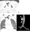
Fig. 2
29-year-old man with main bronchial diverticula.
A. 1.25 mm axial scan shows small air cyst (arrow) bulging medial side of left main bronchus.
B. 3D volume-rendered image shows two tiny bugling air cysts (arrow) in inferior wall of left main bronchus.
C. Virtual bronchoscopy image shows two holes (arrow) in left main bronchus.
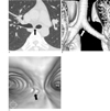
Fig. 3
64-year-old man with left main bronchial diverticula and SPN.
A. 5 mm thickness axial scan shows left main bronchial diverticulum (arrow).
B. In 8 months ago, same size of left main bronchial diverticulum (arrow) is seen on 5 mm thickness axial scan.
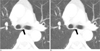
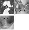
Fig. 4
52-year-old man with multiple main bronchial diverticula and lung cancer.
A. 1.25 mm axial image shows irregular air cyst (arrow) in left main bronchus.
B. Virtual bronchoscopy shows multiple holes (arrows) in both main bronchi.
C. Photograph of bronchoscopy shows multiple holes (arrows) in both main bronchi.

References
1. Goo JM, Im JG, Ahn JM, Moon WK, Chung JW, Park JH, et al. Right paratracheal air cysts in the thoracic inlet: clinical and radiologic significance. AJR Am J Roentgenol. 1999; 173(1):65–70.
2. Polverosi R, Carloni A, Poletti V. Tracheal and main bronchial diverticula: the role of CT. Radiol Med. 2008; 113(2):181–189.
3. Soto-Hurtado EJ, Peñuela-Ruiz L, Rivera-Sánchez I, Torres-Jiménez J. Tracheal diverticulum: a review of the literature. Lung. 2006; 184(6):303–307.
4. Sanford MF, Broderick LS. Multidetector computed tomography detection of bronchial diverticula. J Thorac Imaging. 2007; 22(3):265–267.
5. Endo S, Saito N, Hasegawa T, Sato Y, Sohara Y. Tracheocele: surgical and thoracoscopic findings. Ann Thorac Surg. 2005; 79(2):686–687.
6. Early EK, Bothwell MR. Congenital tracheal diverticulum. Otolaryngol Head Neck Surg. 2002; 127(1):119–121.
7. Ghaye B, Szapiro D, Fanchamps JM, Dondelinger RF. Congenital bronchial abnormalities revisited. Radiographics. 2001; 21(1):105–119.
8. Boiselle PM, Reynolds KF, Ernst A. Multiplanar and three-dimensional imaging of the central airways with multidetector CT. AJR Am J Roentgenol. 2002; 179(2):301–308.
9. Boiselle PM. Multislice helical CT of the central airways. Radiol Clin North Am. 2003; 41(3):561–574.
10. Park KJ, Bergin CJ, Clausen JL. Quantitation of emphysema with three-dimensional CT densitometry: comparison with two-dimensional analysis, visual emphysema scores, and pulmonary function test results. Radiology. 1999; 211(2):541–547.
11. Levin TL, Weingart L, Adam HM, Vicencio AG. Congenital HIV and tracheal diverticulosis. AJR Am J Roentgenol. 2004; 183(4):1115–1116.
12. Barbato A, Novello A Jr, Zanolin D, Corner P, Talenti E. Diverticulosis of the main bronchi: a rare cause of recurrent bronchopneumonia in a child. Thorax. 1993; 48(2):187–188.
13. Cluroe A, Beasley R, Holloway L. Bronchial diverticulitis: complication of bronchial asthma. J Clin Pathol. 1988; 41(8):921–922.
14. Wang NS, Ying WL. Morphogenesis of human bronchial diverticulum. A scanning electron microscopic study. Chest. 1976; 69(2):201–204.
15. MacKinnon D. Tracheal diverticula. J Pathol Bacteriol. 1953; 65(2):513–517.
16. Saito W, Kobayashi H, Motoyoshi K. A case of tracheobronchial diverticula. J Bronchol. 2003; 10:48–50.
 XML Download