

 PDF
PDF ePub
ePub Citation
 Print
Print


INTRODUCTION
Variations in branching pattern of the popliteal artery are common. Imaging that reveals these potential anatomical variants is vital to planning and successful execution of popliteal and tibial artery interventions such as transluminal angioplasty, thromboembolectomy, or reconstruction in the setting of occlusive disease or arterial injury (12). Several variants have been reported with the incidence ranging from 7.2% to 12% (3). Popliteal artery agenesis is one of the congenital arterial malformations around the knee. It is an embryonic tissue remnant of the arterial system following the arrest of the embryogenesis in its truncular stage, which failed to involute properly. To our knowledge, the only reported case of popliteal artery agenesis was discovered during operative exploration (4). Herein, the authors report one additional case of this anatomic variant detected by CT angiography in a 48-year-old male patient with intermittent claudication. The authors review imaging of this entity with an emphasis on the salient findings necessary to distinguish it from other causes of occlusive disease. In addition, the authors also review the embryologic development of the lower extremity vasculature. The institutional review board of our hospital approved this case report.
CASE REPORT
A 48-year-old man was admitted with intermittent pain in his right calf. He had a 10-pack-year smoking history and was recently diagnosed with diabetes mellitus. Initial laboratory examination showed the following values: total cholesterol, 235.9 mg/dL (normal range, less than 200.0 mg/dL); triglyceride, 185 mg/dL (normal range, less than 150 mg/dL); fasting glucose, 133 mg/dL (normal range, 70–120 mg/dL); Hb A1c, 7.5% A1c (normal range, 4.3–6.0% A1c). Physical examination demonstrated absent popilteal pulses bilaterally in the setting of normal femoral, posterior tibial, and dorsal pedal pulses. Resting ankle brachial index (ABI) was 1.14 on the right and 0.94 on the left.
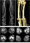
Lower extremity CT angiography demonstrated normal aortoiliac and femoral vessels. The popliteal arteries, however, were laterally deviated. They terminated into prominent muscular arteries as well as a cluster of well formed collaterals with distal reconstitution of the P3 segment (Fig. 1A, B). Axial CT angiographic images showed absent popliteal artery above the knee on both sides. Aberrant muscle or muscular attachment was not delineated in the popliteal fossa (Fig. 1C). In the setting of intermittent claudication in a patient with well-collateralized popliteal artery occlusions, we opted for continued follow-up over intervention.
DISCUSSION
Popliteal artery agenesis is an extremely rare anomaly with the only reported case in the literature describing unilateral popliteal artery agenesis discovered during operative exploration (4). Our case report describes bilateral popliteal artery agenesis of the P2 segment with extensive collateralization resulting in distal reconstitution of the P3 vessel.
Development of the lower extremity arterial system begins with the embryo of 9-mm in size and is completed by the third month (5). The lower extremity vessels arise from two sources: axial artery and the femoral artery. The axial artery, which is also called the sciatic artery or ischiopopliteal artery, is a continuation of the internal iliac artery communicating with the popliteal artery and the tibial arteries distally in the early embryo. It develops from the dorsal root of the umbilical artery and passes into the developing limb along its dorsal surface. Later, the femoral artery develops from the external iliac artery and descends on the ventral aspect of the developing limb. By the time the embryo is 14 mm, the femoral artery anastomoses with the sciatic artery at the level of the adductor canal and becomes the major arterial supply of the lower extremity. As the femoral artery starts developing, the proximal sciatic artery regresses while the middle and distal portions persist to become the popliteal and peroneal arteries respectively (5). The adult arterial system is completed by the third month of gestation. Popliteal artery agenesis, therefore, would be due to abnormal regression of the middle portion of the sciatic artery.
Imaging is necessary to evaluate for significant anatomic variants before surgery or interventions. Digital subtraction angiography (DSA) has long been the mainstay in the diagnosis of vascular disease but has the disadvantages of its invasiveness, radiation exposure, patient's discomfort and a low risk of complications (6). With the introduction of the multidetector technique, modern CT angiography can provide a minimally invasive and viable alternative to DSA for most vascular indications including vascular anomalies (7). CT can also demonstrate the anatomic relationship between popliteal artery and adjacent structures, which is beneficial in planning subsequent surgical intervention (8). In our case, CT with 3-dimensional angiographic reconstruction coupled with strategic cross sections provided exceptional clarity in diagnosing this rare anomaly.
Congenital anomalies of the popliteal artery are asymptomatic and usually discovered incidentally most often at autopsy because this congenital arterial agenesis gives rise to extensive collateral vessels sufficient to supply increased metabolic demands, as in our case. His recent onset of right sided unilateral intermittent claudication was fortuitous in triggering our imaging but is totally inconsistent with the excellent collateralization circumventing his bilateral popliteal agenesis. Although we are not sure of sudden development of his symptom, we suggest that it was not related to the agenesis, because his ABI was within normal limit, even higher on the right than on the left.
The popliteal artery can be affected by a unique set of pathologic conditions (9). It is important to be able to distinguish popliteal artery agenesis from atherosclerosis obliterans, popliteal artery entrapment syndrome and Buerger's disease. Atherosclerosis is the most common cause of popliteal artery occlusion or stenosis (9). In our case, the abundant collateral network suggested a chronic process; however, no atherosclerotic changes were noted throughout the arterial tree. Atherosclerotic occlusion exclusive to the popliteal artery is not likely. Popliteal artery entrapment syndrome is a developmental abnormality that results from an abnormal anatomic relationship between the popliteal artery and the gastrocnemius muscle (910). The abnormal position of the muscle causes medial deviation especially in type I to type III and compression or even occlusion of the artery (10). In our case, however, aberrant muscle or muscular attachment was not delineated in the popliteal fossa. Moreover, the P1 segment of the popliteal artery was deviated laterally. Buerger's disease is one of the most common types of arteritis affecting small and medium-sized arteries. There is a high association of the disease with smoking. On the contrary to popliteal artery agenesis embryologically involving P2 segment as in our case, the patients with Buerger's disease typically have occlusive disease in the distal femoral and popliteal arteries, extending into the proximal tibioperoneal vessels with well-developed collateral circulation (10).
In conclusion, popliteal artery agenesis is extremely rare, but should be included in the anatomic variations around the knee. Awareness of this variant is important for those performing surgical or percutaneous vascular reconstruction in the lower extremity. CT angiography can provide a minimally invasive and valuable tool for the evaluation of these anatomic variations.
 XML Download