

 PDF
PDF ePub
ePub Citation
 Print
Print


INTRODUCTION
Barium peritonitis is a rare but life-threatening complication associated with gastrointestinal (GI) contrast investigation. After the first report of barium peritonitis by Himmelmann (1) in 1932, the incidence of peritonitis following barium examination has been reported to be 0.2–0.8% (23). To date, there are about 30 reported cases of barium peritonitis following an upper GI series in the literature, which focused on clinical findings and management (3). However, to the best of our knowledge, there has been no report of barium peritonitis following an upper GI series that deals with radiologic findings. In this report, we described a rare case of barium peritonitis following an upper GI series with its imaging findings and facilitated a discussion on the prevention and management of this disease entity.
CASE REPORT
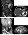
A 74-year-old female presented to the emergency department with a history of sudden abdominal pain immediately after swallowing half a cup of barium for an upper GI series at an outside hospital about three hours ago. During the examination, intraperitoneal barium leakage was identified, representing intestinal perforation. An upper GI series was planned for her general health check-up and she had no clinical symptom prior to the examination. The patient's past medical history was unremarkable. Unenhanced abdominopelvic computed tomography (CT) was performed at an outside hospital and she was diagnosed as having barium peritonitis. Physical examination, which was performed in our hospital, showed diffuse abdominal tenderness and involuntary abdominal guarding. Plain abdominal radiograph and imported outside unenhanced abdominopelvic CT images (Sensation 64 multi-detector scanner; Siemens Medical System, Erlangen, Germany) revealed extensive intraperitoneal contrast spillage with pneumoperitoneum (Fig. 1A, B). At emergency laparotomy, a perforated gastric ulcer was noted on the anterior wall of gastric antrum, along with barium contamination throughout the intraperitoneal cavity. Distal gastrectomy with loop gastrojejunostomy was performed, followed by vigorous peritoneal toilet with warm saline and removal of barium. However, complete removal of intraperitoneal barium contamination was impractical. Postoperatively, the patient was admitted to the surgical intensive care unit for three days and was then transferred to the department of internal medicine for ventilatory support for concurrent pneumonia. The plain abdominal radiograph and contrast-enhanced abdomen CT images (Discovery 750 HD 128 multi-detector scanner; GE Healthcare, Waukesha, WI, USA) obtained after the operation (Fig. 1C, D) demonstrated a significant quantity of barium left behind. Also, CT images revealed not only the residual barium adhering along the peritoneum but also diffuse mesenteric fat infiltration with ascites, suggesting barium peritonitis. She remained in the hospital for 152 days to maintain prolonged nutritional support. The pathologic specimen of the stomach was finally diagnosed as adenocarcinoma with perforation.
DISCUSSION
Although the frequency of investigation of the upper GI tract by ingestion of oral contrast material has decreased with wide-spread availability of endoscopy and development of CT gastroscopy, examination of the upper GI tract by barium ingestion is still performed as a screening tool due to its easy accessibility and safety. However, some complications of barium studies have been reported, including intestinal perforation, barium impaction and aspiration into the airway.
The relative risks of single and double contrast studies have not been reported, although, theoretically, double contrast study may have an increased risk of perforation due to greater gastric distension. However, for barium enema, previous studies have shown that there is no difference in the intraluminal pressure between double contrast and single column techniques (456). In addition, use of an intestinal relaxant has not been shown to have a significant effect on intraluminal pressure, although it decreased the spasm (56).
Most of the reported cases of intestinal perforation during barium enema might be related to some underlying disease or trauma including ischemia, inflammatory bowel disease, neoplasm, and prior endoscopic procedure that makes the bowel wall vulnerable to perforation (4). We think that this inference can be applied to an upper GI series as well as in our case. Therefore, careful review of the clinical history, such as endoscopic procedure including biopsy or endoscopic mucosal resection prior to the examination, and review of previous radiographs are important to prevent complications (4).
Because of the high morbidity and mortality associated with barium peritonitis (7), prompt recognition and management are vital for reducing the morbidity and mortality. When perforation occurs, spread of barium into the peritoneal cavity may be identified radiographically during the examination (4), and it was instantaneously diagnosed in our case. If perforation is clinically suspected, review of overhead radiographs or obtaining decubitus or upright films could be helpful (4). On subsequent abdominal plain radiography and CT, linear or aggregated barium with high density is seen outlining the parietal and visceral surfaces of the peritoneal cavity. Beam hardening artifact may be observed on a CT scan.
Barium contamination in the peritoneal cavity causes marked chemical peritonitis, which leads to exudation of extracellular fluid with albumin, resulting in hypovolemia and shock (4). Therefore, prompt fluid replacement is important. Moreover, spilled barium quickly agglomerates together and the clumps adhere to the parietal and visceral surfaces of the peritoneal cavity because of its mucosal coating properties, which cannot be easily removed (34). If consequent spillage of bowel contents occurs, it has known to be associated with poor outcome (8). Therefore, early laparotomy with thorough irrigation is considered as the first-line management and such management has been shown to diminish the severity of peritonitis and reduce morbidity and mortality (3). Also, consecutive surgical resection or repair of the perforated bowel should be performed (3). Postoperatively, critical care support, such as fluid balance and administration of broad-spectrum antibiotics, and nutritional support are required. In our case, the above mentioned surgical and medical management options were performed in a sequence and we assume that such type of management contributed to diminution of the intensity of peritonitis and led to a relatively better prognosis.
However, complete removal of barium is clinically impractical. The remaining barium clumps undergo stages of chemical inflammatory reaction including phagocytosis, fibrosis and subsequent adhesion causing small bowel obstruction (34). Postoperative small bowel obstruction has been reported in up to 30% of patients who survive barium peritonitis (9). Fortunately, our patient did not suffer from bowel obstruction during the recovery period, even though a significant quantity of barium was left behind after the operation. The prognosis of barium peritonitis has been regarded as poor and the mortality rate has been reported to be as high as 35–50% (47). Minimal barium spillage is associated with better prognosis.
In conclusion, we have presented a case of barium peritonitis following an upper GI series with its imaging findings. Because of its rarity and high mortality rate, we suggest that there is a need for clinical awareness about barium peritonitis following intestinal perforation as a possible complication of an upper GI series. Careful review of clinical history, previous endoscopic procedure, and preliminary radiographs could prevent this fatal complication. Once perforation occurs, prompt recognition and management should be performed to decrease the morbidity and mortality.
 XML Download