

 PDF
PDF ePub
ePub Citation
 Print
Print


INTRODUCTION
Normally, the ophthalmic artery (OA) arises from the supraclinoid segment of the internal carotid artery (ICA) and enters the orbit via the optic canal. Rarely, the OA originates from the cavernous segment of the ICA and enters the orbit via the superior orbital fissure (SOF); this variation is termed as persistent dorsal OA (1). Also, the middle meningeal artery, the anterior cerebral artery, the basilar artery, and the ICA bifurcation have rarely been observed as variations in the origin of OA (1234567).
The prevalence of persistent dorsal OA was 0.42%, and the prevalence of OA arising from the middle meningeal artery was 1.45% with a tendency toward right-side predominance (2).
To the best of our knowledge, persistent dorsal OA has not been described in the Korean literature. In this report, we present three cases of persistent dorsal OA arising from the cavernous segment of the ICA.
CASE REPORT
Case 1
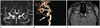
A 65-year-old woman with a history of dizziness underwent brain magnetic resonance (MR) imaging and MR angiography. She had no history of head trauma and there were no significant abnormal findings on physical examination. Maximum intensity projection and volume rendered three-dimensional time-of-flight MR angiography showed an anomalous artery arising from the cavernous segment of the right ICA (Fig. 1A, B). Source image of MR angiography showed the artery running anterolaterally and entering the orbit via the SOF (Fig. 1C).
MR image demonstrated no remarkable findings. At this time, the presence of a right persistent dorsal OA was noted. In the left ICA, the OA normally arose from the supraclinoid segment of the ICA and entered the orbit via the optic canal.
Case 2
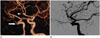
A 43-year-old man was hospitalized with a history of headache and mental change. Outside computed tomography (CT) showed subarachnoid hemorrhage and intraventricular hemorrhage. On outside brain CT angiography, a small outpouching lesion was noted on the anterior communicating artery, and an anomalous artery running anterolaterally to the cavernous segment of the right ICA (Fig. 2A).
To confirm the lesion, digital subtraction angiography was performed. Right internal carotid angiography showed an anomalous OA arising from the cavernous segment of the right ICA (Fig. 2B). Left internal carotid angiography showed an anterior communicating artery aneurysm and normal OA origin. Left pterional craniotomy with aneurysmal clipping was performed.
Case 3
A 67-year-old man presented with a history of headache. He had no history of head trauma and there were no specific findings on examinations including neurologic evaluation. CT angiography revealed an anomalous artery arising from the cavernous segment of the left ICA (Fig. 3A). Sagittal multiplanar reformatted images showed a persistent dorsal OA running inferolaterally, and entering the orbit via the SOF (Fig. 3B, C).
DISCUSSION
Embryogenesis of the OA is highly complex, as elegantly depicted by Padget (8) in detailed embryonic diagrams about 70 years ago. According to Padget (8), normal embryogenesis of the OA begins with emergence of the dorsal OA and the ventral OA at the 4–8 mm stage of fetal development. The dorsal OA and ventral OA are the two arteries supplying the orbit in the embryo (89).
But, the origin site and embryogenesis of OA are controversial. There are two main theories about the embryologic origin of the OA (8910). According to the theory proposed by Padget (8), the dorsal OA originates at the opposite side of the bifurcation of the primitive ICA (in front of the posterior communicating artery), giving off the hyaloid artery (future central retinal artery) and the lateral posterior ciliary arteries. The ventral OA will arise from the primitive ICA near the anterior choroidal artery origin, giving off the medial posterior ciliary arteries. In a later phase, the dorsal OA will migrate to an anterior position to the final adult OA position. The proximal ventral OA will regress and will not descend to the cavernous portion. The stapedial artery will give rise to the maxillomandibular artery and the supraorbital artery which will later enter the orbit through the SOF and fuse with the dorsal OA and ventral OA (810).
In contrast, Lasjaunias et al. (9) and Komiyama (10) proposed that the OA arises from the fusion of two primitive arteries: the dorsal OA and the ventral OA. The ventral OA arises from the anterior cerebral artery and will become the primitive OA, passing through the optic canal. The dorsal OA originates from the ICA at its horizontal portion in the cavernous segment and enters the orbit through the SOF. Later, two fusions occur, one between the ventral OA and the ICA and the other between the ventral OA and the dorsal OA. The process continues with a combination of regression and fusion that will lead to the final OA emerging from the supraclinoid segment of the ICA (910).
Normally, the OA arises from the supraclinoid segment of the ICA and enters the orbit via the optic canal. Rarely, the OA originates from the cavernous segment of the ICA and enters the orbit via SOF; this variation is termed as persistent dorsal OA (1). The middle meningeal artery is the most common ectopic origin of the OA. Other origins of the OA include the middle meningeal artery the anterior cerebral artery, the basilar artery, and the ICA bifurcation (123456). Extremely rarely, both primitive dorsal and ventral OAs persist and form double OAs that arise from the ICA (7).
A persistent dorsal OA is dangerous during surgery of the parasellar region, and it is important to be aware of the OA arising from the middle meningeal artery during transarterial embolization or infusion chemotherapy in the ECA territory because ischemic complication of the central retinal artery or retinal damage may occur (45). The occurrence of such an anatomical variation in the vasculature warrants important considerations for surgical procedures and vascular intervention (45). Thus, recognition and reporting of anomalous OA are important when evaluating CT and MR angiography, and digital subtraction angiography.
In conclusion, detailed knowledge of congenital vascular variations/anomalies is very important in planning and executing surgery and interventional procedures.

 XML Download