

 PDF
PDF ePub
ePub Citation
 Print
Print


Persistent trigeminal artery (PTA) is the rare anastomosis between the precavernous portion of the internal carotid artery (ICA) and basilar artery, with an incidence between 0.1% and 0.3% (1). Variation of PTA that originates directly from the precavernous portion of the ICA to the cerebellar arteries without joining the basilar artery is extremely rare (23). We presented the case of variation of PTA that was incidentally found directly terminating as the ipsilateral anterior inferior cerebellar artery (AICA) without joining the basilar artery.
CASE REPORT
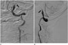
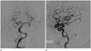
A 36-year-old man presented at an outside institution with acute onset dizziness and left facial numbness. The patient had no significant past or family medical history except gout for 1 year. He denied any history of recent head trauma. Diffusion weighted image indicated restricted diffusion consistent with acute infarction in the left lateral medulla and cerebellar hemisphere. He was subsequently transferred to our institution for further evaluation. Computed tomography angiography of the head and neck demonstrated occlusion of distal left vertebral artery and suspicious intimal flap, without other vascular abnormality. Digital subtraction angiography (DSA) was performed under suspicion of left vertebral artery dissection. DSA showed multifocal luminal irregularity and narrowing of the left distal vertebral artery, suggesting vertebral artery dissection (Fig. 1). Incidentally, the variation of PTA was identified arising from cavernous segment of the left ICA and running to the posterior fossa without joining the basilar artery. This anomalous artery supplied the anterior inferior part of the left cerebellar hemisphere, suggesting territories of the AICA. DSA of the bilateral vertebral artery showed absence of left AICA. The variation of PTA was contributed to the ipsilateral AICA without joining the basilar artery (Saltzman type IIIb) (Fig. 2). No other vascular abnormalities, including arteriovenous malformation and aneurysms of the circle of Willis, were encountered. The patient was treated conservatively with anticoagulants and antiplatelet agents for vertebral artery dissection. His symptoms gradually improved 6 weeks later.
DISCUSSION
According to the human embryogenesis of the carotid-basilar anastomoses described by Padget (4), the two longitudinal neural arteries are provided by four temporary anastomoses between carotid and the vertebrobasilar systems at approximately 35 days of gestational age i.e., the trigeminal, otic, hypoglossal and proatlantal arteries (4). In general, these anastomoses begin to regress during the formation of the posterior communicating artery and fusion of the paired longitudinal neural arteries into the basilar artery (4). Failure of regression of the fetal carotid-vertebrobasilar anastomoses results in their persistence into adulthood. The PTA is the most common carotid-vertebrobasilar anastomosis (1). Park et al. (5) reported a 0.14% approximate incidence of PTA variants on conventional cerebral angiography and magnetic resonance angiography (MRA); of the total 5 cases of PTA variants, 4 terminated directly as AICA without connection of the basilar artery (0.11%) and only 1 terminated directly as PICA. Usually, PTA originates from the precavernous ICA and joins the upper third of basilar artery. Haughton et al. (6) reported variants of PTA terminating as cerebellar arteries without joining basilar artery. During embryogenesis, incomplete fusion of the longitudinal neural arteries may disable a direct connection of the PTA with the basilar artery and results in its termination as one of the cerebellar arteries (67).
Saltzman (8) first described the angiographic appearance of PTA and its classification into three types according to angio-graphic appearance. The Saltzman type I PTA connects the basilar artery at the level between superior cerebellar artery (SCA) and AICA. The proximal basilar artery and posterior communicating artery are usually hypoplastic and subsequently both posterior cerebral arteries and SCAs are supplied through the PTA. The Saltzman type II PTA connects the basilar artery above the origin of the SCAs. The posterior communicating arteries are present and provide the posterior cerebral arteries. The Saltzman type III PTA is considered a combination of types I and II. Ali et al. (9) reviewed Saltzman classification and included variations of PTA in type III, in which there is no connection of the basilar artery. The variations of PTA type III arise from the internal carotid and terminate directly as the SCA (type IIIa), AICA (type IIIb), and PICA (type IIIc) without interposition of the basilar artery (9). The reported incidence of variations of PTA is extremely rare, with approximately 0.18% on DSA and 0.76% on MRA (10). Our case was consistent with type IIIb variation of PTA.
In almost every case, PTA and its variations have been found incidentally. Nevertheless, numerous vascular anomalies are associated with PTA, such as arteriovenous malformation, aneurysms of the circle of Willis, carotid and vertebral artery agenesis, carotid-cavernous fistula, and Moyamoya disease (39). In our case of PTA, aneurysm or other vascular malformation could not be detected and the correlation of vertebral artery dissection with PTA was unclear. However, neuroradiologists should be aware that PTA could be accompanied by other vascular anomalies.
In conclusion, we presented the extremely rare case of variation of PTA, which directly terminates in the AICA. Although PTA and variation of PTA are rare, appropriate diagnosis is necessary to avoid complications, such as unexpected hemorrhage and ischemia in brain stem and cerebellum due to vascular injury or uncontrolled emboli passing through the anomalous vessels during interventional radiologic procedures or head and neck surgeries.
 XML Download