

 PDF
PDF ePub
ePub Citation
 Print
Print


Adrenal myelolipoma is a rare, benign tumor that is commonly small and non-functioning. It generally comprises adipocytes and mature hematopoietic elements. It occurs unilaterally and originates from the adrenal cortices in most cases (1). Most adrenal myelolipomas are asymptomatic and detected incidentally by imaging modalities such as ultrasonography (US), computed tomography (CT), and magnetic resonance imaging (MRI) (12). However, in rare cases, they may be symptomatic when the tumor size is large enough to compress adjacent structures or when tumor rupture causes hemorrhage (13). Spontaneous retroperitoneal hemorrhaging from an adrenal myelolipoma is uncommon (1). Herein, we reported a case of adrenal myelolipoma with spontaneous retroperitoneal hemorrhage and briefly reviewed the relevant literature.
CASE REPORT
A 36-year-old male patient visited a local clinic with the complaint of right flank pain for 10 days prior; he was referred to our hospital for further evaluation and treatment. At presentation, his blood pressure was 150/80 mm Hg. Regular checkups for the previous 8 years had indicated high blood pressure for which, he had never taken hypertensive medication. Laboratory tests indicated that hemoglobin, aspartate aminotransferase, and alanine transaminase levels were 12.9 g/dL, 122 IU/L, and 145 IU/L, respectively. Renal function test results were unremarkable.
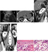
US and contrast-enhanced CT performed a week earlier at the outside hospital showed a right retroperitoneal mass. On US, a very large, well-circumscribed, heterogeneous, echogenic mass was visible in the right suprarenal area (Fig. 1A). Axial pre-contrast CT showed a heterogeneous, 11.9 × 10 cm diameter, fatcontaining mass between the right kidney and liver (Fig. 1B). The right adrenal gland could not be identified. The CT attenuation values of the tumor were -110 to -95 Hounsfield units (HU) and 45–60 HU for areas in the central portion of the mass, suggestive of recent hemorrhage. On sagittal reformatted contrast-enhanced CT, high attenuation areas were observed in the posterior of the mass extending inferiorly and partially around the right kidney, concordant with both intratumoral and peritumoral hemorrhages (Fig. 1C). Coronal and sagittal reformatted CT images showed an inferiorly displaced right kidney but intact upper pole (Fig. 1C, D). Retroperitoneal bleeding due to spontaneous rupture of a large adrenal myelolipoma was suspected on the basis of these findings.
The tumor was symptomatic and required differentiation from other retroperitoneal tumors; hence, an operation was performed a week later. The resected specimen revealed a well-circumscribed tumor with multiple hemorrhagic areas and fat tissue, measuring 12 × 13 × 4 cm. The tumor comprised mature adipose tissue, focal hematopoietic cells, and hemorrhagic and necrotic areas (Fig. 1E, F). These findings were histologically compatible with adrenal myelolipoma. The patient recovered without major complications and was discharged 2 weeks postoperatively.
DISCUSSION
Adrenal myelolipoma is an uncommon and asymptomatic tumor generally found incidentally in < 1% of the general population via autopsy or imaging performed for other purposes (4). It is a benign tumor comprising mature adipocytes interspersed with hematopoietic cells, resembling bone marrow (15). Adrenocortical metaplasia secondary to necrosis, infection, or stress is a possible precipitating factor in the development of adrenal myelolipoma (156).
The incidence of adrenal myelolipoma is similar between the sexes (7). It occurs in patients aged 40–70 years, although cases have been reported in all age groups (8). Although adrenal myelolipomas are usually small, they can range from several mm to > 30 cm in size (4). Of the 64 adrenal myelolipomas identified in a retrospective review of CT images, 9 (14%) were > 4 cm (9). Adrenal myelolipomas are not hormonally functional. However, very rare myelolipomatous foci are associated with adrenal functional disorders such as Cushing syndrome (13). These are usually asymptomatic unless they grow large (> 5 cm) enough to exert mass effect or cause internal hemorrhaging (13). Therefore, tumor size exceeding 7 cm is an indication for removal owing to mass effect and increased risk of bleeding (3). Hypertension may occur coincidentally or due to direct compression of the adjacent kidney or renal artery (7). Sudden onset pain, nausea, and vomiting are the commonly reported symptoms of acute internal bleeding from the tumor (2). In the present case, the patient complained of sudden onset right flank pain due to retroperitoneal hemorrhage.
Imaging is an integral part of the investigation of patients with symptomatic myelolipomas. On US, the appearance varies depending on the tissue component of the tumor i.e., it appears hyperechoic if it contains more fatty tissue, and hypoechoic if myeloid cells are predominant (4). The typical appearance of an adrenal myelolipoma on non-contrast CT images is an adrenal mass with mixed regions of soft tissue (20–50 HU) and fat (-115 to -30 HU) attenuation. Focal calcifications are commonly detected and may be single, multiple, or peripheral in distribution (7). After intravenous contrast medium administration, softtissue enhancement may be absent or significant (up to a 60 HU increase) according to the hematopoietic elements (7). Hemorrhage within the mass may enhance soft-tissue density and rapidly increase the tumor size (6). On MRI, the fatty component of adrenal myelolipomas exhibits increased signal intensity on T1-weighted images and decreased signal intensity on fat-saturated T2-weighted images. Opposed-phase imaging demonstrates low signal intensity in the voxels containing both fat tissue and water (3).
Differential diagnostic considerations for adrenal myelolipoma include other fat-containing retroperitoneal masses such as exophytic renal angiomyolipoma, retroperitoneal liposarcoma, and retroperitoneal teratoma (8). Invasive tissue sampling is rarely required for a definitive diagnosis. However, if imaging results are unclear, percutaneous needle biopsy may be required. A tissue sample exhibiting mature adipocytes and hematopoietic elements is suggestive for diagnosis (45).
Adrenal tumor associated with spontaneous hemorrhage is a very rare condition that includes metastases, pheochromocytomas and adrenocortical cancers (10). In case of benign tumors such as adrenal myelolipoma, spontaneous hemorrhage is even rarer with fewer than 15 cases reported, including the present case (1). In the present case, despite the retroperitoneal hemorrhage, there were no remarkable abnormalities on laboratory examination or life-threatening conditions besides right flank pain. However, large myelolipomas with acute hemorrhaging may present with anemia and hemodynamic compromise. Therefore, diagnosis as well as management of ruptured tumors should be performed earlier if a large hemorrhagic suprarenal mass is detected by imaging. Even small myelolipomas that are asymptomatic and < 5 cm in size, should be followed-up regularly with imaging such as CT or MRI in order to prevent an emergent condition (46).
Although adrenal myelolipoma presenting with spontaneous rupture is relatively rare, it should be suspected in patients with a large suprarenal mass containing fat and hemorrhages. Knowledge of imaging findings of ruptured adrenal myelolipomas is crucial for improving diagnostic accuracy and reducing mortality caused by spontaneous bleeding.
 XML Download