

 PDF
PDF ePub
ePub Citation
 Print
Print


Abstract
Purpose
To evaluate the variable factors affecting the results of percutaneous needle biopsies for infectious spondylitis.
Materials and Methods
In all, 249 patients who underwent both MRI and percutaneous needle biopsies due to a suspicion of infectious spondylitis were evaluated with respect to the following factors: the usage of antibiotics before the procedure, the location of the biopsy, the guiding equipment used, the experience level of the operators, and the number of biopsies performed.
Results
The positivity of culture in cases of treated with antibiotics (16.3%) before the biopsy was lower than in the untreated cases (30.5%) (p = 0.004). Biopsies performed at the abscess (43.5%) and with fluoroscopic guidance (27.8%) showed higher culture positivity as well. The experience level of the operators and the number of biopsies had no effect on culture positivity.
Figures and Tables
Fig. 1
A 70-year-old man with back pain.
A. A sagittal T2-weighted MR image shows the loss of fatty marrow signal intensity and subchondral bone destruction at the T11 and T12 vertebral bodies. A burst fracture of the T12 vertebral body is also noted.
B. Contrast-enhanced T1-weighted image shows diffuse enhancement of the T11 and T12 vertebral bodies and phlegmon at the anterior epidural space of the T12 level.
C. A percutaneous biopsy was performed at the T12 vertebral body under fluoroscopic guidance. The isolated organism was Staphylococcus aureus.
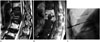
Fig. 2
A 68-year-old man with back pain.
A. The T2-weighted axial image shows an intraosseous abscess in the L4 vertebral body with an anterior epidural extension (arrowheads). There is also a direct extension into the left psoas muscle (arrow).
B. A percutaneous biopsy and aspiration were performed under CT guidance in the left psoas abscess. The isolated causative organism was Mycobacterium tuberculosis.
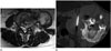
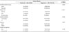
Table 1
Multivariate Logistic Regression Results for Culture Results
References
1. Butler JS, Shelly MJ, Timlin M, Powderly WG, O'Byrne JM. Nontuberculous pyogenic spinal infection in adults: a 12-year experience from a tertiary referral center. Spine (Phila Pa 1976). 2006; 31:2695–2270.
2. Hwang CM, Shin MJ, Kim SM, Lee SH, Lee SM, Shin JH, et al. The diagnostic usefulness of CT-guided needle biopsy or aspiration in infectious spondylitis. J Korean Radiol Soc. 2003; 48:497–504.
3. Rankine JJ, Barron DA, Robinson P, Millner PA, Dickson RA. Therapeutic impact of percutaneous spinal biopsy in spinal infection. Postgrad Med J. 2004; 80:607–609.
4. Sehn JK, Gilula LA. Percutaneous needle biopsy in diagnosis and identification of causative organisms in cases of suspected vertebral osteomyelitis. Eur J Radiol. 2012; 81:940–946.
5. Mylona E, Samarkos M, Kakalou E, Fanourgiakis P, Skoutelis A. Pyogenic vertebral osteomyelitis: a systematic review of clinical characteristics. Semin Arthritis Rheum. 2009; 39:10–17.
6. Bhagat S, Mathieson C, Jandhyala R, Johnston R. Spondylodiscitis (disc space infection) associated with negative microbiological tests: comparison of outcome of suspected disc space infections to documented non-tuberculous pyogenic discitis. Br J Neurosurg. 2007; 21:473–477.
7. Kim CJ, Song KH, Park WB, Kim ES, Park SW, Kim HB, et al. Microbiologically and clinically diagnosed vertebral osteomyelitis: impact of prior antibiotic exposure. Antimicrob Agents Chemother. 2012; 56:2122–2124.
8. Wall SD, Fisher MR, Amparo EG, Hricak H, Higgins CB. Magnetic resonance imaging in the evaluation of abscesses. AJR Am J Roentgenol. 1985; 144:1217–1221.
9. Runge VM, Williams NM, Lee C, Timoney JF. Magnetic resonance imaging in a spinal abscess model. Preliminary report. Invest Radiol. 1998; 33:246–225.
10. Thrush A, Enzmann D. MR imaging of infectious spondylitis. AJNR Am J Neuroradiol. 1990; 11:1171–1180.
11. Kwon JW, Yoon YC, Choi SH, Jung JY, Choe BK. MR imaging for the differentiation of early infectious spondylitis and modic type I change in the lumbar spine. J Korean Soc Radiol. 2010; 62:563–570.
12. Colmenero JD, Jiménez-Mejías ME, Sánchez-Lora FJ, Re-guera JM, Palomino-Nicás J, Martos F, et al. Pyogenic, tuberculous, and brucellar vertebral osteomyelitis: a descriptive and comparative study of 219 cases. Ann Rheum Dis. 1997; 56:709–715.
13. Hadjipavlou AG, Kontakis GM, Gaitanis JN, Katonis PG, Lander P, Crow WN. Effectiveness and pitfalls of percutaneous transpedicle biopsy of the spine. Clin Orthop Relat Res. 2003; 54–60.
14. Chew FS, Kline MJ. Diagnostic yield of CT-guided percutaneous aspiration procedures in suspected spontaneous infectious diskitis. Radiology. 2001; 218:211–214.
15. Marschall J, Bhavan KP, Olsen MA, Fraser VJ, Wright NM, Warren DK. The impact of prebiopsy antibiotics on pathogen recovery in hematogenous vertebral osteomyelitis. Clin Infect Dis. 2011; 52:867–872.
16. Yang SC, Fu TS, Chen LH, Chen WJ, Tu YK. Identifying pathogens of spondylodiscitis: percutaneous endoscopy or CT-guided biopsy. Clin Orthop Relat Res. 2008; 466:3086–3092.
17. Nam KH, Song GS, Han IH, Choi BK, Cha SH. Diagnostic value of biopsy techniques in lumbar spondylodiscitis: percutaneous needle biopsy and open biopsy. Korean J Spine. 2011; 8:267–271.
18. Mellado JM, Pérez del Palomar L, Camins A, Salvadó E, Ramos A, Saurí A. MR imaging of spinal infection: atypical features, interpretive pitfalls and potential mimickers. Eur Radiol. 2004; 14:1980–1989.
19. de Lucas EM, González Mandly A, Gutiérrez A, Pellón R, Martín-Cuesta L, Izquierdo J, et al. CT-guided fine-needle aspiration in vertebral osteomyelitis: true usefulness of a common practice. Clin Rheumatol. 2009; 28:315–320.
20. Kim BJ, Lee JW, Kim SJ, Lee GY, Kang HS. Diagnostic yield of fluoroscopy-guided biopsy for infectious spondylitis. AJNR Am J Neuroradiol. 2013; 34:233–238.
21. Michel SC, Pfirrmann CW, Boos N, Hodler J. CT-guided core biopsy of subchondral bone and intervertebral space in suspected spondylodiskitis. AJR Am J Roentgenol. 2006; 186:977–980.
 XML Download